Traumatic Dental Injuries: Concussions, Luxations, Fractures
•Download as PPT, PDF•
452 likes•154,064 views

The document provides information on traumatic injuries to teeth, including concussions, luxations, and fractures. It describes the clinical signs, radiographic findings, and treatment approaches for each type of injury. Concussions involve inflammation of the periodontal ligament without tooth displacement. Luxations occur when a tooth is displaced from its socket, sometimes with alveolar bone fractures. Fractures are classified as enamel fractures, enamel-dentin fractures, enamel-dentin-pulp fractures, or root fractures. Treatment depends on the specific injury but may include repositioning displaced teeth, pulpotomies, root canals, extractions, or orthodontic/surgical repositioning.

Recommended
More Related Content
What's hot
What's hot (20)
Viewers also liked
Viewers also liked (20)
Similar to Traumatic Dental Injuries: Concussions, Luxations, Fractures
Similar to Traumatic Dental Injuries: Concussions, Luxations, Fractures (20)
More from Chelsea Mareé
Recently uploaded
Recently uploaded (20)
Traumatic Dental Injuries: Concussions, Luxations, Fractures
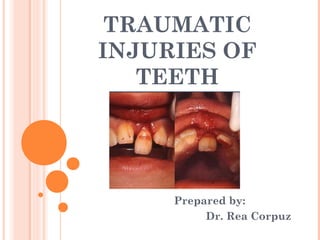
- 1. TRAUMATIC INJURIES OF TEETH Prepared by: Dr. Rea Corpuz
- 2. Traumatic Injuries of Teeth Case History Chief complaint History of present illness Medical History
- 3. Traumatic Injuries of Teeth Clinical Examination External Examination Soft Tissues Facial Skeleton Teeth and Supporting Structures
- 4. Traumatic Injuries of Teeth Radiographic Examination Periapical Occlusal Panoramic
- 5. Traumatic Injuries of Teeth (1) Concussion (2) Luxation (3) Fracture
- 6. Concussion tooth is not mobile not displaced periodontal ligament (PDL) absorbs injury + inflammed leaves tooth tender to biting pressure + percussion
- 7. Concussion Visual sign: not displaced Percussion test: tender to touch or tapping Mobility test: no increased mobility
- 8. Concussion Pulp Sensibility Test: positive result it is important in assessing future risk of healing complications lack of response to the test indicates an increased risk of later pulp necrosis
- 9. Concussion Radiographic findings: no radiographic abnormalities Radiographs: occlusal periapical lateral view from mesial + distal aspect of tooth in question
- 10. Concussion Treatment Objectives: usually there is no treatment Treatment: monitor pulpal condition for at least 1 year
- 11. Concussion Patient Instructions: soft food for 1 week brush with soft bristle rinse with chlorhexidine 0.1% to prevent plaque accumulation
- 12. Luxation tooth is displaced in a labial, lingual or lateral direction PDL is usually torn fractures of supporting alveolus may occur
- 13. Luxation similar to extrusion injuries partial or total separation of periodontal ligament
- 14. Luxation Visual sign: displaced, usually in a palatal/lingual or labial direction Percussion test: usually gives a metallic (ankylotic) sound Mobility test:
- 15. Luxation Pulp Sensibility Test: likely give a lack of response except for teeth with minor displacement test is important in assessing risk of healing complications positive result at the initial examination indicates a reduced risk of future pulp necrosis
- 16. Luxation Radiographic findings: widened periapical ligament space best seen on occlusal or eccentric exposures Radiographs: occlusal periapical lateral view from mesial + distal aspect of tooth in
- 17. Luxation Treatment Objective: reposition + splint a displaced tooth to facilitate pulp + periodontal ligament healing
- 18. Luxation Treatment: rinse the exposed part of root surface with saline before repositioning apply local anesthesia reposition tooth with forceps or with digital pressure to disengage it from its bony socket
- 19. Luxation Treatment: gently reposition it into its original position stabilize the tooth for 4 weeks using a flexible splint 4 weeks is indicated due to associated bone fracture
- 20. Luxation Patient Instructions: soft food for 1 week brush with soft bristle rinse with chlorhexidine 0.1% to prevent plaque accumulation
- 21. Fracture Ellis and Davey classification of crown fracture is useful in recording extent of damage to crown Class I – simple fracture of crown involving little or no dentin Class II – extensive fracture of crown involving considerable dentin but not dental pulp
- 22. Fracture Class III – extensive fracture of crown with an exposure of dental pulp Class IV – loss of entire crown
- 23. Fracture Enamel Fracture Enamel-Dentin Fracture Enamel-Dentin-Pulp Fracture Root Fracture
- 24. Enamel Fracture fracture confined to the enamel with loss of tooth structure
- 25. Enamel Fracture Visual sign: visible loss of enamel no visible sign of exposed dentin Percussion test: not tender if tenderness is observed evaluate tooth for a possible luxation or root fracture injury
- 26. Enamel Fracture Mobility test: normal mobility Sensibility test: usually positive test may be negative initially indicating transient pulpal damage
- 27. Enamel Fracture Sensibility test: monitor pulpal response until definitive pulpal diagnosis can be made test is important in assessing risk of future healing complications lack of response at initial examination indicates an increased risk of later pulpal necrosis
- 28. Enamel Fracture Radiographic findings: enamel lost is visible Radiographs: occlusal periapical recommended to rule out possible presence of root fracture or a luxation injury
- 29. Enamel Fracture Treatment: if tooth fragment is available, it can be bonded to the tooth grinding or restoration with composite resin depending on extent + location of fracture
- 30. Enamel-Dentin Fracture fracture confined to enamel + dentin with loss of tooth structure, but not involving pulp
- 31. Enamel-Dentin Fracture Visual sign: visible loss of enamel + dentin no visible sign of exposed pulp tissue Percussion test: not tender if tenderness is observed evaluate tooth for a possible luxation or root fracture injury
- 32. Enamel-Dentin Fracture Mobility test: normal mobility Sensibility test: usually positive test may be negative initially indicating transient pulpal damage
- 33. Enamel-Dentin Fracture Sensibility test: monitor pulpal response until definitive pulpal diagnosis can be made test is important in assessing risk of future healing complications lack of response at initial examination indicates an increased risk of later pulpal necrosis
- 34. Enamel-Dentin Fracture Radiographic findings: enamel-dentin lost is visible Radiographs: occlusal periapical recommended to rule out displacement or possible presence of root fracture
- 35. Enamel-Dentin Fracture Treatment: if tooth fragment is available, it can be bonded to the tooth otherwise perform provisional treatment by covering exposed dentin with glass ionomer or a permanent restoration using a bonding agent + composite resin
- 36. Enamel-Dentin-Pulp Fracture (Complicated Crown Fracture) a fracture involving enamel + dentin with loss of tooth structure + exposure of pulp
- 37. Enamel-Dentin-Pulp Fracture Visual sign: visible loss of enamel + dentin exposed pulp tissue Percussion test: not tender if tenderness is observed evaluate tooth for a possible luxation or root fracture injury
- 38. Enamel-Dentin-Pulp Fracture Mobility test: normal mobility Sensibility test: usually positive
- 39. Enamel-Dentin-Pulp Fracture Sensibility test: test is important in assessing risk of future healing complications lack of response at initial examination indicates an increased risk of later pulpal necrosis
- 40. Enamel-Dentin-Pulp Fracture Radiographic findings: lost of tooth substance is visible Radiographs: occlusal periapical recommended to rule out displacement or possible presence of luxation or root fracture
- 41. Enamel-Dentin-Pulp Fracture Treatment: if young patients with open apices, it is very important to preserve pulp vitality by pulp capping or partial pulpotomy in order to secure further root development this treatment is also treatment of choice in patients with closed apices
- 42. Enamel-Dentin-Pulp Fracture Treatment: Calcium hydroxide compunds + MTA are suitable materials for such procedures in older patients with closed apices + luxation injury with displacement, root canal treatment is usually treatment of choice
- 43. Crown-Root Fracture without pulp involvement fracture involving: enamel dentin cementum with loss of tooth structure but not exposing pulp
- 44. Crown-Root Fracture without pulp involvement Visual sign: crown fracture extending below gingival margin Percussion test: tender
- 45. Crown-Root Fracture without pulp involvement Mobility test: coronal fragment mobile Sensibility test: usually positive for apical fragment
- 46. Crown-Root Fracture without pulp involvement Radiographic findings: apical extension of fracture usually not visible Radiographs: occlusal periapical recommended to detect fracture lines in root cone beam exposure can reveal whole fracture extension
- 47. Crown-Root Fracture without pulp involvement Treatment: Fragment removal only • removal of superficial coronal crown-root fragment • subsequent restoration of exposed dentin above gingival level
- 48. Crown-Root Fracture without pulp involvement Treatment: Fragment removal + gingivectomy (sometimes ostectomy) • removal of coronal segment with subsequent endodontic treatment + restoration with a post-retained crown
- 49. Crown-Root Fracture without pulp involvement Treatment: Orthodontic extrusion of apical fragment • removal of coronal segment with subsequent endodontic treatment + orthodontic extrusion of remaining root with sufficient length after extrusion to support a post- retained crown
- 50. Crown-Root Fracture without pulp involvement Treatment: Surgical extrusion • removal of mobile fractured fragment • subsequent surgical repositioning of root in a more coronal position
- 51. Crown-Root Fracture without pulp involvement Treatment: Decoronation (root submergence) • implant solution is planned, root fragment may be left in situ after in order to avoid alveolar bone resorption • thereby maintaining volume of alveolar process for later implant installation
- 52. Crown-Root Fracture without pulp involvement Treatment: Extraction • with immediate or delayed implant-retained crown restoration or a coventional bridge • fractures with severe apical extension, the extreme being a vertical fracture
- 53. Crown-Root Fracture with pulp involvement fracture involving: enamel dentin cementum with loss of tooth structure exposure of pulp
- 54. Crown-Root Fracture with pulp involvement Visual sign: crown fracture extending below gingival margin Percussion test: tender
- 55. Crown-Root Fracture with pulp involvement Mobility test: coronal fragment mobile Sensibility test: usually positive for apical fragment
- 56. Crown-Root Fracture without pulp involvement Radiographic findings: apical extension of fracture usually not visible Radiographs: occlusal periapical cone beam exposure can reveal whole fracture extension
- 57. Crown-Root Fracture with pulp involvement Treatment: Fragment removal + gingivectomy (sometimes ostectomy) • removal of coronal segment with subsequent endodontic treatment + restoration with a post-retained crown
- 58. Crown-Root Fracture with pulp involvement Treatment: Orthodontic extrusion of apical fragment • removal of coronal segment with subsequent endodontic treatment + orthodontic extrusion of remaining root with sufficient length after extrusion to support a post- retained crown
- 59. Crown-Root Fracture with pulp involvement Treatment: Surgical extrusion • removal of mobile fractured fragment • subsequent surgical repositioning of root in a more coronal position
- 60. Crown-Root Fracture with pulp involvement Treatment: Decoronation (root submergence) • implant solution is planned, root fragment may be left in situ after in order to avoid alveolar bone resorption • thereby maintaining volume of alveolar process for later implant installation
- 61. Crown-Root Fracture with pulp involvement Treatment: Extraction • with immediate or delayed implant-retained crown restoration or a coventional bridge • fractures with severe apical extension, the extreme being a vertical fracture
- 62. Root Fracture fracture confined to the root of tooth involving: cementum dentin pulp
- 63. Root Fracture Visual sign: coronal segment may be mobile some cases displaced transient crown discoloration (red or gray) may occur bleeding from gingival sulcus may be noted
- 64. Root Fracture Percussion test: tooth may be tender Mobility test: coronal segment may be mobile
- 65. Root Fracture Sensibility test: the test is important in assessing risk of healing complications a positive sensibility test at the initial examination indicates a significantly reduced risk of later pulpal necrosis
- 66. Root Fracture Sensibility test: may give negative results initially indicating transient or permanent neural damage pulp sensibility test is usually negative for root fractures except for teeth with minor displacements
- 67. Root Fracture Radiographic findings: root fracture line is usually visible fracture involves root of the tooth in a horizontal or diagonal plane
- 68. Root Fracture Treatment: rinse exposed root surface with saline before repositioning if displaced, reposition the coronal segment of the tooth as soon as possible check that correct position has been reached radiographically
- 69. Root Fracture Treatment: stabilize the tooth with flexible splint for 4 weeks if the root fracture is near cervical area of the tooth stabilization is beneficial for a longer period of time (upto 4 months)
- 70. Root Fracture Treatment: monitor healing for at least 1 year to determine pulpal status if pulp necrosis develops, then root canal treatment of the coronal tooth segment to the fracture is indicated
- 71. References: Books McDonald, Avery et al: Dentistry for the Child and Adolescent • (pages 458-459) Internet http://www.dentaltraumaguide.org