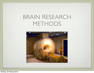
2. BRAIN RESEARCH METHODS
BRAINS ARE HARD TO STUDY BECAUSE THEY ARE
SURROUNDED BY A SCULL
IN THE PAST RESEARCH’S USED THE BRAINS OF DEAD PEOPLE
AND ANIMALS. BUT THIS HAD MAJOR LIMITATIONS!!
RESEARCH’S ALSO USED LIVING ANIMALS BUT THIS WAS
HIGHLY UNETHICAL.
IT HAS ONLY BEEN IN THE LATER HALF OF THE 20TH CENTURY
THAT RESEARCH’S HAVE HAD THE TECHNOLOGY TO STUDY
THE BRAIN EFFECTIVELY AND ETHICALLY.
Monday, 22 February 2010
3. CASE STUDIES
WE CAN’T GO BUSTING HEADS IN ORDER TO SEE WHAT
HAPPENS TO THE BRAIN. THIS IS UNETHICAL!
THE ETHICAL WAY TO GO ABOUT STUDYING A PERSON WITH
SOME DAMAGE TO THEIR BRAIN IS TO WAIT FOR THEM TO HAVE
AN ACCIDENT AND THEN HOPE THEY ARE PREPARED TO ALLOW
YOU TO STUDY THEM.
STUDYING MEANS DOING ALL SORTS OF MEASUREMENTS
LIKE, DIAGNOSTIC TESTS, INTERVIEWS, OBSERVATIONS, AND
LOOKING AT MEDICAL RECORDS
Monday, 22 February 2010
4. ADVANTAGES
• LOTS OF INDEPTH DETAIL
• RICH AMOUNT OF INFO
LIMITATIONS
• TAKE A LONG TIME TO GET INFORMATION
• INDIVIDUAL DIFFERENCES ARE NOT MEASURED
• PLASTICITY - THE BRAIN CAN ADAPT TO
• AS THEY ARE QUITE UNIQUE TO THE INDIVIDUAL, THE RESULTS CAN’T
BE GENERALISED FAIRLY
Monday, 22 February 2010
5. ELECTRICAL STIMULATION OF THE
BRAIN (ESB)
ELECTRICAL STIMULATION OF THE BRAIN INVOLVES USING AN
ELECTRODE TO DELIVER AN ELECTRIC CURRENT TO THE BRAIN,
THEN THAT PART OF THE BRAIN THAT RECEIVES THE CURRENT
RESPONDS ACCORDINGLY.
EXAMPLE: STIMULATING THE HYPOTHALAMUS IN ANIMALS
EVOKES FEEDING, DRINKING, SEXUAL AROUSAL AND
AGGRESSION DEPENDING ON THE AREA STIMULATED
Monday, 22 February 2010
6. ADVANTAGES
• CAN PINPOINT LOCATION AND FUNCTION OF CERTAIN BRAIN
STRUCTURES.
• CAN HELP IDENTIFY HEMISPHERIC SPECIALISATION
LIMITATIONS
• EXTREMELY INVASIVE - MUST APPLY DIRECTLY TO THE BRAIN
• CRUDE - NOT EASY TO TELL HOW FAR THE STIMULATION HAS SPREAD
• CAN’T ALWAYS BE GENERALISED TO ALL PEOPLE, AS ESB IS USUALLY
DONE ON NOT PROPERLY FUNCTIONING BRAINS
• CAN ONLY BE USED ON THOSE UNDERGOING BRAIN SURGERY
Monday, 22 February 2010
7. ELECTROENCEPHALOGRAPH (EEG)
THE EEG MEASURES ELECTRICAL ACTIVITY IN LOCALISED
AREAS OF THE BRAIN. ELECTRODES ARE ATTACHED TO THE
SURFACE OF THE SCALP AND THE ACTIVITY OF THE NEURONS
ARE MEASURED.
Monday, 22 February 2010
8. ADVANTAGES
• NON-INVASIVE
• INEXPENSIVE AND SAFE CAN BE USED ON EVERYONE
• CAN BE USED TO MEASURE OVER A LONG PERIOD OF TIME EG.
SLEEP
DISADVANTAGES
• GENERAL MEASURE OF ACTIVITY
• NOT AN IMAGING TECHNIQUE
• SKULL CAN INTERFERE WITH ELECTRODE READING
Monday, 22 February 2010
9. COMPUTERISED TOMOGRAPHY
(CT OR CAT )
CAT SCANS WERE THE FIRST NEURO-IMAGING TECHNIQUES
CAT SCANS TAKE A NUMBER OF X-RAYS FROM DIFFERENT
ANGLES TO PRODUCE A COMPTUTER IMAGE OF SLICES OF THE
BRAIN
Monday, 22 February 2010
10. ADVANTAGES
• NON-INVASIVE
• PROVIDE VERY DETAILED IMAGE OF LIVING BRAIN
• CAN FORM 3D IMAGE
DISADVANTAGES
• WILL ONLY SHOW STRUCTURE AND NOT FUNCTION
• EXPENSIVE AND NEEDS HIGHLY QUALIFIED STAFF
• MUST REMAIN STILL IN AN ENCLOSED SPACE
Monday, 22 February 2010
11. POSITRON EMISSION
TOMOGRAPHY
A PET CONSTRUCTS A COMPUTER IMAGE OF BRAIN FUNCTION AND
ACTIVITY. IT IS USED TO VIEW COGNITIVE AND BEHAVIORAL
ACTIVITY WHEN PATIENTS ARE GIVEN A RANGE OF TASKS. RED
INDICATES AREAS OF HIGH ACTIVITY, WHERE AS PURPLE
INDICATES AREAS OF LOW ACTIVITY. A HARMLESS RADIOACTIVE
SUBSTANCE IS INJECTED INTO THE PATIENT WHICH EMITS
SIGNALS WHEN PRESENT IN THE BRAIN. A COMPUTER THEN READS
THIS INFORMATION TO HELP COLOUR CODE THE IMAGE.
Monday, 22 February 2010
12. ADVANTAGES
• YOU CAN SEE HOW THE BRAIN FUNCTIONS
• YOU CAN SEE WHICH WHICH AREAS OF THE BRAIN ARE ACTIVE
FOR CERTAIN TYPES OF TASKS
• COLOR CODE MAKES THINGS EASY TO UNDERSTAND
LIMITATIONS
• NEED TO INJECT RADIOACTIVE MATERIAL INTO THE PERSON, SO
SESSIONS MUST BE KEPT SHORT
• ONLY ABLE TO VIEW THE BRAIN FOR A SHORT PERIOD OF TIME,
AS IT TAKES 30 SECONDS TO SCAN AND YOU MUST REST FOR
40 SECONDS BETWEEN SCANS
Monday, 22 February 2010
13. MAGNETIC RESONANCE IMAGING
MAGNETIC AND RADIO WAVES BOUNCE OFF NEURONS IN THE
BRAIN AND THESE VIBRATIONS ARE DETECTED BY A HUGE MAGNET
IN THE MACHINE AND FED INTO A COMPUTER. THE COMPUTER
THEN PROCESSEs THIS INFO AND CREATES A COLOUR CODED 3D
IMAGE. IT IS MORE DETAILED THAN A CT SCAN.
IT IS USED TO DIAGNOSE STRUCTURAL ABNORMALITIES AND
SMALL CHANGES OVER TIME. FOR EXAMPLE YOU CAN STUDY THE
BRAIN OF A SCHIZOPHRENIA PATIENT AND LOOK THE BRAIN
CELLS CHANGE OVER TIME. IT IS MORE EXACT THAN A CT SCAN.
Monday, 22 February 2010
14. ADVANTAGES
• NON INVASIVE
• DETAILED AND CLEAR IMAGES OF THE BRAIN.
LIMITATIONS
• THE INABILITY TO USE IT ON PEOPLE WITH METAL IN THEIR
BODIES
• DOES NOT SHOW YOU THE FUNCTIONING ON THE BRAIN.
• CONFINED SPACE A PROBLEM
Monday, 22 February 2010
15. FUNCTIONAL MAGNETIC
RESONANCE IMAGING (FMRI)
THE FMRI SHOWS HOW THE BRAIN IS FUNCTIONING BY
MONITORING THE LEVELS OF OXYGEN IN THE BRAIN. THE HIGHER
THE FUNCTIONING, THE HIGHER THE BLOOD FLOW.
THE MAJOR DIFFERENCE BETWEEN AN FMRI AND A PET IS THAT IT
CAN PICK UP CHANGES RAPIDLY, OVER SECONDS AS OPPOSED TO
MINUTES. FMRI HAS ALSO BECOME 3D IN RECENT YEARS.
Monday, 22 February 2010
16. ADVANTAGES
• NO RADIATION
• SHOWS FUNCTIONING OF BRAIN
• COLOUR CODING MAKES IT EASY TO UNDERSTAND
• CAN TRACK BRAIN CHANGES MOMENT TO MOMENT
LIMITATIONS
• ACTIVITY MAY BE A RESULT OF SOMETHING ELSE, OTHER THAN
THE TASK GIVEN
Monday, 22 February 2010
17. CLARIFICATIONS
INVASIVE: IN RELATION TO BRAIN RESEARCH, ANY TECHNIQUE
THAT INVOLVES OPENING THE BRAIN UP.
DIFFERENCE BETWEEN PET AND CT INJECTIONS.
• CT - THE PARTICIPANT MUST BE INJECTED WITH A SUBSTANCE
IN THE ARM OR HAND. THE SUBSTANCE IS BASED ON IODINE
AND IS NOT RADIOACTIVE
• PET - PRIOR TO THE PROCEDURE A RADIOACTIVE SUBSTANCE IS
IS INJECTED INTO THE PATIENTS BLOOD VESSEL.
Monday, 22 February 2010
19. PRACTICE EXAM QUESTIONS
Arthur has agreed to participate in a research study. In the study a small
amount of radioactive substance will be injected into his blood vessels and
transported to his brain. At the same time as the injection he will be asked to
think of as many words as he can beginning with the letter L.
Monday, 22 February 2010
20. PRACTICE EXAM QUESTIONS
Arthur has agreed to participate in a research study. In the study a small
amount of radioactive substance will be injected into his blood vessels and
transported to his brain. At the same time as the injection he will be asked to
think of as many words as he can beginning with the letter L.
What is this recording procedure known as?
Monday, 22 February 2010
21. PRACTICE EXAM QUESTIONS
Arthur has agreed to participate in a research study. In the study a small
amount of radioactive substance will be injected into his blood vessels and
transported to his brain. At the same time as the injection he will be asked to
think of as many words as he can beginning with the letter L.
What is this recording procedure known as?
A. EEG
Monday, 22 February 2010
22. PRACTICE EXAM QUESTIONS
Arthur has agreed to participate in a research study. In the study a small
amount of radioactive substance will be injected into his blood vessels and
transported to his brain. At the same time as the injection he will be asked to
think of as many words as he can beginning with the letter L.
What is this recording procedure known as?
A. EEG
B. PET
Monday, 22 February 2010
23. PRACTICE EXAM QUESTIONS
Arthur has agreed to participate in a research study. In the study a small
amount of radioactive substance will be injected into his blood vessels and
transported to his brain. At the same time as the injection he will be asked to
think of as many words as he can beginning with the letter L.
What is this recording procedure known as?
A. EEG
B. PET
C. CT
Monday, 22 February 2010
24. PRACTICE EXAM QUESTIONS
Arthur has agreed to participate in a research study. In the study a small
amount of radioactive substance will be injected into his blood vessels and
transported to his brain. At the same time as the injection he will be asked to
think of as many words as he can beginning with the letter L.
What is this recording procedure known as?
A. EEG
B. PET
C. CT
D. MRI
Monday, 22 February 2010
25. PRACTICE EXAM QUESTIONS
Arthur has agreed to participate in a research study. In the study a small
amount of radioactive substance will be injected into his blood vessels and
transported to his brain. At the same time as the injection he will be asked to
think of as many words as he can beginning with the letter L.
What is this recording procedure known as?
A. EEG
B. PET
C. CT
D. MRI
Monday, 22 February 2010
26. PRACTICE EXAM QUESTIONS
Arthur has agreed to participate in a research study. In the study a small
amount of radioactive substance will be injected into his blood vessels and
transported to his brain. At the same time as the injection he will be asked to
think of as many words as he can beginning with the letter L.
What is this recording procedure known as?
A. EEG
B. PET
C. CT
D. MRI
Monday, 22 February 2010
27. PRACTICE EXAM QUESTIONS
Arthur has agreed to participate in a research study. In the study a small
amount of radioactive substance will be injected into his blood vessels and
transported to his brain. At the same time as the injection he will be asked to
think of as many words as he can beginning with the letter L.
What is this recording procedure known as?
A. EEG
B. PET
C. CT
D. MRI
Monday, 22 February 2010
28. PRACTICE EXAM QUESTIONS
Arthur has agreed to participate in a research study. In the study a small
amount of radioactive substance will be injected into his blood vessels and
transported to his brain. At the same time as the injection he will be asked to
think of as many words as he can beginning with the letter L.
What is this recording procedure known as?
A. EEG
B. PET
C. CT
D. MRI
Monday, 22 February 2010
29. Arthur has agreed to participate in a research study. In the study a small amount of
radioactive substance will be injected into his blood vessels and transported to his
brain. At the same time as the injection he will be asked to think of as many words
as he can beginning with the letter L.
What is this recording procedure known as?
A. EEG
B. PET
C. CT
D. MRI
Answer: B. PET
Monday, 22 February 2010
30. The brain scanning methods that provide the most comprehensive information
on the functioning brain are _________ and __________.
A. PET; fMRI
B. CT; MRI
C. MRI; fMRI
D. CT; PET
Monday, 22 February 2010
31. The brain scanning methods that provide the most comprehensive information
on the functioning brain are _________ and __________.
A. PET; fMRI
B. CT; MRI
C. MRI; fMRI
D. CT; PET
Answer: A. PET; fMRI
Monday, 22 February 2010
36. One disadvantage of using an electroencephalograph (EEG) to investigate the
brain is that
Monday, 22 February 2010
37. One disadvantage of using an electroencephalograph (EEG) to investigate the
brain is that
A. it cannot be used with a range of patients such as infants.
Monday, 22 February 2010
38. One disadvantage of using an electroencephalograph (EEG) to investigate the
brain is that
A. it cannot be used with a range of patients such as infants.
B. it is invasive.
Monday, 22 February 2010
39. One disadvantage of using an electroencephalograph (EEG) to investigate the
brain is that
A. it cannot be used with a range of patients such as infants.
B. it is invasive.
C. it does not provide detailed information about brain function compared to
positron emission tomography (PET).
Monday, 22 February 2010
40. One disadvantage of using an electroencephalograph (EEG) to investigate the
brain is that
A. it cannot be used with a range of patients such as infants.
B. it is invasive.
C. it does not provide detailed information about brain function compared to
positron emission tomography (PET).
D. it is expensive compared to functional magnetic resonance imaging
(fMRI).
Monday, 22 February 2010
41. One disadvantage of using an electroencephalograph (EEG) to investigate the
brain is that
A. it cannot be used with a range of patients such as infants.
B. it is invasive.
C. it does not provide detailed information about brain function compared to
positron emission tomography (PET).
D. it is expensive compared to functional magnetic resonance imaging
(fMRI).
Monday, 22 February 2010
42. One disadvantage of using an electroencephalograph (EEG) to investigate the
brain is that
A. it cannot be used with a range of patients such as infants.
B. it is invasive.
C. it does not provide detailed information about brain function compared to
positron emission tomography (PET).
D. it is expensive compared to functional magnetic resonance imaging
(fMRI).
Monday, 22 February 2010
43. One disadvantage of using an electroencephalograph (EEG) to investigate the
brain is that
A. it cannot be used with a range of patients such as infants.
B. it is invasive.
C. it does not provide detailed information about brain function compared to
positron emission tomography (PET).
D. it is expensive compared to functional magnetic resonance imaging
(fMRI).
Monday, 22 February 2010
44. One disadvantage of using an electroencephalograph (EEG) to investigate the
brain is that
A. it cannot be used with a range of patients such as infants.
B. it is invasive.
C. it does not provide detailed information about brain function compared to
positron emission tomography (PET).
D. it is expensive compared to functional magnetic resonance imaging
(fMRI).
Monday, 22 February 2010
45. One disadvantage of using an electroencephalograph (EEG) to investigate the brain
is that
A. it cannot be used with a range of patients such as infants.
B. it is invasive.
C. it does not provide detailed information about brain function compared to
positron emission tomography (PET).
D. it is expensive compared to functional magnetic resonance imaging (fMRI).
C. it does not provide detailed information about brain function compared to
positron emission tomography (PET).
Monday, 22 February 2010
46. Which of the following is incorrect about computerised tomography (CT)
scans?
A. CT scans provide information about the function and extent of damage to
the brain.
B. The risks to the patient associated with CT scans are negligible.
C. CT scans usually require an injection into the bloodstream.
D. CT scans use X-rays taken at various angles to create an image of the brain.
Monday, 22 February 2010
47. Which of the following is incorrect about computerised tomography (CT)
scans?
A. CT scans provide information about the function and extent of damage to
the brain.
B. The risks to the patient associated with CT scans are negligible.
C. CT scans usually require an injection into the bloodstream.
D. CT scans use X-rays taken at various angles to create an image of the brain.
A. CT scans provide information about the function and extent of damage
to the brain.
Monday, 22 February 2010
48. A PET scan provides information
A. in very high resolution pictures that show brain structure and function.
B. in a colour-coded map that reveals areas of high activity in the brain.
C. about abnormalities in the structure of the brain.
D. about the number of cognitive tasks that were performed correctly.
Monday, 22 February 2010
49. A PET scan provides information
A. in very high resolution pictures that show brain structure and function.
B. in a colour-coded map that reveals areas of high activity in the brain.
C. about abnormalities in the structure of the brain.
D. about the number of cognitive tasks that were performed correctly.
B. in a colour-coded map that reveals areas of high activity in the brain.
Monday, 22 February 2010
51. 1. In terms of the potential risks to participants, describe one disadvantage of
PET scanning in comparison with CT scanning. (1 mark)
Monday, 22 February 2010
52. 1. In terms of the potential risks to participants, describe one disadvantage of
PET scanning in comparison with CT scanning. (1 mark)
Monday, 22 February 2010
53. 1. In terms of the potential risks to participants, describe one disadvantage of
PET scanning in comparison with CT scanning. (1 mark)
Monday, 22 February 2010
54. 1. In terms of the potential risks to participants, describe one disadvantage of
PET scanning in comparison with CT scanning. (1 mark)
Monday, 22 February 2010
55. 1. In terms of the potential risks to participants, describe one disadvantage of
PET scanning in comparison with CT scanning. (1 mark)
2. If a person has a metallic implant in their body, such as a heart pacemaker
or a pin in a bone, they are advised not to undertake a certain brain scanning
technique. What is the name of this technique? (1 mark)
Monday, 22 February 2010
56. 1. In terms of the potential risks to participants, describe one disadvantage of
PET scanning in comparison with CT scanning. (1 mark)
2. If a person has a metallic implant in their body, such as a heart pacemaker
or a pin in a bone, they are advised not to undertake a certain brain scanning
technique. What is the name of this technique? (1 mark)
Monday, 22 February 2010
57. 1. In terms of the potential risks to participants, describe one disadvantage of
PET scanning in comparison with CT scanning. (1 mark)
2. If a person has a metallic implant in their body, such as a heart pacemaker
or a pin in a bone, they are advised not to undertake a certain brain scanning
technique. What is the name of this technique? (1 mark)
Monday, 22 February 2010
58. 1. In terms of the potential risks to participants, describe one disadvantage of PET
scanning in comparison with CT scanning. (1 mark)
The PET scan:
• is more invasive
• involves injecting a radioactive substance into the body.
2. If a person has a metallic implant in their body, such as a heart pacemaker
or a pin in a bone, they are advised not to undertake a certain brain scanning
technique. What is the name of this technique? (1 mark)
MRI or fMRI scanning
Monday, 22 February 2010
59. In positron emission tomography (PET) scans, a substance is tagged with a radioactive marker and then monitored. This substance is a form of
A. iodine.
B. kryptonite.
C. uranium.
D. glucose.
D. glucose.
Which procedure results in a high-quality three-dimensional picture of the brain?
A. functional magnetic resonance imaging (fMRI) scan
B. electrical stimulation of the brain (ESB) scan
C. positron emission topography (PET) scan
D. electroencephalograph (EEG) scan
Which two procedures allow researchers to view changes in brain activity over time?
A. positron emission topography (PET) scan and computerised tomography (CT) scan
B. positron emission topography (PET) scan and functional magnetic resonance imaging (fMRI) scan
C. magnetic resonance imaging (MRI) scan and functional magnetic resonance imaging (fMRI) scan
D. computerised tomography (CT) scan and magnetic resonance imaging (MRI) scan
Monday, 22 February 2010
61. a. What information about the brain does positron emission tomography (PET) provide? (1 mark)
Monday, 22 February 2010
62. a. What information about the brain does positron emission tomography (PET) provide? (1 mark)
Monday, 22 February 2010
63. a. What information about the brain does positron emission tomography (PET) provide? (1 mark)
the PET scan shows which areas of the brain are active while certain tasks are performed
Monday, 22 February 2010
64. a. What information about the brain does positron emission tomography (PET) provide? (1 mark)
the PET scan shows which areas of the brain are active while certain tasks are performed
Monday, 22 February 2010
65. a. What information about the brain does positron emission tomography (PET) provide? (1 mark)
the PET scan shows which areas of the brain are active while certain tasks are performed
• the PET scan gives information about brain functioning.
Monday, 22 February 2010
66. a. What information about the brain does positron emission tomography (PET) provide? (1 mark)
the PET scan shows which areas of the brain are active while certain tasks are performed
• the PET scan gives information about brain functioning.
Monday, 22 February 2010
67. a. What information about the brain does positron emission tomography (PET) provide? (1 mark)
the PET scan shows which areas of the brain are active while certain tasks are performed
• the PET scan gives information about brain functioning.
Monday, 22 February 2010
68. a. What information about the brain does positron emission tomography (PET) provide? (1 mark)
the PET scan shows which areas of the brain are active while certain tasks are performed
• the PET scan gives information about brain functioning.
b. When compared to computerised tomography (CT), outline one advantage of using positron emission tomography
(PET). (1 mark)
Monday, 22 February 2010
69. a. What information about the brain does positron emission tomography (PET) provide? (1 mark)
the PET scan shows which areas of the brain are active while certain tasks are performed
• the PET scan gives information about brain functioning.
b. When compared to computerised tomography (CT), outline one advantage of using positron emission tomography
(PET). (1 mark)
Advantages of PET include:
Monday, 22 February 2010
70. a. What information about the brain does positron emission tomography (PET) provide? (1 mark)
the PET scan shows which areas of the brain are active while certain tasks are performed
• the PET scan gives information about brain functioning.
b. When compared to computerised tomography (CT), outline one advantage of using positron emission tomography
(PET). (1 mark)
Advantages of PET include:
• CT is static while PET shows the brain in action
Monday, 22 February 2010
71. a. What information about the brain does positron emission tomography (PET) provide? (1 mark)
the PET scan shows which areas of the brain are active while certain tasks are performed
• the PET scan gives information about brain functioning.
b. When compared to computerised tomography (CT), outline one advantage of using positron emission tomography
(PET). (1 mark)
Advantages of PET include:
• CT is static while PET shows the brain in action
• CT shows structural features and location of damage while PET shows functional areas
Monday, 22 February 2010
72. a. What information about the brain does positron emission tomography (PET) provide? (1 mark)
the PET scan shows which areas of the brain are active while certain tasks are performed
• the PET scan gives information about brain functioning.
b. When compared to computerised tomography (CT), outline one advantage of using positron emission tomography
(PET). (1 mark)
Advantages of PET include:
• CT is static while PET shows the brain in action
• CT shows structural features and location of damage while PET shows functional areas
• PET is more sensitive than CT in detecting areas of brain damage
Monday, 22 February 2010
73. a. What information about the brain does positron emission tomography (PET) provide? (1 mark)
the PET scan shows which areas of the brain are active while certain tasks are performed
• the PET scan gives information about brain functioning.
b. When compared to computerised tomography (CT), outline one advantage of using positron emission tomography
(PET). (1 mark)
Advantages of PET include:
• CT is static while PET shows the brain in action
• CT shows structural features and location of damage while PET shows functional areas
• PET is more sensitive than CT in detecting areas of brain damage
• unlike CT scans, PET scans are colour-coded, which aids interpretation.
Monday, 22 February 2010
74. a. What information about the brain does positron emission tomography (PET) provide? (1 mark)
the PET scan shows which areas of the brain are active while certain tasks are performed
• the PET scan gives information about brain functioning.
b. When compared to computerised tomography (CT), outline one advantage of using positron emission tomography
(PET). (1 mark)
Advantages of PET include:
• CT is static while PET shows the brain in action
• CT shows structural features and location of damage while PET shows functional areas
• PET is more sensitive than CT in detecting areas of brain damage
• unlike CT scans, PET scans are colour-coded, which aids interpretation.
Monday, 22 February 2010
75. a. What information about the brain does positron emission tomography (PET) provide? (1 mark)
the PET scan shows which areas of the brain are active while certain tasks are performed
• the PET scan gives information about brain functioning.
b. When compared to computerised tomography (CT), outline one advantage of using positron emission tomography
(PET). (1 mark)
Advantages of PET include:
• CT is static while PET shows the brain in action
• CT shows structural features and location of damage while PET shows functional areas
• PET is more sensitive than CT in detecting areas of brain damage
• unlike CT scans, PET scans are colour-coded, which aids interpretation.
This question demanded a speci7ic comparison; however, many students mistakenly compared PET scans with
other forms of brain‐imaging.
Monday, 22 February 2010
76. a. What information about the brain does positron emission tomography (PET) provide? (1 mark)
the PET scan shows which areas of the brain are active while certain tasks are performed
• the PET scan gives information about brain functioning.
b. When compared to computerised tomography (CT), outline one advantage of using positron emission tomography
(PET). (1 mark)
Advantages of PET include:
• CT is static while PET shows the brain in action
• CT shows structural features and location of damage while PET shows functional areas
• PET is more sensitive than CT in detecting areas of brain damage
• unlike CT scans, PET scans are colour-coded, which aids interpretation.
This question demanded a speci7ic comparison; however, many students mistakenly compared PET scans with
other forms of brain‐imaging.
Monday, 22 February 2010
78. c. What is the difference in the information provided by magnetic resonance imaging (MRI) and functional
magnetic resonance imaging (fMRI)? (1 mark)
Monday, 22 February 2010
79. c. What is the difference in the information provided by magnetic resonance imaging (MRI) and functional
magnetic resonance imaging (fMRI)? (1 mark)
DIFFERENCES INCLUDE:
Monday, 22 February 2010
80. c. What is the difference in the information provided by magnetic resonance imaging (MRI) and functional
magnetic resonance imaging (fMRI)? (1 mark)
DIFFERENCES INCLUDE:
• fMRI uses a magnetic field to create a three dimensional image of the brain where any two dimensional slice can be
selected, and also displays brain activity on the screen
Monday, 22 February 2010
81. c. What is the difference in the information provided by magnetic resonance imaging (MRI) and functional
magnetic resonance imaging (fMRI)? (1 mark)
DIFFERENCES INCLUDE:
• fMRI uses a magnetic field to create a three dimensional image of the brain where any two dimensional slice can be
selected, and also displays brain activity on the screen
• MRI shows only the structure of the brain.
Monday, 22 February 2010
82. c. What is the difference in the information provided by magnetic resonance imaging (MRI) and functional
magnetic resonance imaging (fMRI)? (1 mark)
DIFFERENCES INCLUDE:
• fMRI uses a magnetic field to create a three dimensional image of the brain where any two dimensional slice can be
selected, and also displays brain activity on the screen
• MRI shows only the structure of the brain.
Monday, 22 February 2010