

Recommended
Recommended
More Related Content
What's hot
What's hot (20)
Similar to Acute Cholecystitis
Similar to Acute Cholecystitis (20)
More from Jack Frost
More from Jack Frost (20)
Recently uploaded
Recently uploaded (20)
Acute Cholecystitis
- 1. Dave Jay S. Manriquez RN. Acute Cholecystitis I. Introduction Cholecystitis is an inflammation of the gallbladder wall and nearby abdominal lining. Cholecystitis is usually caused by a gallstone in the cystic duct, the duct that connects the gallbladder to the hepatic duct. The presence of gallstones in the gallbladder is called cholelithiasis. Cholelithiasis is the pathologic state of stones or calculi within the gallbladder lumen. A common digestive disorder worldwide, the annual overall cost of cholelithiasis is approximately $5 billion in the United States, where 7580% of gallstones are of the cholesterol type, and approximately 1025% of gallstones are bilirubinate of either black or brown pigment. In Asia, pigmented stones predominate, although recent studies have shown an increase in cholesterol stones in the Far East. Gallstones are crystalline structures formed by concretion (hardening) or accretion (adherence of particles, accumulation) of normal or abnormal bile constituents. According to various theories, there are four possible explanations for stone formation. First, bile may undergo a change in composition. Second, gallbladder stasis may lead to bile stasis. Third, infection may predispose a person to stone formation. Fourth, genetics and demography can affect stone formation. Risk factors associated with development of gallstones include heredity, obesity, rapid weight loss, through diet or surgery, age over 60, Native American or Mexican American racial makeup, female gender where gallbladder disease is more common in women than in men. Women with high estrogen levels, as a result of pregnancy, hormone replacement therapy, or the use of birth control pills, are at particularly high risk for gallstone formation. Diet with very low calorie diets, prolonged fasting, and lowfiber/highcholesterol/highstarch diets all may contribute to gallstone formation. Sometimes, persons with gallbladder disease have few or no symptoms. Others, however, will eventually develop one or more of the following symptoms; (1) Frequent bouts of indigestion, especially after eating fatty or greasy foods, or certain vegetables such as cabbage, radishes, or pickles, (2) Nausea and bloating (3) Attacks of sharp pains in the upper right part of the abdomen. This pain occurs when a gallstone causes a blockage that prevents the gallbladder from emptying (usually by obstructing the cystic duct). (4) Jaundice (yellowing of the skin) may occur if a gallstone becomes stuck in the common bile duct, which leads into the intestine blocking the flow of bile from both the gallbladder and the liver. This is a serious complication and usually requires immediate treatment. The only treatment that cures gallbladder disease is surgical removal of the gallbladder, called cholecystectomy. Generally, when stones are present and causing symptoms, or when the gallbladder is infected and inflamed, removal of the organ is usually necessary. When the
- 2. gallbladder is removed, the surgeon may examine the bile ducts, sometimes with Xrays, and remove any stones that may be lodged there. The ducts are not removed so that the liver can continue to secrete bile into the intestine. Most patients experience no further symptoms after cholecystectomy. However, mild residual symptoms can occur, which can usually be controlled with a special diet and medication. II. Epidemiology Frequency An estimated 1020% of Americans have gallstones, and as many as one third of these people develop acute cholecystitis. Cholecystectomy for either recurrent biliary colic or acute cholecystitis is the most common major surgical procedure performed by general surgeons, resulting in approximately 500,000 operations annually. Cholelithiasis, the major risk factor for cholecystitis, has an increased prevalence among people of Scandinavian descent, Pima Indians, and Hispanic populations, whereas cholelithiasis is less common among individuals from subSaharan Africa and Asia. Mortality/Morbidity Most patients with acute cholecystitis have a complete remission within 14 days. However, 25 30% of patients either require surgery or develop some complication. Patients with acalculous cholecystitis have a mortality rate ranging from 1050%, which far exceeds the expected 4% mortality rate observed in patients with calculous cholecystitis. Emphysematous cholecystitis has a mortality rate approaching 15%. Perforation occurs in 1015% of cases. Race Pima Indian and Scandinavian people have the highest prevalence of cholelithiasis and, consequently, cholecystitis. Populations at the lowest risk reside in subSaharan Africa and Asia. In the United States, white people have a higher prevalence than black people. Sex Gallstones are 23 times more frequent in females than in males, resulting in a higher incidence of calculous cholecystitis in females. Elevated progesterone levels during pregnancy may cause biliary stasis, resulting in higher rates of gallbladder disease in pregnant females. Acalculous cholecystitis is observed more often in elderly men. Age The incidence of cholecystitis increases with age. The physiologic explanation for the increasing incidence of gallstone disease in the elderly population is unclear. The increased incidence in elderly men has been linked to changing androgentoestrogen ratios.
- 3. III. Anatomy and Physiology Gallbladder, a muscular organ that serves as a reservoir for bile, is usually present in most vertebrates. In humans, it is a pearshaped membranous sac on the undersurface of the right lobe of the liver just below the lower ribs. It is generally about 7.5 cm (about 3 in) long and 2.5 cm (1 in) in diameter at its thickest part; it has a capacity varying from 1 to 1.5 fluid ounces. The body (corpus) and neck (collum) of the gallbladder extend backward, upward, and to the left. The wide end (fundus) points downward and forward, sometimes extending slightly beyond the edge of the liver. Structurally, the gallbladder consists of an outer peritoneal coat (tunica serosa); a middle coat of fibrous tissue and unstriped muscle (tunica muscularis); and an inner mucous membrane coat (tunica mucosa) The function of the gallbladder is to store bile, secreted by the liver and transmitted from that organ via the cystic and hepatic ducts, until it is needed in the digestive process. The gallbladder, when functioning normally, empties through the biliary ducts into the duodenum to aid digestion by promoting peristalsis and absorption, preventing putrefaction, and emulsifying fat. Digestion of fat occurs mainly in the small intestine, by pancreatic enzymes called lipases. The purpose of bile is to; help the lipases to work, by emulsifying fat into smaller droplets to increase access for the enzymes, Enable intake of fat, including fatsoluble vitamins: Vitamin A, D, E, and K, rid the body of surpluses and metabolic wastes cholesterol and bilirubin.
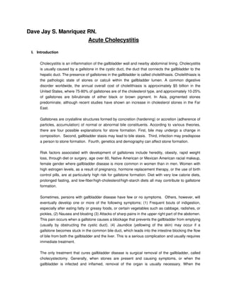
- 4. IV. Pathophysiology RISK FACTORS Heredity Obesity Rapid Weight Loss, through diet or surgery Age over 60 Bile must become The solute precipitate Crystals must come supersaturated with from solution as solid together and fuse to cholesterol and calcium crystals form stones Gallstones Obstruction of the cystic duct and common bile duct Sharp pain in the right part Jaundice of the abdomen Distention of the gall bladder Localized cellular Venous and lymphatic Areas of ischemia may Proliferation of bacteria irritation or infiltration or drainage is impaired occur both take place Inflammation of the gall bladder CHOLECYSTITIS
- 5. V. Signs and Symptoms • intense and sudden pain in the upper right part of the abdomen • recurrent painful attacks for several hours after meals • pain (often worse with deep breaths, and extending to the lower part of the right shoulder blade) • nausea • vomiting • rigid abdominal muscles on right side • slight fever • chills • jaundice yellowing of the skin and eyes • loose, lightcolored bowel movements • abdominal bloating VI. Diagnostic and Laboratory Procedures a. Complete Blood Count This is to determine blood components and the response to inflammatory process and streptococcal infection b. Fasting Blood Sugar This is to measure the blood glucose levels c. Creatinine This is the indicator of the renal function d. Blood Urea Nitrogen (BUN) This is an indicator of renal function and perfusion, dietary intake of protein and the level of protein metabolism e. Urinalysis Urinalysis yields a large amount of information about possible disorders of the kidney and lower urinary tract, and systematic disorders that alter urine composition. Urinalysis data include color, specific gravity, pH, and the presence of protein, RBC’s, WBC’s, bacteria, leukocyte, esterase, bilirubin, glucose, ketones, casts and crystals f. Chest Xray This is used to rule out respiratory causes of referred pain. g. Ultrasound/Sonography
- 6. A diagnostic imaging technique which uses highfrequency sound waves to create an image of the internal organs. Ultrasounds are used to view internal organs of the abdomen such as the liver spleen, and kidneys and to assess blood flow through various vessels. h. Hepatobiliary Scintigraphy An imaging technique of the liver, bile ducts, gallbladder, and upper part of the small intestine. i. Cholangiography Xray examination of the bile ducts using an intravenous (IV) dye (contrast). j. Percutaneous Transhepatic Cholangiography (PTC) A needle is introduced through the skin and into the liver where the dye (contrast) is deposited and the bile duct structures can be viewed by xray. k. Endoscopic Retrograde Cholangiopancreatography (ERCP) A procedure that allows the physician to diagnose and treat problems in the liver, gallbladder, bile ducts, and pancreas. The procedure combines xray and the use of an endoscope. A long, flexible, lighted tube is used. The scope is guided through the patient's mouth and throat, then through the esophagus, stomach, and duodenum. The physician can examine the inside of these organs and detect any abnormalities. A tube is then passed through the scope, and a dye is injected which will allow the internal organs to appear on an xray. l. Computed Tomography Scan (CT Scan) A diagnostic imaging procedure using a combination of xrays and computer technology to produce crosssectional images (often called slices), both horizontally and vertically, of the body. A CT scan shows detailed images of any part of the body, including the bones, muscles, fat, and organs. CT scans are more detailed than general xrays. VII. Medical / Surgical Interventions 1. Intake and Output – I&O measurement provide another means of assessing fluid balance. This data provide insight into the cause of imbalance such as decrease fluid intake or increase fluid loss. This measurement is not that accurate as body weight, however, because of relative risk of errors in recording. 2. Electrocardiogram – The ECG is an essential tool in evaluating cardiac rhythm. Electrocardiography detects and amplifies the very small electrical potential changes
- 7. between different points on the surface of the body as a myocardial cell depolarize and repolarize, causing the heart to contract. 3. O2 Inhalation – Oxygen therapies are used to provide more oxygen to the body into order to promote healing and health. 4. Intravenous Rehydration – when the fluid loss is severe or life threatening, intravenous (IV) fluids are used for replacement. 5. Cholecystectomy – removal of the gallbladder. This procedure may be performed to treat chronic or acute cholecystitis, with or without cholelithiasis, to remove a malignancy or to remove polyps. 6. Cholecystotomy – the establishment of an opening into the gallbladder to allow drainage of the organ and removal of stones. A tube is then placed in the gallbladder to established external drainage. This is performed when the patient cannot tolerate cholecystectomy. 7. Choledochoscopy – the insertion of a choledochoscope into the common bile duct in order to directly visualize stones and facilitate their extraction. VIII. Nursing Management A. Pain Management ACTIONS / INTERVENTIONS RATIONALE >assists in differentiating cause of pain and 1. Observe and document location, severity (0–10 provides information about disease scale), and character of pain (e.g., steady, progression/resolution, development of intermittent, colicky). complications, and effectiveness of interventions >severe pain not relieved by routine measures 2. Note response to medication, and report to may indicate developing complications/need for physician if pain is not being relieved. further intervention >bed rest in lowFowler’s position reduces intra 3. Promote bed rest, allowing patient to assume abdominal pressure; however, patient will position of comfort. naturally assume least painful position 4. Use soft/cotton linens; calamine lotion, oil (Alpha >reduces irritation/dryness of the skin and itching Keri) bath; cool/moist compresses as indicated. sensation >cool surroundings aid in minimizing dermal 5. Control environmental temperature. discomfort
- 8. 6. Encourage use of relaxation techniques, e.g., >promotes rest, redirects attention, may enhance guided imagery, visualization, deepbreathing coping exercises. Provide diversional activities. 7. Make time to listen to and maintain frequent >helpful in alleviating anxiety and refocusing contact with patient. attention, which can relieve pain B. Maintain Adequate Fluid Balance ACTIONS / INTERVENTIONS RATIONALE 1. Maintain accurate I&O, noting output less than intake, increased urine specific gravity. Assess >provides information about fluid skin/mucous membranes, peripheral pulses, and status/circulating volume and replacement needs capillary refill. 2. Monitor for signs/symptoms of increased/continued nausea or vomiting, abdominal cramps, weakness, >prolonged vomiting, gastric aspiration, and twitching, seizures, irregular heart rate, restricted oral intake can lead to deficits in paresthesia, hypoactive or absent bowel sounds, sodium, potassium, and chloride depressed respirations. 3. Eliminate noxious sights/smells from environment. >reduces stimulation of vomiting center 4. Perform frequent oral hygiene with alcoholfree >decreases dryness of oral mucous membranes; mouthwash; apply lubricants. reduces risk of oral bleeding 5. Use smallgauge needles for injections and apply >reduces trauma, risk of bleeding/hematoma firm pressure for longer than usual after formation venipuncture. 6. Assess for unusual bleeding, e.g., oozing from >prothrombin is reduced and coagulation time injection sites, epistaxis, bleeding gums, prolonged when bile flow is obstructed, increasing ecchymosis, petechiae, and hematemesis/melena. risk of bleeding/hemorrhage C. Nutrition Management ACTIONS / INTERVENTIONS RATIONALE >identifies nutritional deficiencies/needs. 1. Estimate/calculate caloric intake. Keep comments Focusing on problem creates a negative about appetite to a minimum. atmosphere and may interfere with intake 2. Weigh as indicated. >monitors effectiveness of dietary plan 3. Consult with patient about likes/dislikes, foods that >involving patient in planning enables patient to cause distress, and preferred meal schedule. have a sense of control and encourages eating 4. Provide a pleasant atmosphere at mealtime; >useful in promoting appetite/reducing nausea remove noxious stimuli.
- 9. 5. Provide oral hygiene before meals. >a clean mouth enhances appetite >may lessen nausea and relieve gas. 6. Offer effervescent drinks with meals, if tolerated. Note: may be contraindicated if beverage causes gas formation/gastric discomfort 7. Assess for abdominal distension, frequent belching, >nonverbal signs of discomfort associated with guarding, and reluctance to move. impaired digestion, gas pain >helpful in expulsion of flatus, reduction of abdominal distension. Contributes to overall 8. Ambulate and increase activity as tolerated. recovery and sense of wellbeing and decreases possibility of secondary problems related to immobility (e.g., pneumonia, thrombophlebitis) D. Teaching the Disease Process ACTIONS / INTERVENTIONS RATIONALE 1. Provide explanations of/reasons for test procedures >information can decrease anxiety, thereby and preparation needed. reducing sympathetic stimulation 2. Review disease process/prognosis. Discuss >provides knowledge base from which patient hospitalization and prospective treatment as can make informed choices. Effective indicated. Encourage questions, expression of communication and support at this time can concern. diminish anxiety and promote healing >Gallstones often recur, necessitating longterm therapy. Development of diarrhea/cramps during chenodiol therapy may be dose related or 3. Review drug regimen, possible side effects. correctable. Note: Women of childbearing age should be counseled regarding birth control to prevent pregnancy and risk of fetal hepatic damage >obesity is a risk factor associated with 4. Discuss weight reduction programs if indicated. cholecystitis, and weight loss is beneficial in medical management of chronic condition 5. Instruct patient to avoid food/fluids high in fats (e.g., whole milk, ice cream, butter, fried foods, nuts, gravies, pork), gas producers (e.g., cabbage, >prevents/limits recurrence of gallbladder attacks beans, onions, carbonated beverages), or gastric irritants (e.g., spicy foods, caffeine, citrus). 6. Review signs/symptoms requiring medical >indicative of progression of disease intervention, e.g., recurrent fever; persistent process/development of complications requiring nausea/vomiting, or pain; jaundice of skin or eyes, further intervention itching; dark urine; claycolored stools; blood in urine, stools; vomitus; or bleeding from mucous
- 10. membranes. 7. Recommend resting in semiFowler’s position after >promotes flow of bile and general relaxation meals. during initial digestive process. 8. Suggest patient limit gum chewing, sucking on >promotes gas formation, which can increase straw/hard candy, or smoking. gastric distension/discomfort 9. Discuss avoidance of aspirincontaining products, forceful blowing of nose, straining for bowel >reduces risk of bleeding related to changes in movement, contact sports. Recommend use of soft coagulation time, mucosal irritation, and trauma toothbrush, electric razor.
- 11. References Books Black, J.M. & Hawks, H.H. (2004). Medicalsurgical nursing: clinical management for positive outcomes (7th ed.). Singapore: Elsevier Saunders., Vol. 1, pp.13111313. Doenges, M.E., et. al. (2002). Nursing care plans: guidelines for individualizing patient care (6th ed.). Philadelphia: F.A. Davis Co. pp.351361. Online Resources Brunetti, J.C. (2005). eMedicine specialties: cholelithiasis. Retrieved December 17, 2008 at http://emedicine.medscape.com/article/366246overview Lee, F.M., et. al. (2006). eMedicine specialties: cholelithiasis. Retrieved December 17, 2008 at http://emedicine.medscape.com/article/774352overview Medical Encyclopedia (2008). Gall bladder. Retrieved December 17, 2008 at http://www.nlm.nih.gov/medlineplus/ency/imagepages/8732.htm Sharma, R., et. al. (2007). eMedicine specialties: cholecystitis and biliary colic. Retrieved December 17, 2008 at http://emedicine.medscape.com/article/774352overview University of Virginia Health System (2004). Liver, biliary, & pancreatic disorders: cholecystitis. Retrieved December 17, 2008 at http://www.healthsystem.virginia.edu/uvahealth/adult_liver/chole.cfm