2. Ab are glycoprotein molecule produced
by plasma cells in response to an Ag
and react specifically in an observable
manner
FUNCTIONS-Ag binding
Effector functions- Complement
fixtn,other cells fixation
3. › Soluble: secreted in blood and tissue
› Membrane-bound: found on surface of B-
cell, also known as a B-cell receptor (BCR)
- BCR binds circulating antigen, activating the
B-cell and forming plasma cells or memory
B-cells
- Epitope-Ag
- Paratope-Ab
- Idiotype-Antigenic determinant on paratope
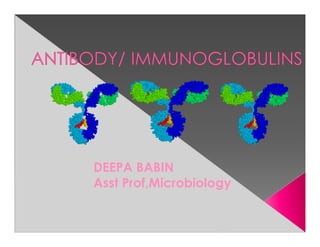
6. Monomer: A flexible Y-shaped molecule with
four protein chains:
2 identical light chains
2 identical heavy chains
Each heavy and light chain has a constant
and variable region
The variable region binds the antigen in a
“lock-and-key” manner
L chains : 2 forms – kappa (κ) & lambda (λ)
Each molecule of Ig can have either κ or λ,
but never both.
7. Antibodies can also be divided into two
regions based on their function
› Fab (fragment, antigen binding) region.
Tip of the antibody
Binds the antigen
› Fc (fragment, crystallizable) region
› Determines biological properties of Ig
molecule.
Base of the antibody
Can bind cell receptors, complement
proteins and other molecules
8.
9. H chain designated by Greek letter.
– 5 different types: IgA, IgD, IgE, IgG, IgM
› IgM µ (mu)
› IgD δ (delta)
› IgG γ (gamma)
› IgA α (alpha)
› IgE ε (epsilon)
H chain also divided into VH & CH regions; the CH region
is further divided into CH1, CH2 & CH3.
Regions also called as DOMAINS :
- globular in shape
- stabilized by intrachain disulphide bonds
Ag binding sites are located in the variable domains.
10.
11. IgG- PROTECTS BODY FLUIDS
IgA- PROTECTS BODY SURFACE
IgM- PROTECTS BLOOD STREAM
IgE- MEDIATES REAGINIC HYPERSENSITIVITY
IgD- Recognisation receptor for Ag
12. Structure: Monomer
Percentage serum antibodies: 80%
Location: Blood, lymph, intestine
Half-life in serum: 23 days
Complement Fixation: Yes
Placental Transfer: Yes only Ab
Major Ab of secondary response, found both in
serum & body fluids.
4 subclasses found in humans – IgG1, IgG2,
IgG3 & IgG4, each having a distinct type of
gamma chain
Functions: Enhances phagocytosis,
neutralizes toxins and viruses, protects fetus
and newborn.
13. Structure: Pentamer
Percentage serum antibodies: 5-10%
Location: Blood, lymph, B cell surface
(monomer)
Half-life in serum: 5 days
Complement Fixation: Yes
Placental Transfer: No
primary immune response.
Functions: First antibodies produced during
an infection. Effective against microbes and
agglutinating antigens. Useful in the diagnosis
of congenital infections like syphilis, rubella, HIV,
dengue,toxoplasmosis etc.
14. Structure: Dimer second most abundunt Ab
Location: Secretions (colostrum,tears, saliva,
intestine, milk), blood and lymph.
Half-life in serum: 6 days
Complement Fixation: No
Placental Transfer: No
Occur in 2 forms : IgA1 & IgA2
Secretory IgA is always in dimeric form – composed
of 2 basic chain units, a J chain & the secretory
component.
Secretory component helps to transport the dimer
from the submucosa to the mucosal cell surface
Functions: Localized protection of mucosal
surfaces. Provides immunity to infant digestive
tract.
15. Structure: Monomer resemble Ig G
Percentage serum antibodies: 0.2%
Location: B-cell surface, blood, and
lymph
Half-life in serum: 3 days
Complement Fixation: No
Placental Transfer: No
Functions: In serum function is unknown.
Occurs along with Ig M on the surface of B
cell- initiate immune response.
16. Structure: Monomer LOW LEVEL IN SERUM
Percentage serum antibodies: 0.002%
Location: linings of respiratory & intestinal
tracts.Bound to mast cells and basophils
throughout body. Blood.
Half-life in serum: 2 days
Complement Fixation: No
Placental Transfer: No
Functions: anaphylactic type of
hypersensitivity ,Allergic reactions.
Possibly lysis of worms.
17. › B cells develop from stem cells in the bone
marrow of adults (liver of fetuses).
› After maturation B cells migrate to lymphoid
organs (lymph node or spleen).
› Clonal Selection: When a B cell encounters
an antigen it recognizes, it is stimulated and
divides into many clones called plasma
cells, which actively secrete antibodies.
› Each B cell produces antibodies that will
recognize only one antigenic determinant.
18. Programmed cell death (“Falling away”).
› Human body makes 100 million lymphocytes
every day. If an equivalent number doesn’t
die, will develop leukemia.
› B cells that do not encounter stimulating
antigen will self-destruct and send signals to
phagocytes to dispose of their remains.
› Many virus infected cells will undergo
apoptosis, to help prevent spread of the
infection.
19. Structurally similar proteins in serum seen
in certain pathological conditions.
Bence Jones protein in multiple myeloma
– light chains of Igs.
Cryoglobulinemia – formation of gel or ppt
on cooling the serum which redissolves
on warming – in myelomas, SLE etc.
20. An individual produces a large number of
Abs to cope with the vast number of different
Ags.
This Ab diversity is due to the Ig genes.
Genes coding for the variable & constant
portions of the chains are separate
One or only few genes code for C region
whereas many genes code for the V region.
21. Multiple V- region genes.
V-J & V-D-J recombination.
Junctional diversity
1. Nucleotide addition – extra nucleotides may
get inserted between VH & D, and between
D & JH segments
Somatic mutation – point mutation in the
genes for V domain.