SlideShare une entreprise Scribd logo
1  sur  10
DISEASE OTHER
TERM
DESCRIPTION EPIDEMIOLOGY PATHOGENESIS CLINICAL SIGNS LESIONS DIAGNOSIS DIFFERENTIAL
DIAGNOSIS
TREATMENT CONTROL &
PREVENTION
SPORADIC
EXERTIONAL
RHABDOMYOLYSIS
Tying-up syndrome of
muscle fatigue,
pain, or cramping
associated with
exercise.
All breeds of
horses are
susceptible to
sporadic exertional
rhabdomyolysis.
It is likely due to
abnormal
regulation of
intra-cellular
calcium in
skeletal muscles.
In
Thoroughbreds, it
is likely inherited
as an autosomal
dominant trait.
Excessive
sweating,
tachypnea,
tachycardia,
muscle
fasciculations,
reluctance or
refusal to move,
and firm, painful
lumbar and
gluteal
musculature.
subclinical to
severe muscle
necrosis
muscle cramping
and stiffness
following
exercise, and
moderate to
marked
elevations in
serum CK and
AST
-reestablish fluid
balance and
induce diuresis
-Horses should
be stall rested on
a hay diet with a
dietary vitamin
and mineral
ration balancer
supplement for a
few days.
Endurance
horses should
be encouraged
to drink
electrolyte-
supplemented
water during an
endurance ride
and monitored
particularly
closely during
hot, humid
conditions.
CHRONIC
EXERTIONAL
RHABDOMYOLYSIS
1. Type 1
polysaccharide
storage myopathy
1 PSSM Glycogen storage
disorder
characterized by
accumulation of
nonbioavailable
polysaccharide in
the muscle
seen frequently in
Quarter horse-
related breeds
Morgans, and draft
horses
Horses with type
1 PSSM have
constitutively
active glycogen
synthase that is
further stimulated
by elevated blood
insulin
concentrations,
resulting in high
muscle glycogen
concentrations.
When fed a
starch meal,
these horses take
up a higher
proportion of the
absorbed glucose
in their muscles
compared with
healthy horses.
tucked-up
abdomen, a
camped-out
stance, muscle
fasciculations,
sweating, gait
asymmetry,
hindlimb stiffness,
and reluctance to
move
Subsarcolemma
l vacuoles,
glycogen
storage and
abnormal PAS
positive
inclusions in
fast twich fibers
identification of
the GYS1 mutati
on and/or the
presence of
muscle fibers
with
subsarcolemmal
vacuoles, dark
periodic acid-
Schiff (PAS)
staining for
glycogen, and
most notably,
amylase-resistant
abnormal
complex
polysaccharide
accumulation
Clostridial
myositis
Influenza myositis
Sarcocystis
myositis
Immune-
mediated
myopathy
Gradual training
and providing a
high fat diet
Hay should be
fed at 1.5–2%
of body weight
and high-fat,
low-starch
concentrates
should be
selected that
provide ≤20%
of daily
digestible
energy as
nonstructural
carbohydrate
and 20–25% of
digestible
energy as fat.
2. Type 2
polysaccharide
storage myopathy
2 PSSM Horses with type
2 PSSM also
have excessive
glycogen storage.
occurs in light
breeds such as
Arabians,
Morgans,
Thoroughbreds, a
variety of
Warmblood
breeds and some
Quarter horses
Horses with Type
2 PSSM lack the
mutation that is
specific for Type
1 PSSM. At
present there is
not a specific
genetic test for
type 2 PSSM and
we do not have
conclusive
Chronic episodes
of muscle
stiffness,
soreness, and
muscle atrophy
with modest
elevations in
serum CK
Subsarcolemma
l vacuoles,
glycogen
storage and
abnormal PAS
positive
inclusions in
fast twich fibers
absence of
the GYS1 mutati
on and the
presence of
muscle fibers
with aggregates
of amylase-
sensitive PAS
positive staining
glycogen and
occasionally
Clostridial
myositis
Influenza myositis
Sarcocystis
myositis
Immune-
mediated
myopathy
Gradual training
and providing a
high fat diet
Hay should be
fed at 1.5–2%
of body weight
and high-fat,
low-starch
concentrates
should be
selected that
provide ≤20%
of daily
digestible
evidence that it is
inherited.
small amounts of
amylase-resistant
PAS positive
material.
energy as
nonstructural
carbohydrate
and 20–25% of
digestible
energy as fat
3. Recurrent
exertional
rhabdomyolysis
RER Recurrent
exertional
rhabdomyolysis
(RER) is caused
by an abnormality
in intracellular
calcium regulation
It is seen
frequently in
Thoroughbreds,
Standardbreds,
and Arabian
horses.
It appears there
is intermittent
disruption of
muscle
contraction,
particularly when
horses
susceptible to the
condition are fit
and have a
nervous
temperament.
horses sweat
profusely, breathe
rapidly and
become stiff and
reluctant to move.
The cells from
the affected
horse have
nuclei located
toward the
center of
mature muscle
fibers rather
than
underneath the
cell membrane
as they are in
the healthy
muscle cells.
history, clinical
signs, elevations
in serum CK and
AST, and muscle
biopsy.
Clostridial
myositis
Influenza myositis
Sarcocystis
myositis
Immune-
mediated
myopathy
Phenytoin (1.4–
2.7 mg/kg, PO,
bid)
decreasing the
triggering
factors for
excitement and
pharmacologic
alteration of
intracellular
calcium flux
with
contraction
SARCOCYSTIS
MYOSITIS
Cysts of the
sporozoan
parasite
Sarcocystis are
present in 90% of
esophageal
muscles from
horses >8 yr of
age and in 6% of
gluteal muscle
biopsies from
healthy horses
Occasionally,
heavy infestations
occur through
contamination of
feed with canine
feces
After ingestion of
sporocysts by a
suitable
intermediate host,
sporozoites are
liberated and
initiate
development of
schizonts
Merozoites are
liberated from the
mature schizonts
and produce a
second
generation of
endothelial
schizonts in
capillaries from
several organs.
Merozoites from
this second
generation
subsequently
invade the
muscle fibers and
develop into the
typical
sarcocysts.
fever, anorexia,
stiffness, weight
loss, muscle
fasciculations,
and weakness.
Muscle atrophy history, clinical
signs, laboratory
evaluation, and
the
demonstration of
an inflammatory
reaction to
immature cysts in
muscle biopsies.
NSAID and drugs
such as
trimethoprim
sulfa and
pyrimethamine or
ponazuril.
Supplies of
grain and feed
should be kept
covered; dogs
and cats
should not be
allowed in
buildings used
to store feed or
house animals
STREPTOCOCCUS Severe It is not clear A stiff gait is the At postmortem, hematologic vitamin IV penicillin
EQUI
RHABDOMYOLYSIS
rhabdomyolysis
can occur in
horses with
Streptococcus
equi equi
submandibular
lymphadenopathy
and/or guttural
pouch empyema.
whether
myonecrosis is a
direct toxic effect
of S equi on
muscle cells or is
due to profound
nonspecific T cell
stimulation by
streptococcal
superantigens
and the release
of high levels of
inflammatory
cytokines.
initial clinical sign,
which progresses
rapidly to severely
painful, firm,
swollen, epaxial
and gluteal
muscles.
large, pale
areas of
necrotic muscle
are evident in
hindlimb and
lumbar
muscles.
histopathologic
lesions are
characterized
by severe acute
myonecrosis
with a degree of
macrophage
infiltration.
Sublumbar
muscles often
show the most
severe and
chronic necrosis
as indicated by
greater
macrophage
infiltration of
myofibers.
abnormalities
typical of S equi
infection, marked
elevations in CK
(>100,000 U/L),
and PCR or
bacterial culture.
E/selenium
deficiency
ionophore toxicity
PSSM
combined with an
antimicrobial that
inhibits protein
synthesis, such
as rifampin.
Flushing infected
guttural pouches
and draining
abscessed lymph
nodes
NSAID and
possibly high
doses of short-
acting
corticosteroids
CLOSTRIDIAL
MYOSITIS
myonecrosi
s
A variety of
clostridial bacteria
can sporulate at
the site of an
injection or deep
wound causing
focal muscle
swelling and
systemic toxemia
in horses
Clostridium
septicum,
Clostridium
chauvoei,
Clostridium
sporogenes, and
mixed infections
are associated
with a high fatality
rate, whereas
Clostridium
perfringens type A
has a mortality
rate of 20% with
early and
aggressive
treatment.
Clostridial spores
may lie dormant
in skeletal
muscle, or spore
deposition directly
into the tissue
may occur in
association with
penetration. If
suitable necrotic
conditions exist,
the spores
convert to the
vegetative form
releasing
powerful
exotoxins.
Within 48 hr,
horses show
depression, fever,
toxemia,
tachypnea, and
swelling and
variable crepitus
at the injection
site. Tremors,
ataxia, dyspnea,
recumbency,
coma, and death
may occur in the
next 12–24 hr.
At postmortem,
swelling,
crepitus, and
autolysis are
rapid, and
bloodstained
fluid is often
observed
discharging
from body
orifices.
Ultrasonographic
evaluation
Aspirates of
affected tissues
examined via
direct smears or
fluorescent
antibody staining
Anaerobic
bacterial culture
Wound
fenestration and
aggressive
surgical
debridement over
the entire
affected area
Additional
treatment
includes high
doses of IV
potassium
penicillin every
2–4 hr until the
animal is stable
(1 to 5 days),
combined with or
followed by oral
metronidazole
along with
supportive fluid
therapy and anti-
inflammatory
agents
MUSCLE Staphylococcus develops following Initially there is an depends on its There may be Ultrasonography poulticing,
ABSCESSES aureus,
Streptococcus
equi, and
Corynebacterium
pseudotuberculos
is are common
causes of skeletal
muscle
abscessation
penetrating
injuries or by
hematogenous or
local spread of
infection
ill-defined
cellulitis, which
may heal or
progress to a
well-defined
abscess. An
abscess may
heal, expand, or
fistulate, usually
to the skin
surface with
potential for a
chronic
granuloma with
intermittent
discharge
location and can
vary from mild
stiffness to severe
lameness.
an elevated
fibrinogen and
nucleated WBC.
and culture of
aspirated fluid
lancing, flushing,
and draining
IMMUNE
MEDIATED
MYOPATHIES
1. INFARCTIVE
HEMORRHAGIC
PURPURA
Purpura
hemorrhagica is a
noncontagious,
type III immune-
mediated
vasculitis of
horses that is
characterized by
subcutaneous
edema of the
head, ventral
abdomen, and
limbs and by
petechial
hemorrhages of
the mucous
membranes.
most commonly
affects young adult
horses, although
the disease has
been identified in
yearlings as well
as geriatric horse.
There appears to
be no breed or sex
predilection.
most often occurs
as a rare
complication of
Streptococcus
equi infection but
can also develop
after infection
with other
bacterial and viral
organisms,
particularly those
that cause
formation of
purulent or
necrotic foci.
mild elevations in
serum CK activity
unilateral
lameness
variable edema,
acute colic, firm
swellings within
muscle and
under the skin
based on clinical
signs, a
leukocytoclastic
vasculitis in skin
and affected
tissues, and very
high S equi M
protein titer
early detection,
penicillin for 14
days, and
prolonged high
doses of
dexamethasone
(0.12–0.2 mg/kg)
for at least 10
days, followed by
tapering doses of
prednisolone at
an initial dosage
of 2 mg/kg.
2. IMMUNE-
MEDIATED
MYOSITIS
It occurs in
Quarter Horses,
although other
breeds may be
affected. The
condition shows a
bimodal age
distribution
affecting horses
<8 yr or >16 yr of
age
In approximately
one-third of
affected horses, a
triggering factor
appears to have
been exposure to
S equi or a
respiratory
disease. Rapid
onset of atrophy
of the back and
croup muscles is
accompanied by
stiffness and
croup stiffness
and malaise
mild to moderate
elevations in
serum CK and
AST
Muscle biopsy
of epaxial and
gluteal muscles
shows
lymphocytic
vasculitis,
anguloid
atrophy,
myofiber
infiltration with
lymphocytes,
fiber necrosis
with
macrophage
biopsy antibiotic therapy
for horses with
concurrent signs
of infection and
administration of
dexamethasone
(0.05 mg/kg for 3
days), followed
by prednisolone
(1 mg/kg for 7–10
days) tapered by
100 mg/wk over 1
mo
malaises. infiltration, and
regeneration in
acute stages.
NUTRITIONAL
MYODEGENERATI
ON
White
muscle
disease
nutritional
myodegeneration
of foals is a
peracute to
subacute
myodegenerative
disease affecting
skeletal and
cardiac muscle
Young, rapidly
growing foals born
to dams that
consumed
selenium deficient
diets during
gestation can
develop nutritional
myodegeneration
Selenium is an
essential
component of 5
antioxidant
selenoproteins
and vitamin E
acts as an
antioxidant within
lipid bilayers.
Muscle
degeneration is
the result of
oxidant damage
to cell
membranes and
proteins leading
to a loss of
cellular integrity.
Clinical signs in
foals include
dyspnea; a rapid,
irregular
heartbeat; and
sudden death in
those with
myocardial
involvement.
Dysphagia,
muscle stiffness,
trembling, firm
muscles, difficulty
rising, and
myoglobinuria
may also be seen.
moderate to
marked
elevated serum
CK and AST,
combined with
low whole blood
selenium
concentrations
(<0.07 ppm) or
vitamin E (<2
ppm).
severe
rhabdomyolysis
spinal cord
compression,
cerebellar
disease,
suppurative and
nonsuppurative
meningitis/myeliti
s, polyarthritis,
neurotoxins such
as
organophosphate
s, tetanus, pelvic
fractures,
parasitic myositis,
clostridial
myositis, and
traumatic injuries
M injection of
selenium (0.055–
0.067 mg/kg) and
either injectable
or oral vitamin E
(0.5–1.5 IU/kg)
administration
of antibiotics to
combat
secondary
pneumonia and
feeding via
nasogastric
tube, provision
of adequate
energy intake,
and attention to
the fluid and
electrolyte
balance.
PLANTS CAUSING
MYOPATHIES
1. TREMATONE
CONTAINING
PLANTS
Horses ingesting
0.5–2% body
weight of
trematone-
containing plants
are likely to die
from skeletal
muscle and
cardiac muscle
necrosis
Trematone is
hypothesized to
interfere with
glucose
metabolism
marked
depression,
weakness, low
head posture, and
increased cardiac
and respiratory
rate
Serum AST and
CK are often
markedly
elevated, and
serum electrolyte
abnormalities
such as
hypocalcemia,
hyponatremia,
hypochloremia,
hyperkalemia, and
hyperphospha-
temia may be
present
skeletal muscle
and cardiac
muscle necrosis
Supplemental
feeding with high
quality forage,
fluid therapy with
glucose as
needed, and rest.
Trematone
remains active
in the hay and
in the stalks of
the dead plants
on pasture, so
both the fresh
and the dried
form of the
plants should
be kept from
horses.
2. ATYPICAL
MYOGLOBINURIA
Pasture
myopathy
Atypical
myoglobinuria
occurs
sporadically in
horses kept on
pasture, usually
It occurs most
often in spring and
autumn and is
often associated
with a sudden
deterioration in
The cause is
unknown but
appears to
involve disruption
in lipid
metabolism due
Affected horses
are reluctant to
move, have
muscle weakness
and
fasciculations,
pulmonary
edema may be
present
Necropsy
reveals
clinical signs and
a blood test
The disease can
look similar to a
severe case of
colic.
Supportive
therapy including
antioxidants (eg,
vitamin C, vitamin
E, riboflavin) and
IV fluids
Provision of
supplementary
feeding,
especially
during periods
of inclement
with no
supplemental
feeding.
weather
conditions.
to exposure to a
toxin in well
grazed, cool, wet
pastures.
and may become
recumbent. Choke
may be present,
and gut sounds
may be reduced,
with reduction in
feces production,
although appetite
may be
unaffected.
widespread
myodegeneratio
n in postural
and respiratory
skeletal
muscles and
the
myocardium.
containing
dextrose are
recommended
weather
Clean, fresh
water supply
Traumatic and
Anesthetic
Myopathies
1. POST-
ANESTHETIC
MYOPATHY
Following
anesthesia,
horses may
develop severe
muscle pain and
weakness in one
or more muscle
groups.
Generalized
myopathies may
develop in horses
that were
hypotensive during
anesthesia.
Hypoperfusion of
compressed
muscle groups
with resultant
high
intracompartment
al pressure is the
most important
causative factor
for focal muscle
involvement
Horses may
appear
distressed, with
profuse sweating,
tachycardia, and
tachypnea.
A dropped elbow
stance typical of
radial nerve
paralysis
characterizes
triceps myopathy.
Gluteal myopathy
results in
unwillingness to
bear weight on
the hindlimbs.
Affected
muscles may
feel very hard
and show
localized
swelling.
History and
clinical signs
Pain relief is
provided by
sedation with
detomidine,
combined with
opiate analgesics
and NSAID.
Fluid therapy
helps maintain
renal perfusion
and urine output
and ensure
adequate muscle
perfusion.
minimizing the
time under
general
anesthesia,
careful
positioning of
the horse on
the operating
table, and
maintenance of
arterial blood
pressure above
60 mm Hg
using fluid
therapy and
inotropic
agents such as
dobutamine
2. FIBROTIC
MYOPATHY
Fibrotic myopathy
describes a
classic gait
abnormality that
develops when
horses injure their
semitendinosus
and
semimembranosu
s muscles at the
point of a
tendinous
insertion during
exercise that
requires abrupt
turns and sliding
stops
Trauma (eg,
catching a foot in a
fence), IM
injections, and a
congenital form
are other potential
causes of fibrotic
myopathy.
develops when
horses injure their
semitendinosus
and
semimembranosu
s muscles at the
point of a
tendinous
insertion during
exercise that
requires abrupt
turns and sliding
stops
characterized by
an abrupt
cessation of the
anterior phase of
the stride of the
affected limb,
causing the leg to
jerk suddenly to
the ground rather
than continue its
forward motion.
The stride has a
short anterior
phase with a
characteristic
hoof-slapping gait.
Chronically,
hardened areas
within the
muscle may
represent
fibrosis and
ossification.
ultrasonography,
thermography, or
scintigraphy
palpation
acute cases may
benefit from rest
and cold therapy
followed by deep
heating
ultrasound and
controlled
stretching.
Chronic cases
may require
surgical excision
or transection of
the fibrotic part of
the muscle or
tenotomy of the
tibial insertion of
the semimembra-
nosus tendon.
MUSCLE
CRAMPING
1. ELECTROLYTE
DISTURBANCES
Muscle cramps
can be induced
by forceful
contraction of a
shortened
muscle, by
changes in the
electrolyte
composition of
extracellular fluid,
and by ear tick
infestations in
horses.
most frequent in
hot, humid
weather.
Horses may lose
up to 15 L/hr of
fluids in the form
of sweat and
develop
remarkable
deficits in sodium,
potassium,
chloride
magnesium, and
calcium.
exhausted horses
are often dull,
depressed, and
clinically
dehydrated with
elevated heart
and respiratory
rates and
persistently
elevated body
temperature.
muscle stiffness
and periodic
spasms of
muscle groups.
Mild muscle
cramping is self-
limiting, and the
signs abate with
rest or light
exercise.
Exhausted
horses with
metabolic
derangements,
however, require
immediate
treatment
including plasma
volume
expansion with
oral or IV isotonic
polyionic fluids
and cooling
(using water and
fans).
Daily direct
addition of 2 oz
of sodium
chloride and 1
oz of
potassium
chloride to the
feed is
recommended
for horses with
recurrent
cramping, in
addition to
electrolyte
supplementatio
n before and
after
endurance
rides.
2. HYPOCALCEMIA Transport
tetany,
Lactation
tetany,
Eclampsia
an uncommon
condition
associated with
acute depletion of
serum ionized
calcium and
sometimes with
alterations in
serum
concentrations of
magnesium and
phosphate.
It occurs after
prolonged physical
exertion or
transport
(transport tetany)
and in lactating
mares (lactation
tetany).
. Corticosteroids
inhibit vitamin D
activity, which
leads to
decreased
intestinal
absorption and
skeletal
mobilization of
calcium. Stress
and lack of
calcium intake
have been
associated with
transport tetany.
synchronous
diaphragmatic
flutter ,anxious
appearance, and
signs of tetany,
including
increased muscle
tone, stiffness of
gait, muscle
tremors, prolapse
of the third eyelid,
inability to chew,
trismus,
salivation,
recumbency,
convulsions, and
cardiac
arrhythmias.
signs, history,
and response to
treatment
tetanus,
endotoxemia,
colic, exertional
rhabdomyolysis
or other muscle
disorder, seizure
disorder,
laminitis, and
botulism
IV administration
of calcium
solutions, such
as 20% calcium
borogluconate
A balanced
feed ration
should be
provided to
supply
adequate
amounts and
ratios of
calcium and
phosphorus
throughout
gestation.
Stress and
fasting during
transport
should be
minimized
3. SYNCHRONOUS
DIAPHRAGMATIC
FLUTTER
Thumps Synchronous
diaphragmatic
flutter is due to
firing of the
phrenic nerve in
synchrony with
atrial
depolarization,
causing the
Inciting causes
include endurance
exercise,
hypocalcemia,
hypoparathyroidis
m, digestive
disturbances, and
repeated
administration of
The most
consistently
reported
metabolic
derangement is
low serum ionized
calcium
concentrations
usually
rhythmic twitching
or "thumping' in
the flank area, in
time with the
beating of the
heart
Clinical signs calcium solutions
IV
For horses with
chronic
diaphragmatic
flutter, provision
of chloride,
potassium,
Limiting alfalfa
hay, which has
a relatively
high calcium
concentration,
may be
indicated in
chronically
affected
diaphragm to
contract with each
heartbeat.
calcium-containing
fluids to
performance
horses.
associated with
hypochloremic
metabolic
alkalosis.
Metabolic
alkalosis may
alter the ratio of
free to bound
calcium which
possibly induces
diaphragmatic
flutter.
sodium, calcium,
and magnesium
during prolonged
exercise may
help reduce fluid
losses and the
metabolic
alkalosis
horses.
4. EAR TICK-
ASSOCIATED
MUSCLE
CRAMPING
Otobius megnini
infestations in the
ear canal can
produce
remarkably
painful
intermittent
muscle cramps
O megnini is found
in the
southwestern USA
Otobius megnini
infestations in the
ear canal can
produce
remarkably
painful
intermittent
muscle cramps
not associated
with exercise that
last from minutes
to a few hours
and often
resemble colic.
Horses may fall
over when
stimulated.
Percussion of
triceps, pectoral,
or semi-
tendinosus
muscles results in
a typical myotonic
cramp. Horses
have elevated
serum CK ranging
from 4,000–
170,000 IU/L.
Tick bites Numerous ear
ticks can be
identified in the
external ear
canal of affected
horses.
local treatment of
the ear ticks
using pyrethrins
and piperonyl
butoxide results
in recovery within
12–36 hr.
Acepromazine
may be helpful to
relieve painful
cramping.
Keeping
animals away
from tick-prone
areas
SHIVERS “Shivers” is a
spastic condition
of the hind- and
occasionally
forelimbs of
horses that is
usually only
evident when
horses are
backing or having
their feet picked
up
It is most common
in adult draft horse
breeds,
Warmbloods,
Warmblood
crosses, and
Thoroughbreds
The condition is
characterized by
periodic,
involuntary
spasms of the
muscles in the
pelvic region,
pelvic limbs, and
tail that are
exacerbated by
backing or picking
up the hindlimbs.
The affected limb
is elevated,
abducted, and
may actually
shake and shiver;
the tail head is
usually elevated
concurrently and
trembles.
Clinical signs There are no
known
treatments, but
avoiding stall rest
and keeping
horses fit
appears helpful.
Myotonic Disorders
1. MYOTONIA
CONGENITA AND
DYSTROPHICA
The initial signs of
myotonia in foals
are well-
developed
musculature and
mild pelvic limb
stiffness. Bilateral
bulging (dimpling)
of the thigh and
Myotonia
congenita usually
does not show
progression of
clinical signs
beyond 6–12 mo
of age, and
muscle stiffness
may improve with
Percussion of
affected muscles
exacerbates the
muscle dimpling
below a large
area of tight
contraction that
can persist for a
minute or more
Foals with
myotonia
dystrophica show
a progression of
signs in the first
1–2 yr of life to
include areas of
muscle atrophy
fibrosis and
ringed fibers,
numerous
centrally
displaced
myonuclei,
sarcoplasmic
masses, and an
increase in
endomysial and
tentative
diagnosis of
myotonia can be
made on the
basis of age and
clinical signs of
stiff gait, muscle
bulging, and
prolonged
dystrophic foals
usually require
euthanasia due
to the severity of
stiffness and
atrophy that
develop over time
rump muscles is
often obvious and
gives the
impression that
the animal is very
well developed.
exercise. with subsequent
slow relaxation.
stiffness that
worsens with
exercise
perimysial
connective
tissue
contractions
following muscle
stimulation.
Definitive
requires
electromyogra-
phic exam.
2. HYPERKALEMIC
PERIODIC
PARALYSIS
HyPP Hyperkalemic
periodic paralysis
(HYPP) is an
inherited disease
of the muscle
which is caused
by a genetic
defect. In the
muscle of
affected horses, a
point mutation
exists in the
sodium channel
gene and is
passed on to
offspring.
HyPP is an
autosomal
dominant trait
affecting Quarter
horses, American
Paint horses,
Appaloosas, and
Quarter horse
crossbreeds
worldwide.
When the
defective sodium
channel gene is
present, the
channel becomes
"leaky" and
makes the
muscle overly
excitable and
contract
involuntarily. The
channel becomes
"leaky" when
potassium levels
fluctuate in the
blood. This may
occur with fasting
followed by
consumption of a
high potassium
feed such as
alfalfa.
asymptomatic to
intermittent
muscle
fasciculations and
weakness
with swaying,
staggering,
dogsitting, or
recumbency may
be seen, with
severe attacks
lasting 15–60 min
or longer. Heart
and respiratory
rates may be
elevated, but
horses remain
relatively bright
and alert
DNA testing of
mane or tail hair
delay before
serum
centrifugation,
hemolysis,
chronic renal
failure, and
severe
rhabdomyolysis.
low-grade
exercise or
feeding grain or
corn syrup to
stimulate insulin-
mediated
movement of
potassium across
cell membranes.
In severe cases,
administration of
calcium
gluconate or IV
dextrose, alone
or combined with
sodium
bicarbonate (1–2
mEq/kg), often
provides
immediate
improvement.
Prevention
requires
decreasing
dietary
potassium to
0.6–1.1% total
potassium
concentration
and increasing
renal losses of
potassium.
GLYCOGEN
BRANCHING
ENZYME
DEFICIENCY
GBED GBED is found in
Quarter Horse or
related bloodlines
that causes late-
term abortion or
death of foals by
a few months of
age.
Nine percent of
Quarter horses
and Paint horses
carry the mutation
and at least 3% of
abortions are
attributed to this
disease in Quarter
horses.
Glycogen
branching
enzyme
deficiency
(GBED) is a
glycogen storage
disorder causing
abortion,
seizures, and
muscle weakness
in Quarter horse-
related breeds
caused by an
autosomal
recessive
mutation in the
glycogen
branching
enzyme (GBE1)
gene.
Most foals
diagnosed with
GBED present
with hypothermia,
weakness, and
flexural
deformities of all
limbs at 1 day of
age.
Persistent
leukopenia,
intermittent
hypoglycemia,
and high serum
CK (1,000–15,000
U/L), AST, and γ-
glutamyltransferas
e activities are
features of
affected foals.
Gross lesions
are not evident,
and routine H &
E stains of
tissues may be
normal or show
basophilic
inclusions in
skeletal muscle
and cardiac
tissues.
A diagnosis is
best obtained by
confirming the
presence of the
genetic mutation
in tissue samples
or by identifying
typical PAS
positive
inclusions in
muscle or cardiac
samples.
no successful
treatment for
GBED.

Contenu connexe

Tendances

Systemic lupus erythematosus.
Systemic lupus erythematosus.Systemic lupus erythematosus.
Systemic lupus erythematosus.Maureen Charagu
 
systemic lupus erythematosus
systemic lupus erythematosussystemic lupus erythematosus
systemic lupus erythematosusMEEQAT HOSPITAL
 
Systemic lupus erythematosis.
Systemic lupus erythematosis.Systemic lupus erythematosis.
Systemic lupus erythematosis.Bimel Kottarathil
 
Veterinary Pathology of Skeletal system
Veterinary Pathology  of Skeletal systemVeterinary Pathology  of Skeletal system
Veterinary Pathology of Skeletal systemMuhammad Amir Sohail
 
Systemic Lupus Erythematosus - Case Management and Literature Review
Systemic Lupus Erythematosus - Case Management and Literature ReviewSystemic Lupus Erythematosus - Case Management and Literature Review
Systemic Lupus Erythematosus - Case Management and Literature ReviewBernard Fiifi Brakatu
 
Mixed connective tissue disorder
Mixed connective tissue disorderMixed connective tissue disorder
Mixed connective tissue disordermadhu sunkara
 
Sle Nephrology Gr
Sle Nephrology GrSle Nephrology Gr
Sle Nephrology GrTejas Desai
 
Treatment of Systemic Lupus
Treatment of Systemic LupusTreatment of Systemic Lupus
Treatment of Systemic LupusAhmed Elshebiny
 
Systemic lupus erythematosis
Systemic lupus erythematosisSystemic lupus erythematosis
Systemic lupus erythematosisPurnima Kartha
 
Systemic Lupus Erythematosis - SLE -Etiopathogenesis, Clinical features, Adva...
Systemic Lupus Erythematosis - SLE -Etiopathogenesis, Clinical features, Adva...Systemic Lupus Erythematosis - SLE -Etiopathogenesis, Clinical features, Adva...
Systemic Lupus Erythematosis - SLE -Etiopathogenesis, Clinical features, Adva...Chetan Ganteppanavar
 
Sle diagnosis &amp; treatment
Sle diagnosis &amp; treatmentSle diagnosis &amp; treatment
Sle diagnosis &amp; treatmentDrSuman Roy
 
Recent advances in diagnosis &amp; management of SLE
Recent advances in diagnosis &amp; management of SLERecent advances in diagnosis &amp; management of SLE
Recent advances in diagnosis &amp; management of SLEShadab Ahmad
 
Rheumatoid arthritis
Rheumatoid arthritisRheumatoid arthritis
Rheumatoid arthritisKemUnited
 
Systemic lupus erythmatosus
Systemic lupus erythmatosusSystemic lupus erythmatosus
Systemic lupus erythmatosusApoorvaMukund
 
Neurometabolic Disorders
Neurometabolic  DisordersNeurometabolic  Disorders
Neurometabolic DisordersAmr Hassan
 

Tendances (20)

Systemic lupus erythematosus.
Systemic lupus erythematosus.Systemic lupus erythematosus.
Systemic lupus erythematosus.
 
systemic lupus erythematosus
systemic lupus erythematosussystemic lupus erythematosus
systemic lupus erythematosus
 
Approach to sle
Approach to sleApproach to sle
Approach to sle
 
Systemic lupus erythematosis.
Systemic lupus erythematosis.Systemic lupus erythematosis.
Systemic lupus erythematosis.
 
Approach to a case of ataxia
Approach to a case of ataxiaApproach to a case of ataxia
Approach to a case of ataxia
 
Veterinary Pathology of Skeletal system
Veterinary Pathology  of Skeletal systemVeterinary Pathology  of Skeletal system
Veterinary Pathology of Skeletal system
 
Systemic Lupus Erythematosus - Case Management and Literature Review
Systemic Lupus Erythematosus - Case Management and Literature ReviewSystemic Lupus Erythematosus - Case Management and Literature Review
Systemic Lupus Erythematosus - Case Management and Literature Review
 
Mixed connective tissue disorder
Mixed connective tissue disorderMixed connective tissue disorder
Mixed connective tissue disorder
 
SLE
SLESLE
SLE
 
SLE & APS for undergraduates: diagnosis & treatment.
SLE & APS for undergraduates: diagnosis & treatment.SLE & APS for undergraduates: diagnosis & treatment.
SLE & APS for undergraduates: diagnosis & treatment.
 
Sle Nephrology Gr
Sle Nephrology GrSle Nephrology Gr
Sle Nephrology Gr
 
Treatment of Systemic Lupus
Treatment of Systemic LupusTreatment of Systemic Lupus
Treatment of Systemic Lupus
 
Systemic lupus erythematosis
Systemic lupus erythematosisSystemic lupus erythematosis
Systemic lupus erythematosis
 
Systemic Lupus Erythematosis - SLE -Etiopathogenesis, Clinical features, Adva...
Systemic Lupus Erythematosis - SLE -Etiopathogenesis, Clinical features, Adva...Systemic Lupus Erythematosis - SLE -Etiopathogenesis, Clinical features, Adva...
Systemic Lupus Erythematosis - SLE -Etiopathogenesis, Clinical features, Adva...
 
Sle diagnosis &amp; treatment
Sle diagnosis &amp; treatmentSle diagnosis &amp; treatment
Sle diagnosis &amp; treatment
 
Recent advances in diagnosis &amp; management of SLE
Recent advances in diagnosis &amp; management of SLERecent advances in diagnosis &amp; management of SLE
Recent advances in diagnosis &amp; management of SLE
 
Rheumatoid arthritis
Rheumatoid arthritisRheumatoid arthritis
Rheumatoid arthritis
 
Systemic lupus erythmatosus
Systemic lupus erythmatosusSystemic lupus erythmatosus
Systemic lupus erythmatosus
 
Neurometabolic Disorders
Neurometabolic  DisordersNeurometabolic  Disorders
Neurometabolic Disorders
 
S Lecture
S LectureS Lecture
S Lecture
 

Similaire à Chronic Exertional Rhabdomyolysis in Horses

Kwashiorkor, marasmus, hypo & hypervitaminoses
Kwashiorkor, marasmus, hypo & hypervitaminosesKwashiorkor, marasmus, hypo & hypervitaminoses
Kwashiorkor, marasmus, hypo & hypervitaminosesArun Ramankutty V
 
Lecture 15 : Animal Diseases
Lecture 15 : Animal DiseasesLecture 15 : Animal Diseases
Lecture 15 : Animal DiseasesWiseAcademy
 
METABOLICDISEASESOFFARMANIMALS.ppt
METABOLICDISEASESOFFARMANIMALS.pptMETABOLICDISEASESOFFARMANIMALS.ppt
METABOLICDISEASESOFFARMANIMALS.pptMaxwellMusora
 
Rickets & osteomalacia
Rickets & osteomalaciaRickets & osteomalacia
Rickets & osteomalaciaNikhil Agarwal
 
Lecture 12 : Animal Diseases
Lecture 12 : Animal DiseasesLecture 12 : Animal Diseases
Lecture 12 : Animal DiseasesWiseAcademy
 
Parturient paresis (milk fever)
Parturient paresis (milk fever)Parturient paresis (milk fever)
Parturient paresis (milk fever)DR ANITA SEVAG
 
Approach to Anemia in children.pptx
Approach to Anemia in children.pptxApproach to Anemia in children.pptx
Approach to Anemia in children.pptxssusere8f40d
 
Gi disorders in horses
Gi disorders in horsesGi disorders in horses
Gi disorders in horsesDevon Avis
 
Diseases of Haemopoietic, and Lymphatic systems Part-I
Diseases of Haemopoietic, and Lymphatic systems Part-I   Diseases of Haemopoietic, and Lymphatic systems Part-I
Diseases of Haemopoietic, and Lymphatic systems Part-I Ajith Y
 
Cellular adaptations & accumulations
Cellular adaptations & accumulationsCellular adaptations & accumulations
Cellular adaptations & accumulationsHabibah Chaudhary
 
Adrenal disorders 1
Adrenal disorders 1Adrenal disorders 1
Adrenal disorders 1KemUnited
 
Metabolic disorders of livestock ppt.
Metabolic disorders of livestock ppt.Metabolic disorders of livestock ppt.
Metabolic disorders of livestock ppt.Dr-Irfan Bhatti
 
The Many Faces of Hyperparathyroidism & Advances in Treatment
The Many Faces of Hyperparathyroidism & Advances in TreatmentThe Many Faces of Hyperparathyroidism & Advances in Treatment
The Many Faces of Hyperparathyroidism & Advances in TreatmentMsccMohamed
 
Rickets & osteomalacia
Rickets & osteomalaciaRickets & osteomalacia
Rickets & osteomalaciaPriyank Uniyal
 
Metacestodal infection
Metacestodal infection Metacestodal infection
Metacestodal infection 94105
 
presentation on CHF,orchitis,shock,anemia
presentation on CHF,orchitis,shock,anemiapresentation on CHF,orchitis,shock,anemia
presentation on CHF,orchitis,shock,anemiaRoshan paudel
 

Similaire à Chronic Exertional Rhabdomyolysis in Horses (20)

Kwashiorkor, marasmus, hypo & hypervitaminoses
Kwashiorkor, marasmus, hypo & hypervitaminosesKwashiorkor, marasmus, hypo & hypervitaminoses
Kwashiorkor, marasmus, hypo & hypervitaminoses
 
Lecture 15 : Animal Diseases
Lecture 15 : Animal DiseasesLecture 15 : Animal Diseases
Lecture 15 : Animal Diseases
 
METABOLICDISEASESOFFARMANIMALS.ppt
METABOLICDISEASESOFFARMANIMALS.pptMETABOLICDISEASESOFFARMANIMALS.ppt
METABOLICDISEASESOFFARMANIMALS.ppt
 
Rickets & osteomalacia
Rickets & osteomalaciaRickets & osteomalacia
Rickets & osteomalacia
 
Lecture 12 : Animal Diseases
Lecture 12 : Animal DiseasesLecture 12 : Animal Diseases
Lecture 12 : Animal Diseases
 
Downer cow syndrome
Downer cow syndromeDowner cow syndrome
Downer cow syndrome
 
CM & R.pptx
CM & R.pptxCM & R.pptx
CM & R.pptx
 
Parturient paresis (milk fever)
Parturient paresis (milk fever)Parturient paresis (milk fever)
Parturient paresis (milk fever)
 
Vitamin C
Vitamin CVitamin C
Vitamin C
 
Approach to Anemia in children.pptx
Approach to Anemia in children.pptxApproach to Anemia in children.pptx
Approach to Anemia in children.pptx
 
calcium.pptx
calcium.pptxcalcium.pptx
calcium.pptx
 
Gi disorders in horses
Gi disorders in horsesGi disorders in horses
Gi disorders in horses
 
Diseases of Haemopoietic, and Lymphatic systems Part-I
Diseases of Haemopoietic, and Lymphatic systems Part-I   Diseases of Haemopoietic, and Lymphatic systems Part-I
Diseases of Haemopoietic, and Lymphatic systems Part-I
 
Cellular adaptations & accumulations
Cellular adaptations & accumulationsCellular adaptations & accumulations
Cellular adaptations & accumulations
 
Adrenal disorders 1
Adrenal disorders 1Adrenal disorders 1
Adrenal disorders 1
 
Metabolic disorders of livestock ppt.
Metabolic disorders of livestock ppt.Metabolic disorders of livestock ppt.
Metabolic disorders of livestock ppt.
 
The Many Faces of Hyperparathyroidism & Advances in Treatment
The Many Faces of Hyperparathyroidism & Advances in TreatmentThe Many Faces of Hyperparathyroidism & Advances in Treatment
The Many Faces of Hyperparathyroidism & Advances in Treatment
 
Rickets & osteomalacia
Rickets & osteomalaciaRickets & osteomalacia
Rickets & osteomalacia
 
Metacestodal infection
Metacestodal infection Metacestodal infection
Metacestodal infection
 
presentation on CHF,orchitis,shock,anemia
presentation on CHF,orchitis,shock,anemiapresentation on CHF,orchitis,shock,anemia
presentation on CHF,orchitis,shock,anemia
 

Plus de Devon Avis

Vm175 digestive system disorders
Vm175 digestive system disordersVm175 digestive system disorders
Vm175 digestive system disordersDevon Avis
 
Respiratory diseases of equine
Respiratory diseases of equineRespiratory diseases of equine
Respiratory diseases of equineDevon Avis
 
Equine reproductive disorders
Equine reproductive disordersEquine reproductive disorders
Equine reproductive disordersDevon Avis
 
Equine medicine my report
Equine medicine my reportEquine medicine my report
Equine medicine my reportDevon Avis
 
Equine med diseases of the urinary system
Equine med diseases of the urinary systemEquine med diseases of the urinary system
Equine med diseases of the urinary systemDevon Avis
 
Equine med. neonatal diseases
Equine med. neonatal diseasesEquine med. neonatal diseases
Equine med. neonatal diseasesDevon Avis
 
Equine med report
Equine med reportEquine med report
Equine med reportDevon Avis
 
Equine colic(digestive disease)
Equine colic(digestive disease)Equine colic(digestive disease)
Equine colic(digestive disease)Devon Avis
 
Dermatologic diseases
Dermatologic diseasesDermatologic diseases
Dermatologic diseasesDevon Avis
 
Cardiovascular and hemolynphatic diseases word printable
Cardiovascular and hemolynphatic diseases word printableCardiovascular and hemolynphatic diseases word printable
Cardiovascular and hemolynphatic diseases word printableDevon Avis
 

Plus de Devon Avis (10)

Vm175 digestive system disorders
Vm175 digestive system disordersVm175 digestive system disorders
Vm175 digestive system disorders
 
Respiratory diseases of equine
Respiratory diseases of equineRespiratory diseases of equine
Respiratory diseases of equine
 
Equine reproductive disorders
Equine reproductive disordersEquine reproductive disorders
Equine reproductive disorders
 
Equine medicine my report
Equine medicine my reportEquine medicine my report
Equine medicine my report
 
Equine med diseases of the urinary system
Equine med diseases of the urinary systemEquine med diseases of the urinary system
Equine med diseases of the urinary system
 
Equine med. neonatal diseases
Equine med. neonatal diseasesEquine med. neonatal diseases
Equine med. neonatal diseases
 
Equine med report
Equine med reportEquine med report
Equine med report
 
Equine colic(digestive disease)
Equine colic(digestive disease)Equine colic(digestive disease)
Equine colic(digestive disease)
 
Dermatologic diseases
Dermatologic diseasesDermatologic diseases
Dermatologic diseases
 
Cardiovascular and hemolynphatic diseases word printable
Cardiovascular and hemolynphatic diseases word printableCardiovascular and hemolynphatic diseases word printable
Cardiovascular and hemolynphatic diseases word printable
 

Chronic Exertional Rhabdomyolysis in Horses

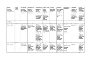
  • 1. DISEASE OTHER TERM DESCRIPTION EPIDEMIOLOGY PATHOGENESIS CLINICAL SIGNS LESIONS DIAGNOSIS DIFFERENTIAL DIAGNOSIS TREATMENT CONTROL & PREVENTION SPORADIC EXERTIONAL RHABDOMYOLYSIS Tying-up syndrome of muscle fatigue, pain, or cramping associated with exercise. All breeds of horses are susceptible to sporadic exertional rhabdomyolysis. It is likely due to abnormal regulation of intra-cellular calcium in skeletal muscles. In Thoroughbreds, it is likely inherited as an autosomal dominant trait. Excessive sweating, tachypnea, tachycardia, muscle fasciculations, reluctance or refusal to move, and firm, painful lumbar and gluteal musculature. subclinical to severe muscle necrosis muscle cramping and stiffness following exercise, and moderate to marked elevations in serum CK and AST -reestablish fluid balance and induce diuresis -Horses should be stall rested on a hay diet with a dietary vitamin and mineral ration balancer supplement for a few days. Endurance horses should be encouraged to drink electrolyte- supplemented water during an endurance ride and monitored particularly closely during hot, humid conditions. CHRONIC EXERTIONAL RHABDOMYOLYSIS 1. Type 1 polysaccharide storage myopathy 1 PSSM Glycogen storage disorder characterized by accumulation of nonbioavailable polysaccharide in the muscle seen frequently in Quarter horse- related breeds Morgans, and draft horses Horses with type 1 PSSM have constitutively active glycogen synthase that is further stimulated by elevated blood insulin concentrations, resulting in high muscle glycogen concentrations. When fed a starch meal, these horses take up a higher proportion of the absorbed glucose in their muscles compared with healthy horses. tucked-up abdomen, a camped-out stance, muscle fasciculations, sweating, gait asymmetry, hindlimb stiffness, and reluctance to move Subsarcolemma l vacuoles, glycogen storage and abnormal PAS positive inclusions in fast twich fibers identification of the GYS1 mutati on and/or the presence of muscle fibers with subsarcolemmal vacuoles, dark periodic acid- Schiff (PAS) staining for glycogen, and most notably, amylase-resistant abnormal complex polysaccharide accumulation Clostridial myositis Influenza myositis Sarcocystis myositis Immune- mediated myopathy Gradual training and providing a high fat diet Hay should be fed at 1.5–2% of body weight and high-fat, low-starch concentrates should be selected that provide ≤20% of daily digestible energy as nonstructural carbohydrate and 20–25% of digestible energy as fat. 2. Type 2 polysaccharide storage myopathy 2 PSSM Horses with type 2 PSSM also have excessive glycogen storage. occurs in light breeds such as Arabians, Morgans, Thoroughbreds, a variety of Warmblood breeds and some Quarter horses Horses with Type 2 PSSM lack the mutation that is specific for Type 1 PSSM. At present there is not a specific genetic test for type 2 PSSM and we do not have conclusive Chronic episodes of muscle stiffness, soreness, and muscle atrophy with modest elevations in serum CK Subsarcolemma l vacuoles, glycogen storage and abnormal PAS positive inclusions in fast twich fibers absence of the GYS1 mutati on and the presence of muscle fibers with aggregates of amylase- sensitive PAS positive staining glycogen and occasionally Clostridial myositis Influenza myositis Sarcocystis myositis Immune- mediated myopathy Gradual training and providing a high fat diet Hay should be fed at 1.5–2% of body weight and high-fat, low-starch concentrates should be selected that provide ≤20% of daily digestible
  • 2. evidence that it is inherited. small amounts of amylase-resistant PAS positive material. energy as nonstructural carbohydrate and 20–25% of digestible energy as fat 3. Recurrent exertional rhabdomyolysis RER Recurrent exertional rhabdomyolysis (RER) is caused by an abnormality in intracellular calcium regulation It is seen frequently in Thoroughbreds, Standardbreds, and Arabian horses. It appears there is intermittent disruption of muscle contraction, particularly when horses susceptible to the condition are fit and have a nervous temperament. horses sweat profusely, breathe rapidly and become stiff and reluctant to move. The cells from the affected horse have nuclei located toward the center of mature muscle fibers rather than underneath the cell membrane as they are in the healthy muscle cells. history, clinical signs, elevations in serum CK and AST, and muscle biopsy. Clostridial myositis Influenza myositis Sarcocystis myositis Immune- mediated myopathy Phenytoin (1.4– 2.7 mg/kg, PO, bid) decreasing the triggering factors for excitement and pharmacologic alteration of intracellular calcium flux with contraction SARCOCYSTIS MYOSITIS Cysts of the sporozoan parasite Sarcocystis are present in 90% of esophageal muscles from horses >8 yr of age and in 6% of gluteal muscle biopsies from healthy horses Occasionally, heavy infestations occur through contamination of feed with canine feces After ingestion of sporocysts by a suitable intermediate host, sporozoites are liberated and initiate development of schizonts Merozoites are liberated from the mature schizonts and produce a second generation of endothelial schizonts in capillaries from several organs. Merozoites from this second generation subsequently invade the muscle fibers and develop into the typical sarcocysts. fever, anorexia, stiffness, weight loss, muscle fasciculations, and weakness. Muscle atrophy history, clinical signs, laboratory evaluation, and the demonstration of an inflammatory reaction to immature cysts in muscle biopsies. NSAID and drugs such as trimethoprim sulfa and pyrimethamine or ponazuril. Supplies of grain and feed should be kept covered; dogs and cats should not be allowed in buildings used to store feed or house animals STREPTOCOCCUS Severe It is not clear A stiff gait is the At postmortem, hematologic vitamin IV penicillin
  • 3. EQUI RHABDOMYOLYSIS rhabdomyolysis can occur in horses with Streptococcus equi equi submandibular lymphadenopathy and/or guttural pouch empyema. whether myonecrosis is a direct toxic effect of S equi on muscle cells or is due to profound nonspecific T cell stimulation by streptococcal superantigens and the release of high levels of inflammatory cytokines. initial clinical sign, which progresses rapidly to severely painful, firm, swollen, epaxial and gluteal muscles. large, pale areas of necrotic muscle are evident in hindlimb and lumbar muscles. histopathologic lesions are characterized by severe acute myonecrosis with a degree of macrophage infiltration. Sublumbar muscles often show the most severe and chronic necrosis as indicated by greater macrophage infiltration of myofibers. abnormalities typical of S equi infection, marked elevations in CK (>100,000 U/L), and PCR or bacterial culture. E/selenium deficiency ionophore toxicity PSSM combined with an antimicrobial that inhibits protein synthesis, such as rifampin. Flushing infected guttural pouches and draining abscessed lymph nodes NSAID and possibly high doses of short- acting corticosteroids CLOSTRIDIAL MYOSITIS myonecrosi s A variety of clostridial bacteria can sporulate at the site of an injection or deep wound causing focal muscle swelling and systemic toxemia in horses Clostridium septicum, Clostridium chauvoei, Clostridium sporogenes, and mixed infections are associated with a high fatality rate, whereas Clostridium perfringens type A has a mortality rate of 20% with early and aggressive treatment. Clostridial spores may lie dormant in skeletal muscle, or spore deposition directly into the tissue may occur in association with penetration. If suitable necrotic conditions exist, the spores convert to the vegetative form releasing powerful exotoxins. Within 48 hr, horses show depression, fever, toxemia, tachypnea, and swelling and variable crepitus at the injection site. Tremors, ataxia, dyspnea, recumbency, coma, and death may occur in the next 12–24 hr. At postmortem, swelling, crepitus, and autolysis are rapid, and bloodstained fluid is often observed discharging from body orifices. Ultrasonographic evaluation Aspirates of affected tissues examined via direct smears or fluorescent antibody staining Anaerobic bacterial culture Wound fenestration and aggressive surgical debridement over the entire affected area Additional treatment includes high doses of IV potassium penicillin every 2–4 hr until the animal is stable (1 to 5 days), combined with or followed by oral metronidazole along with supportive fluid therapy and anti- inflammatory agents MUSCLE Staphylococcus develops following Initially there is an depends on its There may be Ultrasonography poulticing,
  • 4. ABSCESSES aureus, Streptococcus equi, and Corynebacterium pseudotuberculos is are common causes of skeletal muscle abscessation penetrating injuries or by hematogenous or local spread of infection ill-defined cellulitis, which may heal or progress to a well-defined abscess. An abscess may heal, expand, or fistulate, usually to the skin surface with potential for a chronic granuloma with intermittent discharge location and can vary from mild stiffness to severe lameness. an elevated fibrinogen and nucleated WBC. and culture of aspirated fluid lancing, flushing, and draining IMMUNE MEDIATED MYOPATHIES 1. INFARCTIVE HEMORRHAGIC PURPURA Purpura hemorrhagica is a noncontagious, type III immune- mediated vasculitis of horses that is characterized by subcutaneous edema of the head, ventral abdomen, and limbs and by petechial hemorrhages of the mucous membranes. most commonly affects young adult horses, although the disease has been identified in yearlings as well as geriatric horse. There appears to be no breed or sex predilection. most often occurs as a rare complication of Streptococcus equi infection but can also develop after infection with other bacterial and viral organisms, particularly those that cause formation of purulent or necrotic foci. mild elevations in serum CK activity unilateral lameness variable edema, acute colic, firm swellings within muscle and under the skin based on clinical signs, a leukocytoclastic vasculitis in skin and affected tissues, and very high S equi M protein titer early detection, penicillin for 14 days, and prolonged high doses of dexamethasone (0.12–0.2 mg/kg) for at least 10 days, followed by tapering doses of prednisolone at an initial dosage of 2 mg/kg. 2. IMMUNE- MEDIATED MYOSITIS It occurs in Quarter Horses, although other breeds may be affected. The condition shows a bimodal age distribution affecting horses <8 yr or >16 yr of age In approximately one-third of affected horses, a triggering factor appears to have been exposure to S equi or a respiratory disease. Rapid onset of atrophy of the back and croup muscles is accompanied by stiffness and croup stiffness and malaise mild to moderate elevations in serum CK and AST Muscle biopsy of epaxial and gluteal muscles shows lymphocytic vasculitis, anguloid atrophy, myofiber infiltration with lymphocytes, fiber necrosis with macrophage biopsy antibiotic therapy for horses with concurrent signs of infection and administration of dexamethasone (0.05 mg/kg for 3 days), followed by prednisolone (1 mg/kg for 7–10 days) tapered by 100 mg/wk over 1 mo
  • 5. malaises. infiltration, and regeneration in acute stages. NUTRITIONAL MYODEGENERATI ON White muscle disease nutritional myodegeneration of foals is a peracute to subacute myodegenerative disease affecting skeletal and cardiac muscle Young, rapidly growing foals born to dams that consumed selenium deficient diets during gestation can develop nutritional myodegeneration Selenium is an essential component of 5 antioxidant selenoproteins and vitamin E acts as an antioxidant within lipid bilayers. Muscle degeneration is the result of oxidant damage to cell membranes and proteins leading to a loss of cellular integrity. Clinical signs in foals include dyspnea; a rapid, irregular heartbeat; and sudden death in those with myocardial involvement. Dysphagia, muscle stiffness, trembling, firm muscles, difficulty rising, and myoglobinuria may also be seen. moderate to marked elevated serum CK and AST, combined with low whole blood selenium concentrations (<0.07 ppm) or vitamin E (<2 ppm). severe rhabdomyolysis spinal cord compression, cerebellar disease, suppurative and nonsuppurative meningitis/myeliti s, polyarthritis, neurotoxins such as organophosphate s, tetanus, pelvic fractures, parasitic myositis, clostridial myositis, and traumatic injuries M injection of selenium (0.055– 0.067 mg/kg) and either injectable or oral vitamin E (0.5–1.5 IU/kg) administration of antibiotics to combat secondary pneumonia and feeding via nasogastric tube, provision of adequate energy intake, and attention to the fluid and electrolyte balance. PLANTS CAUSING MYOPATHIES 1. TREMATONE CONTAINING PLANTS Horses ingesting 0.5–2% body weight of trematone- containing plants are likely to die from skeletal muscle and cardiac muscle necrosis Trematone is hypothesized to interfere with glucose metabolism marked depression, weakness, low head posture, and increased cardiac and respiratory rate Serum AST and CK are often markedly elevated, and serum electrolyte abnormalities such as hypocalcemia, hyponatremia, hypochloremia, hyperkalemia, and hyperphospha- temia may be present skeletal muscle and cardiac muscle necrosis Supplemental feeding with high quality forage, fluid therapy with glucose as needed, and rest. Trematone remains active in the hay and in the stalks of the dead plants on pasture, so both the fresh and the dried form of the plants should be kept from horses. 2. ATYPICAL MYOGLOBINURIA Pasture myopathy Atypical myoglobinuria occurs sporadically in horses kept on pasture, usually It occurs most often in spring and autumn and is often associated with a sudden deterioration in The cause is unknown but appears to involve disruption in lipid metabolism due Affected horses are reluctant to move, have muscle weakness and fasciculations, pulmonary edema may be present Necropsy reveals clinical signs and a blood test The disease can look similar to a severe case of colic. Supportive therapy including antioxidants (eg, vitamin C, vitamin E, riboflavin) and IV fluids Provision of supplementary feeding, especially during periods of inclement
  • 6. with no supplemental feeding. weather conditions. to exposure to a toxin in well grazed, cool, wet pastures. and may become recumbent. Choke may be present, and gut sounds may be reduced, with reduction in feces production, although appetite may be unaffected. widespread myodegeneratio n in postural and respiratory skeletal muscles and the myocardium. containing dextrose are recommended weather Clean, fresh water supply Traumatic and Anesthetic Myopathies 1. POST- ANESTHETIC MYOPATHY Following anesthesia, horses may develop severe muscle pain and weakness in one or more muscle groups. Generalized myopathies may develop in horses that were hypotensive during anesthesia. Hypoperfusion of compressed muscle groups with resultant high intracompartment al pressure is the most important causative factor for focal muscle involvement Horses may appear distressed, with profuse sweating, tachycardia, and tachypnea. A dropped elbow stance typical of radial nerve paralysis characterizes triceps myopathy. Gluteal myopathy results in unwillingness to bear weight on the hindlimbs. Affected muscles may feel very hard and show localized swelling. History and clinical signs Pain relief is provided by sedation with detomidine, combined with opiate analgesics and NSAID. Fluid therapy helps maintain renal perfusion and urine output and ensure adequate muscle perfusion. minimizing the time under general anesthesia, careful positioning of the horse on the operating table, and maintenance of arterial blood pressure above 60 mm Hg using fluid therapy and inotropic agents such as dobutamine 2. FIBROTIC MYOPATHY Fibrotic myopathy describes a classic gait abnormality that develops when horses injure their semitendinosus and semimembranosu s muscles at the point of a tendinous insertion during exercise that requires abrupt turns and sliding stops Trauma (eg, catching a foot in a fence), IM injections, and a congenital form are other potential causes of fibrotic myopathy. develops when horses injure their semitendinosus and semimembranosu s muscles at the point of a tendinous insertion during exercise that requires abrupt turns and sliding stops characterized by an abrupt cessation of the anterior phase of the stride of the affected limb, causing the leg to jerk suddenly to the ground rather than continue its forward motion. The stride has a short anterior phase with a characteristic hoof-slapping gait. Chronically, hardened areas within the muscle may represent fibrosis and ossification. ultrasonography, thermography, or scintigraphy palpation acute cases may benefit from rest and cold therapy followed by deep heating ultrasound and controlled stretching. Chronic cases may require surgical excision or transection of the fibrotic part of the muscle or tenotomy of the tibial insertion of the semimembra- nosus tendon. MUSCLE CRAMPING
  • 7. 1. ELECTROLYTE DISTURBANCES Muscle cramps can be induced by forceful contraction of a shortened muscle, by changes in the electrolyte composition of extracellular fluid, and by ear tick infestations in horses. most frequent in hot, humid weather. Horses may lose up to 15 L/hr of fluids in the form of sweat and develop remarkable deficits in sodium, potassium, chloride magnesium, and calcium. exhausted horses are often dull, depressed, and clinically dehydrated with elevated heart and respiratory rates and persistently elevated body temperature. muscle stiffness and periodic spasms of muscle groups. Mild muscle cramping is self- limiting, and the signs abate with rest or light exercise. Exhausted horses with metabolic derangements, however, require immediate treatment including plasma volume expansion with oral or IV isotonic polyionic fluids and cooling (using water and fans). Daily direct addition of 2 oz of sodium chloride and 1 oz of potassium chloride to the feed is recommended for horses with recurrent cramping, in addition to electrolyte supplementatio n before and after endurance rides. 2. HYPOCALCEMIA Transport tetany, Lactation tetany, Eclampsia an uncommon condition associated with acute depletion of serum ionized calcium and sometimes with alterations in serum concentrations of magnesium and phosphate. It occurs after prolonged physical exertion or transport (transport tetany) and in lactating mares (lactation tetany). . Corticosteroids inhibit vitamin D activity, which leads to decreased intestinal absorption and skeletal mobilization of calcium. Stress and lack of calcium intake have been associated with transport tetany. synchronous diaphragmatic flutter ,anxious appearance, and signs of tetany, including increased muscle tone, stiffness of gait, muscle tremors, prolapse of the third eyelid, inability to chew, trismus, salivation, recumbency, convulsions, and cardiac arrhythmias. signs, history, and response to treatment tetanus, endotoxemia, colic, exertional rhabdomyolysis or other muscle disorder, seizure disorder, laminitis, and botulism IV administration of calcium solutions, such as 20% calcium borogluconate A balanced feed ration should be provided to supply adequate amounts and ratios of calcium and phosphorus throughout gestation. Stress and fasting during transport should be minimized 3. SYNCHRONOUS DIAPHRAGMATIC FLUTTER Thumps Synchronous diaphragmatic flutter is due to firing of the phrenic nerve in synchrony with atrial depolarization, causing the Inciting causes include endurance exercise, hypocalcemia, hypoparathyroidis m, digestive disturbances, and repeated administration of The most consistently reported metabolic derangement is low serum ionized calcium concentrations usually rhythmic twitching or "thumping' in the flank area, in time with the beating of the heart Clinical signs calcium solutions IV For horses with chronic diaphragmatic flutter, provision of chloride, potassium, Limiting alfalfa hay, which has a relatively high calcium concentration, may be indicated in chronically affected
  • 8. diaphragm to contract with each heartbeat. calcium-containing fluids to performance horses. associated with hypochloremic metabolic alkalosis. Metabolic alkalosis may alter the ratio of free to bound calcium which possibly induces diaphragmatic flutter. sodium, calcium, and magnesium during prolonged exercise may help reduce fluid losses and the metabolic alkalosis horses. 4. EAR TICK- ASSOCIATED MUSCLE CRAMPING Otobius megnini infestations in the ear canal can produce remarkably painful intermittent muscle cramps O megnini is found in the southwestern USA Otobius megnini infestations in the ear canal can produce remarkably painful intermittent muscle cramps not associated with exercise that last from minutes to a few hours and often resemble colic. Horses may fall over when stimulated. Percussion of triceps, pectoral, or semi- tendinosus muscles results in a typical myotonic cramp. Horses have elevated serum CK ranging from 4,000– 170,000 IU/L. Tick bites Numerous ear ticks can be identified in the external ear canal of affected horses. local treatment of the ear ticks using pyrethrins and piperonyl butoxide results in recovery within 12–36 hr. Acepromazine may be helpful to relieve painful cramping. Keeping animals away from tick-prone areas SHIVERS “Shivers” is a spastic condition of the hind- and occasionally forelimbs of horses that is usually only evident when horses are backing or having their feet picked up It is most common in adult draft horse breeds, Warmbloods, Warmblood crosses, and Thoroughbreds The condition is characterized by periodic, involuntary spasms of the muscles in the pelvic region, pelvic limbs, and tail that are exacerbated by backing or picking up the hindlimbs. The affected limb is elevated, abducted, and may actually shake and shiver; the tail head is usually elevated concurrently and trembles. Clinical signs There are no known treatments, but avoiding stall rest and keeping horses fit appears helpful. Myotonic Disorders 1. MYOTONIA CONGENITA AND DYSTROPHICA The initial signs of myotonia in foals are well- developed musculature and mild pelvic limb stiffness. Bilateral bulging (dimpling) of the thigh and Myotonia congenita usually does not show progression of clinical signs beyond 6–12 mo of age, and muscle stiffness may improve with Percussion of affected muscles exacerbates the muscle dimpling below a large area of tight contraction that can persist for a minute or more Foals with myotonia dystrophica show a progression of signs in the first 1–2 yr of life to include areas of muscle atrophy fibrosis and ringed fibers, numerous centrally displaced myonuclei, sarcoplasmic masses, and an increase in endomysial and tentative diagnosis of myotonia can be made on the basis of age and clinical signs of stiff gait, muscle bulging, and prolonged dystrophic foals usually require euthanasia due to the severity of stiffness and atrophy that develop over time
  • 9. rump muscles is often obvious and gives the impression that the animal is very well developed. exercise. with subsequent slow relaxation. stiffness that worsens with exercise perimysial connective tissue contractions following muscle stimulation. Definitive requires electromyogra- phic exam. 2. HYPERKALEMIC PERIODIC PARALYSIS HyPP Hyperkalemic periodic paralysis (HYPP) is an inherited disease of the muscle which is caused by a genetic defect. In the muscle of affected horses, a point mutation exists in the sodium channel gene and is passed on to offspring. HyPP is an autosomal dominant trait affecting Quarter horses, American Paint horses, Appaloosas, and Quarter horse crossbreeds worldwide. When the defective sodium channel gene is present, the channel becomes "leaky" and makes the muscle overly excitable and contract involuntarily. The channel becomes "leaky" when potassium levels fluctuate in the blood. This may occur with fasting followed by consumption of a high potassium feed such as alfalfa. asymptomatic to intermittent muscle fasciculations and weakness with swaying, staggering, dogsitting, or recumbency may be seen, with severe attacks lasting 15–60 min or longer. Heart and respiratory rates may be elevated, but horses remain relatively bright and alert DNA testing of mane or tail hair delay before serum centrifugation, hemolysis, chronic renal failure, and severe rhabdomyolysis. low-grade exercise or feeding grain or corn syrup to stimulate insulin- mediated movement of potassium across cell membranes. In severe cases, administration of calcium gluconate or IV dextrose, alone or combined with sodium bicarbonate (1–2 mEq/kg), often provides immediate improvement. Prevention requires decreasing dietary potassium to 0.6–1.1% total potassium concentration and increasing renal losses of potassium. GLYCOGEN BRANCHING ENZYME DEFICIENCY GBED GBED is found in Quarter Horse or related bloodlines that causes late- term abortion or death of foals by a few months of age. Nine percent of Quarter horses and Paint horses carry the mutation and at least 3% of abortions are attributed to this disease in Quarter horses. Glycogen branching enzyme deficiency (GBED) is a glycogen storage disorder causing abortion, seizures, and muscle weakness in Quarter horse- related breeds caused by an autosomal recessive mutation in the glycogen branching enzyme (GBE1) gene. Most foals diagnosed with GBED present with hypothermia, weakness, and flexural deformities of all limbs at 1 day of age. Persistent leukopenia, intermittent hypoglycemia, and high serum CK (1,000–15,000 U/L), AST, and γ- glutamyltransferas e activities are features of affected foals. Gross lesions are not evident, and routine H & E stains of tissues may be normal or show basophilic inclusions in skeletal muscle and cardiac tissues. A diagnosis is best obtained by confirming the presence of the genetic mutation in tissue samples or by identifying typical PAS positive inclusions in muscle or cardiac samples. no successful treatment for GBED.
  • 10. rump muscles is often obvious and gives the impression that the animal is very well developed. exercise. with subsequent slow relaxation. stiffness that worsens with exercise perimysial connective tissue contractions following muscle stimulation. Definitive requires electromyogra- phic exam. 2. HYPERKALEMIC PERIODIC PARALYSIS HyPP Hyperkalemic periodic paralysis (HYPP) is an inherited disease of the muscle which is caused by a genetic defect. In the muscle of affected horses, a point mutation exists in the sodium channel gene and is passed on to offspring. HyPP is an autosomal dominant trait affecting Quarter horses, American Paint horses, Appaloosas, and Quarter horse crossbreeds worldwide. When the defective sodium channel gene is present, the channel becomes "leaky" and makes the muscle overly excitable and contract involuntarily. The channel becomes "leaky" when potassium levels fluctuate in the blood. This may occur with fasting followed by consumption of a high potassium feed such as alfalfa. asymptomatic to intermittent muscle fasciculations and weakness with swaying, staggering, dogsitting, or recumbency may be seen, with severe attacks lasting 15–60 min or longer. Heart and respiratory rates may be elevated, but horses remain relatively bright and alert DNA testing of mane or tail hair delay before serum centrifugation, hemolysis, chronic renal failure, and severe rhabdomyolysis. low-grade exercise or feeding grain or corn syrup to stimulate insulin- mediated movement of potassium across cell membranes. In severe cases, administration of calcium gluconate or IV dextrose, alone or combined with sodium bicarbonate (1–2 mEq/kg), often provides immediate improvement. Prevention requires decreasing dietary potassium to 0.6–1.1% total potassium concentration and increasing renal losses of potassium. GLYCOGEN BRANCHING ENZYME DEFICIENCY GBED GBED is found in Quarter Horse or related bloodlines that causes late- term abortion or death of foals by a few months of age. Nine percent of Quarter horses and Paint horses carry the mutation and at least 3% of abortions are attributed to this disease in Quarter horses. Glycogen branching enzyme deficiency (GBED) is a glycogen storage disorder causing abortion, seizures, and muscle weakness in Quarter horse- related breeds caused by an autosomal recessive mutation in the glycogen branching enzyme (GBE1) gene. Most foals diagnosed with GBED present with hypothermia, weakness, and flexural deformities of all limbs at 1 day of age. Persistent leukopenia, intermittent hypoglycemia, and high serum CK (1,000–15,000 U/L), AST, and γ- glutamyltransferas e activities are features of affected foals. Gross lesions are not evident, and routine H & E stains of tissues may be normal or show basophilic inclusions in skeletal muscle and cardiac tissues. A diagnosis is best obtained by confirming the presence of the genetic mutation in tissue samples or by identifying typical PAS positive inclusions in muscle or cardiac samples. no successful treatment for GBED.