SlideShare une entreprise Scribd logo
1  sur  14
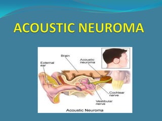
VESTIBULAR SCHWANNOMA/ ACOUSTIC NEUROMA

It’s the most common intracranial schwannoma constituting 80
% of all cerebellopontine angle tumors.

It’s benign encapsulated , extremely slow-growing tumor either
solid or cystic which arises from the Schwann-cells (transition
zone) of the vestibular nerve within the internal auditory canal
are histologically composed of alternating regions of Antony type
A and B areas. It can extend into the cerebellopontine angle.

The number of newly diagnosed cases per year is around 13 per
one million individuals.
Bilateral AC is diagnostic of neurofibromatosis type 2.
ANATOMY OF CEREBELLOPONTINE ANGLE
It’s a triangular area and bounded by:

 1- Laterally: medial portion of posterior surface of temporal
 bone.
 2- Medially: Edge of pons.
 3- Posteriorly: cerebellar hemisphere and flocculus.
 4- Superiorly: trigeminal nerve.

 CONTENTS:
 1- Anterior inferior cerebellar artery. 2- 7th and
 8th Cranial nerves.
FEATURES:
CLINICAL SYMPTOMS
1- Most common ; Progressive Unilateral SNHL (retrocochlear)
present in 95% of cases and often accompanied by tinnitus
which is present in 65% of cases.

2- Marked difficulty in understanding speech out of proportion
to the pure tone hearing loss.
3- May present with sudden hearing loss.
4- True vertigo is seldom seen.
5- Earliest cranial nerve involved is 5th CN.
6- 4th , 6th, 9th , 10th , 11th and 12th can also be involved.
7- Rare presentations include facial numbness or pain,
earache or facial weakness, cerebellar ataxia or symptoms
of hydrocephalus (headache, visual disturbance, mental
status change, nausea, and vomiting).
SIGNS
 Ear: normal otoscopy.
 Cranial nerves:
   1- 5th CN: Earliest sign is impaired corneal reflex.
         Motor functions are affected rarely.

  2- 7th CN: Sensory first; loss of sensation in the
  postero-superior aspect of EAC called Hitselberger
  sign.
   3- 9th and 1oth pulsy: palatal, pharyngeal and
  laryngeal paralysis.

    4- Eyes: Nystagmus.
    5- Cerebellar signs present.
CLASSIFICATION OF AN ACCORDING TO SIZE

    Intameatal   Extrameatal size   Mm
    Grade 1      Small              1-10
    Grade 2      Medium             11-20
    Grade 3      Moderately large   21-30
    Grade 4      Large              31-40
    Grade 5      Giant              More than 40
INVESTIGATIONS
1- Audiological tests: show features of retrocochlear hearing loss
   (high frequency SNHL) ; recruitment negative, poor speech
   discrimination score and presence of roll-over phenomenon.

 2- Acoustic reflex: stapedial reflex decay or missing in up to 70%
  of patients.
 3- Caloric test: diminished or absent normal test finding does not
  eliminate the diagnosis.
 4- Plain x-ray: best view perorbital view ; difference of 1 mm in the
  vertical height of internal acoustic meatus is significant.

 5- CT-scan; can not detect intermeatal tumors.
 6- MRI; MRI with gadolinium enhancement is is gold standard.
Conservative Treatment
• Since especially in the elderly vestibular schwannomas are slowly
growing tumours, a wait and see strategy should be considered
at least in the short term (6–12 months) for patients with a small
vestibular schwannoma (in older patients and individuals in poor
health).
• Radiotherapy: Stereotactic radiosurgery (gamma knife) may stop
vestibular schwannoma growth mainly in small intracanalicular
and extracanalicular lesions. Radiosurgery as the first therapeutic
option is recommended in bilateral vestibular schwannomas (e. g.
morbus Recklinghausen) but only when the tumours are small.
• Annual imaging is recommended for all patients being managed
conservatively for the rest of their life or until vestibular
schwannoma growth is seen to a certain
limit.
Surgical Treatment
 Translabyrinthine approach in patients with medium sized
and large tumours and in patients with small tumours with poor
hearing .
 Middle fossa approach in patients with intracanalicular or
small tumours with good hearing as an attempt to preserve
residual hearing.
 Retrolabyrinthine or retrosigmoidal approaches in
patients with large extracanalicular vestibular schwannomas

Additional Useful Surgical Procedures
• Intraoperative facial nerve monitoring.
• Intraoperative hearing monitoring: may be useful in
Differential Diagnosis
• Meningioma is, in most cases, recognized on radiologic
features (tumour usually extrinsic to internal auditory
canal, sessile with peritumoral dural enhancement and
intratumoral calcifications). An enhanced “dural tail” is a
distinguishing feature.

• Other cerebellopontine angle lesions
(cholesteatoma, arachnoid cyst, lipoma, haemangioma,
glomus tumour, facial neuroma) can in most cases be
recognized by their radiologic aspects and MRI
characteristics
Prognosis
Conservative Therapy With radiotherapy tumour control is
around 96%, tumour regression 35%, lesion of the trigeminal
nerve less than 5.6%, facial weakness 2%, and hearing loss 32%.

There is no CSF leak and no mortality.

If irradiation cannot stop tumour growth (in about 4%
of cases), surgery is technically sometimes extremely difficult.

Malignant transformation after radiotherapy is also valid for
neuromas.
Acoustic neuroma

Contenu connexe

Tendances

Congenital anomalies of inner ear
Congenital anomalies of inner earCongenital anomalies of inner ear
Congenital anomalies of inner earChanmiki Sayoo
 
Juvenile nasopharyngeal angiofibroma
Juvenile nasopharyngeal angiofibromaJuvenile nasopharyngeal angiofibroma
Juvenile nasopharyngeal angiofibromapraneeth koduru
 
Tinnitus and It's Treatment
Tinnitus and It's Treatment Tinnitus and It's Treatment
Tinnitus and It's Treatment Amit kumar
 
Vestibular schwannoma chiru
Vestibular schwannoma chiruVestibular schwannoma chiru
Vestibular schwannoma chiruAditya Roy
 
Otosclerosis dr.p.kartyhikeyan,07.03.2016
Otosclerosis dr.p.kartyhikeyan,07.03.2016Otosclerosis dr.p.kartyhikeyan,07.03.2016
Otosclerosis dr.p.kartyhikeyan,07.03.2016ophthalmgmcri
 
Cholesteatoma
Cholesteatoma Cholesteatoma
Cholesteatoma Razal M
 
Skull base osteomyelitis
Skull base osteomyelitisSkull base osteomyelitis
Skull base osteomyelitiskamalaiims
 
MENIERE'S DISEASE
MENIERE'S DISEASEMENIERE'S DISEASE
MENIERE'S DISEASESuraj Dhara
 
Complications of csom
Complications of csomComplications of csom
Complications of csomAjay Manickam
 
ASSESSMENT OF VESTIBULAR FUNCTION
ASSESSMENT OF VESTIBULAR FUNCTION ASSESSMENT OF VESTIBULAR FUNCTION
ASSESSMENT OF VESTIBULAR FUNCTION TONY SCARIA
 
Maxillary sinus carcinoma
Maxillary sinus carcinomaMaxillary sinus carcinoma
Maxillary sinus carcinomaHarsha Yadav
 
Embryology & anatomy of external ear
Embryology &  anatomy of external earEmbryology &  anatomy of external ear
Embryology & anatomy of external earDr. Pruthvi Raj S
 

Tendances (20)

Tympanosclerosis
TympanosclerosisTympanosclerosis
Tympanosclerosis
 
Congenital anomalies of inner ear
Congenital anomalies of inner earCongenital anomalies of inner ear
Congenital anomalies of inner ear
 
3 (b) pathology,disorders of outer, middle and inner ear
3 (b) pathology,disorders of outer, middle and inner ear3 (b) pathology,disorders of outer, middle and inner ear
3 (b) pathology,disorders of outer, middle and inner ear
 
Juvenile nasopharyngeal angiofibroma
Juvenile nasopharyngeal angiofibromaJuvenile nasopharyngeal angiofibroma
Juvenile nasopharyngeal angiofibroma
 
Tinnitus and It's Treatment
Tinnitus and It's Treatment Tinnitus and It's Treatment
Tinnitus and It's Treatment
 
Vestibular schwannoma chiru
Vestibular schwannoma chiruVestibular schwannoma chiru
Vestibular schwannoma chiru
 
Otosclerosis
OtosclerosisOtosclerosis
Otosclerosis
 
Inverted papilloma
Inverted papillomaInverted papilloma
Inverted papilloma
 
Otosclerosis dr.p.kartyhikeyan,07.03.2016
Otosclerosis dr.p.kartyhikeyan,07.03.2016Otosclerosis dr.p.kartyhikeyan,07.03.2016
Otosclerosis dr.p.kartyhikeyan,07.03.2016
 
Mastoidectomy (by drdhiru456)
Mastoidectomy (by drdhiru456)Mastoidectomy (by drdhiru456)
Mastoidectomy (by drdhiru456)
 
Cholesteatoma
Cholesteatoma Cholesteatoma
Cholesteatoma
 
Skull base osteomyelitis
Skull base osteomyelitisSkull base osteomyelitis
Skull base osteomyelitis
 
Glomus Tumour
Glomus TumourGlomus Tumour
Glomus Tumour
 
MENIERE'S DISEASE
MENIERE'S DISEASEMENIERE'S DISEASE
MENIERE'S DISEASE
 
Complications of csom
Complications of csomComplications of csom
Complications of csom
 
ASSESSMENT OF VESTIBULAR FUNCTION
ASSESSMENT OF VESTIBULAR FUNCTION ASSESSMENT OF VESTIBULAR FUNCTION
ASSESSMENT OF VESTIBULAR FUNCTION
 
Maxillary sinus carcinoma
Maxillary sinus carcinomaMaxillary sinus carcinoma
Maxillary sinus carcinoma
 
Embryology & anatomy of external ear
Embryology &  anatomy of external earEmbryology &  anatomy of external ear
Embryology & anatomy of external ear
 
Glomus tumors
Glomus tumorsGlomus tumors
Glomus tumors
 
Otosclerosis
Otosclerosis Otosclerosis
Otosclerosis
 

En vedette

Surgical management of vestibular schwannoma by drdhiru456
Surgical management of vestibular schwannoma by drdhiru456Surgical management of vestibular schwannoma by drdhiru456
Surgical management of vestibular schwannoma by drdhiru456Dr Dhirendra Patil
 
Accustic neuroma 1
Accustic neuroma 1Accustic neuroma 1
Accustic neuroma 1Verdah Sabih
 
vestibular schwannoma Dr jyoti singh MS ENT
vestibular schwannoma Dr jyoti singh MS ENTvestibular schwannoma Dr jyoti singh MS ENT
vestibular schwannoma Dr jyoti singh MS ENTJyoti Singh
 
Diagnostic Imaging of Cerebellopontine Angle Masses
Diagnostic Imaging of Cerebellopontine Angle MassesDiagnostic Imaging of Cerebellopontine Angle Masses
Diagnostic Imaging of Cerebellopontine Angle MassesMohamed M.A. Zaitoun
 
Acaustic schwannoma
Acaustic schwannomaAcaustic schwannoma
Acaustic schwannomaradiosurgery
 
Brain tumor neuroimaging 4 17th may 02
Brain tumor neuroimaging   4 17th may 02Brain tumor neuroimaging   4 17th may 02
Brain tumor neuroimaging 4 17th may 02PS Deb
 
surgical approaches to frontal sinus ppt
surgical approaches to frontal sinus pptsurgical approaches to frontal sinus ppt
surgical approaches to frontal sinus pptVaibhav Lahane
 
Neurofibromatosis
NeurofibromatosisNeurofibromatosis
Neurofibromatosisshibby5587
 
Otosclerosis 2013
Otosclerosis 2013Otosclerosis 2013
Otosclerosis 2013kamalaiims
 
Anatomy and physiology of oral cavity oropharynx waldeyer’s
Anatomy and physiology of oral cavity oropharynx waldeyer’sAnatomy and physiology of oral cavity oropharynx waldeyer’s
Anatomy and physiology of oral cavity oropharynx waldeyer’sENTDOST
 
Tumores Cerebrais / Sistema Nervoso Central
Tumores Cerebrais / Sistema Nervoso CentralTumores Cerebrais / Sistema Nervoso Central
Tumores Cerebrais / Sistema Nervoso CentralOncoguia
 
Facial nerve ppt roger original
Facial nerve ppt  roger originalFacial nerve ppt  roger original
Facial nerve ppt roger originalRoger Paul
 

En vedette (20)

CP angle 360°
CP angle 360°CP angle 360°
CP angle 360°
 
Surgical management of vestibular schwannoma by drdhiru456
Surgical management of vestibular schwannoma by drdhiru456Surgical management of vestibular schwannoma by drdhiru456
Surgical management of vestibular schwannoma by drdhiru456
 
Accustic neuroma 1
Accustic neuroma 1Accustic neuroma 1
Accustic neuroma 1
 
vestibular schwannoma Dr jyoti singh MS ENT
vestibular schwannoma Dr jyoti singh MS ENTvestibular schwannoma Dr jyoti singh MS ENT
vestibular schwannoma Dr jyoti singh MS ENT
 
Schwannoma
Schwannoma         Schwannoma
Schwannoma
 
Diagnostic Imaging of Cerebellopontine Angle Masses
Diagnostic Imaging of Cerebellopontine Angle MassesDiagnostic Imaging of Cerebellopontine Angle Masses
Diagnostic Imaging of Cerebellopontine Angle Masses
 
Peripheral Nerve Sheath Tumors
Peripheral Nerve Sheath TumorsPeripheral Nerve Sheath Tumors
Peripheral Nerve Sheath Tumors
 
Acoustic Neuroma
Acoustic NeuromaAcoustic Neuroma
Acoustic Neuroma
 
What is acoustic neuroma?
What is acoustic neuroma?What is acoustic neuroma?
What is acoustic neuroma?
 
Neural tumors
Neural tumorsNeural tumors
Neural tumors
 
Schwannoma
SchwannomaSchwannoma
Schwannoma
 
Acaustic schwannoma
Acaustic schwannomaAcaustic schwannoma
Acaustic schwannoma
 
Brain tumor neuroimaging 4 17th may 02
Brain tumor neuroimaging   4 17th may 02Brain tumor neuroimaging   4 17th may 02
Brain tumor neuroimaging 4 17th may 02
 
surgical approaches to frontal sinus ppt
surgical approaches to frontal sinus pptsurgical approaches to frontal sinus ppt
surgical approaches to frontal sinus ppt
 
Neurofibromatosis
NeurofibromatosisNeurofibromatosis
Neurofibromatosis
 
Otosclerosis 2013
Otosclerosis 2013Otosclerosis 2013
Otosclerosis 2013
 
Anatomy and physiology of oral cavity oropharynx waldeyer’s
Anatomy and physiology of oral cavity oropharynx waldeyer’sAnatomy and physiology of oral cavity oropharynx waldeyer’s
Anatomy and physiology of oral cavity oropharynx waldeyer’s
 
Meniere’s disease
Meniere’s diseaseMeniere’s disease
Meniere’s disease
 
Tumores Cerebrais / Sistema Nervoso Central
Tumores Cerebrais / Sistema Nervoso CentralTumores Cerebrais / Sistema Nervoso Central
Tumores Cerebrais / Sistema Nervoso Central
 
Facial nerve ppt roger original
Facial nerve ppt  roger originalFacial nerve ppt  roger original
Facial nerve ppt roger original
 

Similaire à Acoustic neuroma

acousticneuroma-130218031645-phpapp01.pdf
acousticneuroma-130218031645-phpapp01.pdfacousticneuroma-130218031645-phpapp01.pdf
acousticneuroma-130218031645-phpapp01.pdfAhmed307578
 
Tumors of middle and inner ear dr.sithanandha kumar 14.03.2016
Tumors of middle and inner ear  dr.sithanandha kumar 14.03.2016Tumors of middle and inner ear  dr.sithanandha kumar 14.03.2016
Tumors of middle and inner ear dr.sithanandha kumar 14.03.2016ophthalmgmcri
 
Facial nerve paralysis common causes
Facial nerve paralysis common causes Facial nerve paralysis common causes
Facial nerve paralysis common causes Amro1988
 
Vestibular schwannoma and glomus tumors
Vestibular schwannoma  and glomus tumorsVestibular schwannoma  and glomus tumors
Vestibular schwannoma and glomus tumorskrishnakoirala4
 
Vestibular schwannoma and glomus tumors
Vestibular schwannoma and  glomus tumorsVestibular schwannoma and  glomus tumors
Vestibular schwannoma and glomus tumorskrishnakoirala4
 
Cranial nerves anatomy pathology
Cranial nerves anatomy pathologyCranial nerves anatomy pathology
Cranial nerves anatomy pathologyKobee Jai
 
Cranial nerves anatomy & pathology
Cranial nerves anatomy & pathologyCranial nerves anatomy & pathology
Cranial nerves anatomy & pathologyVishal Sankpal
 
Pediatric brain tumors Dr. Muhammad Bin Zulfiqar
Pediatric brain tumors Dr. Muhammad Bin Zulfiqar Pediatric brain tumors Dr. Muhammad Bin Zulfiqar
Pediatric brain tumors Dr. Muhammad Bin Zulfiqar Dr. Muhammad Bin Zulfiqar
 
Acoustic Neuroma causing hearing loss.pptx
Acoustic Neuroma causing hearing loss.pptxAcoustic Neuroma causing hearing loss.pptx
Acoustic Neuroma causing hearing loss.pptximranamc1231
 
Tumours Of The Cp Afinal Power Pressed
Tumours Of The Cp Afinal Power PressedTumours Of The Cp Afinal Power Pressed
Tumours Of The Cp Afinal Power PressedLiew Boon Seng
 
VESTIBULAR SCHWANNOMA.pptx
VESTIBULAR SCHWANNOMA.pptxVESTIBULAR SCHWANNOMA.pptx
VESTIBULAR SCHWANNOMA.pptxKarishmaMishra13
 

Similaire à Acoustic neuroma (20)

acousticneuroma-130218031645-phpapp01.pdf
acousticneuroma-130218031645-phpapp01.pdfacousticneuroma-130218031645-phpapp01.pdf
acousticneuroma-130218031645-phpapp01.pdf
 
ACOUSTIC NEUROMA.pptx
ACOUSTIC NEUROMA.pptxACOUSTIC NEUROMA.pptx
ACOUSTIC NEUROMA.pptx
 
ACOUSTIC NEUROMA.pptx
ACOUSTIC NEUROMA.pptxACOUSTIC NEUROMA.pptx
ACOUSTIC NEUROMA.pptx
 
Vestibular schwanoma
Vestibular schwanoma Vestibular schwanoma
Vestibular schwanoma
 
Tumors of middle and inner ear dr.sithanandha kumar 14.03.2016
Tumors of middle and inner ear  dr.sithanandha kumar 14.03.2016Tumors of middle and inner ear  dr.sithanandha kumar 14.03.2016
Tumors of middle and inner ear dr.sithanandha kumar 14.03.2016
 
CP ANGLE TUMOR.pptx
CP ANGLE TUMOR.pptxCP ANGLE TUMOR.pptx
CP ANGLE TUMOR.pptx
 
Facial nerve paralysis common causes
Facial nerve paralysis common causes Facial nerve paralysis common causes
Facial nerve paralysis common causes
 
Vestibular schwannoma and glomus tumors
Vestibular schwannoma  and glomus tumorsVestibular schwannoma  and glomus tumors
Vestibular schwannoma and glomus tumors
 
Vestibular schwannoma and glomus tumors
Vestibular schwannoma and  glomus tumorsVestibular schwannoma and  glomus tumors
Vestibular schwannoma and glomus tumors
 
Cranial nerves anatomy pathology
Cranial nerves anatomy pathologyCranial nerves anatomy pathology
Cranial nerves anatomy pathology
 
Cranial nerves anatomy & pathology
Cranial nerves anatomy & pathologyCranial nerves anatomy & pathology
Cranial nerves anatomy & pathology
 
Brain stem gliomas
Brain stem gliomasBrain stem gliomas
Brain stem gliomas
 
Pediatric brain tumors Dr. Muhammad Bin Zulfiqar
Pediatric brain tumors Dr. Muhammad Bin Zulfiqar Pediatric brain tumors Dr. Muhammad Bin Zulfiqar
Pediatric brain tumors Dr. Muhammad Bin Zulfiqar
 
Acoustic Neuroma causing hearing loss.pptx
Acoustic Neuroma causing hearing loss.pptxAcoustic Neuroma causing hearing loss.pptx
Acoustic Neuroma causing hearing loss.pptx
 
Tumours Of The Cp Afinal Power Pressed
Tumours Of The Cp Afinal Power PressedTumours Of The Cp Afinal Power Pressed
Tumours Of The Cp Afinal Power Pressed
 
Ear carcinoma
Ear carcinomaEar carcinoma
Ear carcinoma
 
Brain tumor in children
Brain tumor in childrenBrain tumor in children
Brain tumor in children
 
VESTIBULAR SCHWANNOMA.pptx
VESTIBULAR SCHWANNOMA.pptxVESTIBULAR SCHWANNOMA.pptx
VESTIBULAR SCHWANNOMA.pptx
 
Cp angle tumours
Cp angle tumoursCp angle tumours
Cp angle tumours
 
Presentation1
Presentation1Presentation1
Presentation1
 

Acoustic neuroma

  • 1.
  • 2. VESTIBULAR SCHWANNOMA/ ACOUSTIC NEUROMA It’s the most common intracranial schwannoma constituting 80 % of all cerebellopontine angle tumors. It’s benign encapsulated , extremely slow-growing tumor either solid or cystic which arises from the Schwann-cells (transition zone) of the vestibular nerve within the internal auditory canal are histologically composed of alternating regions of Antony type A and B areas. It can extend into the cerebellopontine angle. The number of newly diagnosed cases per year is around 13 per one million individuals. Bilateral AC is diagnostic of neurofibromatosis type 2.
  • 3. ANATOMY OF CEREBELLOPONTINE ANGLE It’s a triangular area and bounded by: 1- Laterally: medial portion of posterior surface of temporal bone. 2- Medially: Edge of pons. 3- Posteriorly: cerebellar hemisphere and flocculus. 4- Superiorly: trigeminal nerve. CONTENTS: 1- Anterior inferior cerebellar artery. 2- 7th and 8th Cranial nerves.
  • 5. CLINICAL SYMPTOMS 1- Most common ; Progressive Unilateral SNHL (retrocochlear) present in 95% of cases and often accompanied by tinnitus which is present in 65% of cases. 2- Marked difficulty in understanding speech out of proportion to the pure tone hearing loss. 3- May present with sudden hearing loss. 4- True vertigo is seldom seen. 5- Earliest cranial nerve involved is 5th CN. 6- 4th , 6th, 9th , 10th , 11th and 12th can also be involved. 7- Rare presentations include facial numbness or pain, earache or facial weakness, cerebellar ataxia or symptoms of hydrocephalus (headache, visual disturbance, mental status change, nausea, and vomiting).
  • 6. SIGNS  Ear: normal otoscopy.  Cranial nerves: 1- 5th CN: Earliest sign is impaired corneal reflex. Motor functions are affected rarely. 2- 7th CN: Sensory first; loss of sensation in the postero-superior aspect of EAC called Hitselberger sign. 3- 9th and 1oth pulsy: palatal, pharyngeal and laryngeal paralysis. 4- Eyes: Nystagmus. 5- Cerebellar signs present.
  • 7. CLASSIFICATION OF AN ACCORDING TO SIZE Intameatal Extrameatal size Mm Grade 1 Small 1-10 Grade 2 Medium 11-20 Grade 3 Moderately large 21-30 Grade 4 Large 31-40 Grade 5 Giant More than 40
  • 8. INVESTIGATIONS 1- Audiological tests: show features of retrocochlear hearing loss (high frequency SNHL) ; recruitment negative, poor speech discrimination score and presence of roll-over phenomenon.  2- Acoustic reflex: stapedial reflex decay or missing in up to 70% of patients.  3- Caloric test: diminished or absent normal test finding does not eliminate the diagnosis.  4- Plain x-ray: best view perorbital view ; difference of 1 mm in the vertical height of internal acoustic meatus is significant.  5- CT-scan; can not detect intermeatal tumors.  6- MRI; MRI with gadolinium enhancement is is gold standard.
  • 9.
  • 10. Conservative Treatment • Since especially in the elderly vestibular schwannomas are slowly growing tumours, a wait and see strategy should be considered at least in the short term (6–12 months) for patients with a small vestibular schwannoma (in older patients and individuals in poor health). • Radiotherapy: Stereotactic radiosurgery (gamma knife) may stop vestibular schwannoma growth mainly in small intracanalicular and extracanalicular lesions. Radiosurgery as the first therapeutic option is recommended in bilateral vestibular schwannomas (e. g. morbus Recklinghausen) but only when the tumours are small. • Annual imaging is recommended for all patients being managed conservatively for the rest of their life or until vestibular schwannoma growth is seen to a certain limit.
  • 11. Surgical Treatment  Translabyrinthine approach in patients with medium sized and large tumours and in patients with small tumours with poor hearing .  Middle fossa approach in patients with intracanalicular or small tumours with good hearing as an attempt to preserve residual hearing.  Retrolabyrinthine or retrosigmoidal approaches in patients with large extracanalicular vestibular schwannomas Additional Useful Surgical Procedures • Intraoperative facial nerve monitoring. • Intraoperative hearing monitoring: may be useful in
  • 12. Differential Diagnosis • Meningioma is, in most cases, recognized on radiologic features (tumour usually extrinsic to internal auditory canal, sessile with peritumoral dural enhancement and intratumoral calcifications). An enhanced “dural tail” is a distinguishing feature. • Other cerebellopontine angle lesions (cholesteatoma, arachnoid cyst, lipoma, haemangioma, glomus tumour, facial neuroma) can in most cases be recognized by their radiologic aspects and MRI characteristics
  • 13. Prognosis Conservative Therapy With radiotherapy tumour control is around 96%, tumour regression 35%, lesion of the trigeminal nerve less than 5.6%, facial weakness 2%, and hearing loss 32%. There is no CSF leak and no mortality. If irradiation cannot stop tumour growth (in about 4% of cases), surgery is technically sometimes extremely difficult. Malignant transformation after radiotherapy is also valid for neuromas.