

Recommandé
Contenu connexe
Tendances
Tendances (20)
En vedette
En vedette (16)
Similaire à Biology m13 human reproductive system
Similaire à Biology m13 human reproductive system (20)
Plus de dionesioable
Plus de dionesioable (14)
Dernier
Dernier (20)
Biology m13 human reproductive system
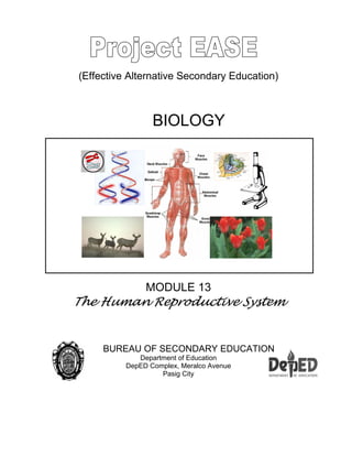
- 1. (Effective Alternative Secondary Education) BIOLOGY MODULE 13 The Human Reproductive System BUREAU OF SECONDARY EDUCATION Department of Education DepED Complex, Meralco Avenue Pasig City
- 2. - 2 - Module 13 The Human Reproductive System What this module is about This module is about the human reproductive system. It will familiarize you with the parts, functions, growth and development of the human embryo. This topic will be of interest to you as it deals with the beginning of human life. There are four lessons prepared for you in this module and they are as follows: Lesson 1 - The Female Reproductive System Lesson 2 - The Male Reproductive System Lesson 3 - From Fertilization to Birth Lesson 4 - Human Reproductive and Developmental Concerns What you are expected to learn As you go over the lesson, you are expected to: 1. Identify the parts of the male and female reproductive systems. 2. Describe the jobs of the parts of the male and female reproductive systems. 3. Compare the stages of the menstrual cycle. 4. Explain the stages of human growth and development. 5. Describe some reproductive technologies. How to learn from this module In order to achieve the objectives of this module successfully, you have to remember the following: 1. Read and follow the instructions carefully. 2. Answer the pretest. 3. Take down notes and record points for clarification.
- 3. - 3 - 4. Take the posttest and check your answers against the key at the end of the module. 5. Try to obtain at least a 70% level of proficiency in the tests. What to do before (Pretest) Test I. Identify the word/phrase being described or defined in each item. __________ 1. Considered as the ovulation period for regular menstrual cycle __________ 2. The stage where organs are formed during fetal development __________ 3. The part of the male reproductive organ that is cut during vasectomy __________ 4. The type of birth control device that is used for safe sex __________ 5. It involves the fertilization of the egg in a “glass” __________ 6. The part of the female reproductive system where the egg is fertilized __________ 7. The technology where the doctor places donated sperm in the woman’s reproductive tract __________ 8. The female sex hormone __________ 9. The periodic shedding of tissues and blood from the inner lining of the uterus __________10. The thick, whitish fluid consisting of sperms and secretions from several glands of the male reproductive tract Test II. Provide the function of the different parts of the male and female reproductive system. Organ Function Testes Uterus Clitoris Vagina Scrotum Vas deferens Seminal vesicle Prostate gland Urethra Penis Key to answers on page 26.
- 4. - 4 - Lesson 1. The Female Reproductive System Humans have many body systems but the system involved in the production of offspring is called the reproductive system. Most organ systems of the body show little difference between male and female except in the case of the reproductive system. There is a striking difference between the male and the female reproductive systems, although they also share a number of similarities. For example, the reproductive organs of male and female are developed from the same embryological structures, and some hormones are the same for the male and female although they produce different responses. The female reproductive system has the following functions: 1. Production of female sex cells 2. Reception of sperm cells from the male 3. Nurturing the development of and providing nourishment for, the new individual Try to examine the diagram on Figure 1a. The figure shows a cut away view of the front of the female reproductive organ. Now, study the job of each part. Part Job Ovary Produces egg cells Oviduct Passageway of eggs from the ovary to the uterus (also the same place where the egg is fertilized. Uterus Place where fertilized egg develops Vagina Receives penis of male during mating The female reproductive organ consists of the ovaries, uterine tubes (fallopian tubes), uterus, vagina, external genitalia, and mammary glands. The internal reproductive organs of the female are located within the pelvis, between the urinary bladder and rectum. The uterus and the vagina are in the middle line, with an ovary on each side of the uterus. Figure 1. The Parts of the Female Reproductive System
- 5. - 5 - The Ovaries There are two ovaries each comparable to the size of an almond nut. It is suspended in the pelvic cavity by a ligament. The ligament extends from each ovary to the lateral body wall, and the ovarian ligament. The mesovarium is the mesentery that suspends the ovary to the body wall. The ovary contains ovarian follicle, which contains an oocyte, the female germ cell. Ovulation When follicles mature, they expand and rupture to release the egg and the process is called ovulation. After ovulation, the remaining cells of the ruptured follicle become transformed into a glandular structure called the corpus luteum. Fallopian Tubes This part extends from the area of the ovaries to the uterus. Long, thin processes called fimbriae surround the opening of each uterine tube. Fertilization usually occurs in the part of the uterine tube near the ovary. Uterus This is as big as a medium-sized pear. The larger rounded part is directed superiorly. The part of the uterus superior to the entrance of the uterine tubes is called the fundus. The main part is called the body, and the narrower part is the cervix, directed inferiorly. Internally, the uterine cavity continues through the cervix as the cervical canal, which opens into the vagina. Vagina This is the female organ for copulation and functions to receive the penis during intercourse. It also allows menstrual flow and childbirth. The vagina extends from the uterus to the outside of the body. In young females, the vaginal opening is covered by a thin mucous membrane called the hymen. This can completely close the vaginal opening, in which case it must be removed to allow menstrual flow. The hymen can be perforated or torn at some earlier time in a young female’s life during a variety of activities including strenuous exercise. The condition of the hymen is therefore not a reliable indicator of virginity.
- 6. - 6 - External Genitalia The female external genitalia consists of the vestibule and its surrounding structures. The vestibule is the space into which the vagina and urethra open. A pair of thin, longitudinal skin folds called the labia minora borders the vestibule. A small erectile structure called the clitoris is located in the anterior margin of the vestibule. The two labia minora unite over the clitoris to form a fold of skin called prepuce skin. Mammary Glands These are the organs of milk production and are located in the breasts. The mammary glands are modified sweat glands. Externally, each breast of both males and females has a raised nipple surrounded by a circular, pigmented, areola. Each breast consists of around 15-20 glandular lobes covered by a considerable amount of fat tissue. It is this fat that gives the breast its form. Figure 3. Mammary Gland Figure 2. The Female External Genitalia
- 7. - 7 - The Menstrual Cycle Figure 4. The Menstrual Cycle We have just discussed that normally, an ovary releases only one egg every 28 days. Now, what controls this timing? Hormones control many of the changes in the reproductive system. Remember that hormones are chemicals that affect certain body organs. The monthly changes that take place in the female reproductive system are called menstruation. This cycle occurs every month starting when a female is 10 to 13 years old. The monthly cycle continues for about 40 years. Refer to figure 4 and figure 5 for a clearer explanation of the different events that take place. Just follow 1-10 in proper order.
- 8. - 8 - In a nutshell, the important events during the menstrual cycle are as follows: 1. The pituitary gland starts the cycle. 2. The pituitary releases hormones that cause the egg in the ovary to mature. The luteinizing hormone (LH) initiates the maturation of the follicles, converts ruptured follicle into corpus luteum and caused the secretion of progesterone. The other hormone, follicle stimulating hormone (FSH) assists in the maturation of the follicles and caused the secretion of estrogen from the follicles. 3. Meanwhile, the ovary itself releases a hormone called estrogen. Estrogen is a hormone that causes changes in the female reproductive system. This hormone also causes the uterus to increase in thickness. The uterus becomes thicker so that the fertilized egg can attach to it. 4. The ovary releases an egg on day 14. Assume that no sperm was present. 5. The egg moves through the oviduct and enters the uterus. 6. Meanwhile the uterus continues to thicken. 7. The egg has not been fertilized, therefore, it will not attach to the uterus. 8. The thick uterus is no longer necessary, so it begins to break apart. The cells of the thickened uterus break off and leave the vagina. The unfertilized egg is lost. Some blood is lost too. This loss of cells from the uterus lining, blood and egg is called menstruation. 9. After menstruation, the cycle starts again. Figure 5. A graph of the menstrual cycle
- 9. - 9 - What you will do Activity 1.1 What is the Menstrual Cycle? Materials: 2 calendar charts diagrams of the male and female reproductive system scissors tape or glue Procedure: Part A - For no fertilization: 1. Get a calendar. It must be marked by the day-to-day changes in the menstrual cycle. 2. Note that certain events are marked on certain days. 3. Make a copy of the diagrams of the menstrual cycle like those found below. Some of the diagrams will show events in the ovary, and some will show events in the uterus. They are not in proper order. Cut out each square. 4. Place the diagram in the space to the right of the corresponding description. 5. Tape or glue your diagrams in right places/dates where they occur. 6. Make sure that they are correctly placed. Part B – With fertilization of Egg 1. Get another calendar marked by the day-to-day changes in the menstrual cycle. 2. Again you will be given a set of diagrams to place on the calendar. The diagrams will not be in proper order. You may not need all the diagrams that show the uterus. Questions: 1. How long does a menstrual cycle last if fertilization does not take place? 2. Describe what happens to an egg during the first 14 days of the cycle in part A. 3. Describe what happens to the egg and the uterus during the last 14 days of the cycle in part B. 4. What takes place after fertilization? Key to answers on page 26.
- 10. - 10 - Did you know? Menstrual cramps are the results of the strong contractions of the uterine wall that occur before and during menstruation. The cramps can result from excessive secretion of prostaglandins. Sloughing of the endometrium of the uterus results in the inflammation in the endometrial layer of the uterus and prostaglandins are produced as part of the inflammation. In some women, menstrual cramps are extremely uncomfortable. Some women take aspirin-like drugs to avoid prostaglandin secretion just before menstruation. These drugs can reduce the pain. Data and Observations :
- 11. - 11 - What you will do Self-Test 1.1 Concept Mapping: Fill-in the concept map of egg release (start with the ovary). Ovary Releases an egg in a process called The egg travels through the To the If the egg is not fertilized, the uterine walls slough off and blood comes out in the process called If egg is fertilized, it is called a Ovary Zygote Key to answers on page 26.
- 12. - 12 - Lesson 2. The Male Reproductive System Examine the diagram on the next page. Shown are the parts of the male reproductive system. Figure 6 shows the main parts of the male reproductive system. Notice that this figure is a cut-away view from the side. Also notice that certain parts of the male reproductive system are also part of the urinary system. Be familiar with the specific functions of the parts using the table below: Part Job Testis Produces sperm cells Scrotum Sac that holds the testis Penis Places sperms into the vagina during mating Tube (vas deferens) Carries sperm from testes to urethra Urethra Carries sperm out of the body Glands a. seminal vesicle b. prostate gland c. Bulbourethral gland Provide liquid in which sperm can swim Secretes one of the components of the semen Secretes a milky fluid that is discharged into the urethra as part of the semen Mucous secreting glands located at the base of the penis Did you know? …that some males do not have descended testes? Ideally, during the seventh and the eighth month of fetal development or in some cases shortly after birth the testes descends. However, in some cases the testes fails to descend into the scrotal sac in a condition called cryptorchidism. It results to sterility because of the inhibiting effect of normal body temperature on sperm cell development. Figure 6. The Male Reproductive System
- 13. - 13 - Scrotum Externally, the scrotum consists of skin. Beneath the skin are a loose connective tissue and a layer of smooth muscle called dartos. In cold temperatures, the dartos muscles contract, causing the skin of the scrotum to become firm and wrinkled, reducing the overall size of the scrotum. Testes The testes are oval organs within the scrotum each about 4-5 cm long. Each testes is composed of cone-shaped lobules that contain seminiferous tubules, in which sperm cells develop. Epididymis This is a tightly coiled series of thread-like tubules that form a comma-shaped structure on the posterior side of the testes. The sperm cells continue to mature along this tube. Vas deferens The vas deferens emerges from the epididymis and ascends along the posterior side of the testis to become associated with the blood vessels and nerves that supply the testes. From the epididymis, sperms move to this tube up to the ampulla of the ductus deferens. The wall of this tube is composed of smooth muscles. Ejaculatory Duct The ejaculatory duct extends into the prostate gland and ends by joining the urethra within the prostate gland. Urethra The male urethra extends from the urinary bladder to the distal end of the penis. The urethra is a passageway for both urine and male reproductive fluids. The two, however, do not exit the urethra at the same time. So, there is no mixing. While seminal fluid passes through the urethra, a reflex causes the urinary sphincter muscles to contract tightly to keep urine from passing the urinary bladder through the urethra. Penis The penis is the male organ of copulation and functions in the transfer of sperm cells from the male to the vagina of the female. It is only an accessory organ of reproduction and not the reproductive organ as most people think of. It is composed of erectile tissues and the engorgement of this erectile tissue with blood causes the penis to enlarge and become firm in a process called erection.
- 14. - 14 - Where Are Sperms Formed? Each testis is partitioned into as many as 300 wedge-shaped lobes. Each contains two to three highly coiled tubes, the seminiferous tubules, and this is where sperms develop. Although a testis is only about 5 cm long, 125 meters of tubes are packed in it! When sperms move out of the testis, they enter a long, coiled duct, the epididymis. The sperms are not fully developed at this time, but secretions from the duct walls help them mature. When they are about to leave the body, they pass through a thick-walled tube the vas deferens, the ejaculatory ducts, and finally, the urethra, where they are ejected. Figure 7. Cross-section of the testis Did you know? …..that prostate cancer is the second most common cause of male deaths from cancer in the United States?
- 15. - 15 - What you will do Self-Test 2.1 Answer the following questions: 1. Why are so many sperms released if only one is needed to fertilize the egg? 2. Trace and label the pathway of the sperm as it moves from the testes to the outside (use arrows). What you will do Activity 2.1 Collect clippings or articles regarding the effect of alcohol and smoking on the male reproductive system. Summarize your work and you can put this in a clear book or portfolio. Lesson 3. From Fertilization to Birth Now that you know the parts and functions of the human reproductive system, let us trace what happens to the ovulated egg. Figure 8 shows the orderly sequence of events from the time an egg is ovulated. It is numbered 1-7. Just follow the numbers and you will be guided accordingly. Figure 8. Fertilization and Implantation in the Uterus Key to answers on page 27.
- 16. - 16 - Below is a summary of the stages of reproduction that can serve as your guide: 1. Egg cells are formed in each ovary. 2. Each month, one ovary releases an egg. Usually, only one egg is released about every 28 days. The ovaries usually take turns releasing the eggs. 3. Once released from the ovary, the egg moves into a tube called oviduct. Oviducts are tube-like organs that connect the ovaries to the uterus. The uterus is a muscular organ in which the fertilized egg develops. 4. Sperms are released into the vagina during mating. The vagina is a muscular tube that leads from outside of the female’s body to the uterus. Sperms swim from the vagina into the uterus and into the oviducts. If an egg is present, fertilization takes place. The fertilized egg moves down the oviduct into the uterus. 5. The fertilized egg then attaches itself to the wall of the uterus implantation. Once attached it will remain there for nine months as it develops into a baby. Ovulation This refers to the release of a mature egg from the ovary. It usually takes place on the 14th day from the first day of menstruation if the cycle is a 28-day cycle. Fertilization When a sperm encounters an egg cell in the fallopian tube, it releases digestive enzymes. Those enzymes clear the path for the sperm nucleus to fuse with the nucleus of the ovum or egg cell. A zygote is now formed. Implantation This occurs before the end of the first week. By this process, the zygote attaches to the uterine lining, and some of its cells send out projections that has been part of the maternal tissue. The inner cell mass becomes the embryonic disc. This disc will give rise to the embryo proper during the week following implantation. Did you know? ... that ectopic pregnancy results if implantation occurs anywhere other than the uterine cavity? The most common site of ectopic pregnancy is the fallopian tube. Implantation in the fallopian tube can be fatal and can cause the tube to rupture. In some cases, implantation can occur in the mesenteries of the abdominal cavity, and the fetus can develop normally, but must be delivered by caesarian section.
- 17. - 17 - Embryonic and Fetal Development Three weeks after fertilization, almost one fourth of the inner surface of the uterus has become a spongy tissue composed of endometrium and embryonic membranes, the chorion. Through this tissue, the placenta and the embryo receive nutrients and oxygen from the mother and send out wastes in return. First trimester The first trimester of the nine months of human development extends from fertilization to the end of the third month. The picture below shows how the embryo looks like during this period. This is the most critical period of embryonic development. Figure 8. First Trimester Embryo http://images.google.com.ph/images?hl=tl&lr=&q= images+of+the+first+trimester+embryo&btnG=Hanapin Second Trimester This extends from the start of the fourth month to the end of the sixth month. All major organs have formed, and the growing individual is now called a fetus. Movements of the facial muscles produce frowns. The sucking reflex is also evident. There are already fetal movements of arms and legs. The heartbeat of the fetus can also be heard from a stethoscope on the mother’s abdomen. Figure 9. Second Trimester Embryo http://images.google.com.ph/images?hl=tl&lr=&q= images+of+the+second+trimester+embryo&btnG=Hanapin Third Trimester This extends from the seventh month until birth. By the middle of the third trimester, the fetus will be able to survive on its own, if born prematurely or removed surgically from the uterus. The advancement in medical science has allowed fetuses as young as 23-25 weeks to survive early delivery. Survival chances increase to about 95 percent on the 9th month. Figure 10. Third Trimester Embryo http://images.google.com.ph/images?q=images%20of%20a%20third%20 trimester%20fetus&hl=tl&lr=&sa=N&tab=wi
- 18. - 18 - Birth or Parturition “Happy birthday!” is a very common greeting to mark the anniversary of a person’s birth. Birth takes place about 39 weeks after fertilization. The birth process begins when the uterus starts to contract. For the next two to eighteen hours, the contraction becomes stronger and more frequent. The cervical canal dilates fully and the amniotic sac ruptures. Birth typically occurs less than an hour after full dilation. Immediately afterward, uterine contraction forces fluid, blood and the placenta from the body. The umbilical cord is now cut, and the newborn embarks on its nurtured existence in the outside world. What you will do Self-Test 3.1 1. What happens when an egg is fertilized? 2. Differentiate ovulation from fertilization. 3. Describe the first, second and third trimester of embryonic and fetal development. 4. What is ectopic pregnancy? 5. Describe the uterus during parturition What you will do Activity 3.1 Measuring the Growth Rate of the Embryo Use the data below to make a graph showing the day of development versus the size of the embryo. When is the fastest period? Week after Fertilization Size 3 3 mm 4 4 mm 6 12 mm 7 20 mm 8 40 mm 9 50 mm Key to answers on page 27. Key to answers on page 27.
- 19. - 19 - Lesson 4. Human Reproductive and Developmental Concerns Have you heard about test tube babies and artificial inseminations? Are you interested to learn more about these topics? Read on. One of the goals of reproductive Biology is to understand human development and to provide new medical technologies. The human reproductive system is unique in that it does not start to function actively until several years after birth. It is also unique because we can survive without it! Isn’t that amazing? As medical science advances, scientists learn more about the complex biological interactions that form a newborn from a fertilized ovum, which, in turn, leads them to discover more ways to prevent conception and treat birth defects, and to heal the fetus or newborn baby. What is Infertility? Infertility is the inability of the sperm nucleus to merge with the egg nucleus. Almost one out of six couples are infertile. What are the causes of infertility? There are several factors that cause infertility but the most common are: 1. Physical difficulties 2. Irregular menstrual cycle 3. Low sperm count in males Infertility in Males Infertility in males is caused by hormonal imbalance. This hormonal imbalance results to lower sperm count. Sometimes man’s immune system produces antibodies that cover the sperm and prevent them from binding to the egg cell. A varicose vein in the scrotum can also cause infertility. This enlarged vein can overheat the developing sperms so they cannot mature. Sperm quality is also a factor. Some sperms are not very motile and so, they could hardly reach the egg cells. Hormone therapy can sometimes solve sperm motility. Infertility in Females Infertility in a woman can arise from abnormalities in any part of the reproductive system. Many women who are infertile have irregular menstrual cycle. A tumor in the ovary or in the brain’s pituitary gland can cause hormonal imbalance that usually results to irregular ovulation. Another could be the abundance of prolactin in non-pregnant women, which prevents ovulation, and therefore, cannot conceive. Fertility drugs can stimulate ovulation but sometimes they may also cause women to “superovulate” and produce more than one egg a month, which could result to triplets, quadruplets, or multiple births!
- 20. - 20 - Another problem could be a blocked fallopian tube. This is a common cause of infertility in females. Blockage can prevent the sperms from reaching the egg cell, or keep the fertilized egg from descending into the uterus, resulting in an ectopic pregnancy in the tube. Figure 11. Ectopic Pregnancy http://images.google.com.ph/images?hl=tl&lr=&q= images+of+ectopic+pregnancy&btnG=Hanapin An excess tissue growing on the uterine lining can also cause female infertility. Benign fibroid tumors or endometriosis can make the uterus not pleasant to the embryo. In endometriosis, the excess tissue bleeds during menstruation causing painful cramps. It can make conception difficult. Infertility is common among older women. They are more likely to produce oocytes with abnormal numbers of chromosomes. Assisted Reproductive Technologies Do you know that some infertility problems are no longer a problem these days? Yes! Certain technologies can help women conceive. What are these technologies? Artificial Insemination This is the oldest reproductive technology, where the doctor places donated sperms in a woman’s reproductive tract. This is needed especially when the partner is infertile or carries a gene for an inherited illness or if she decides to be a single parent. More than 250,000 babies have been born worldwide as a result of this procedure. The first human artificial insemination was born in 1890’s. In 1953, they started to use frozen and stored sperms for later use. Now these are called sperm banks. They freeze and store donated sperms and provide it to physicians who perform artificial insemination. A Donated Uterus - Surrogate Mothers A woman can be both a genetic parent and a gestational parent (or uterus provider). This happens when a man produces healthy sperms but his partner’s uterus is absent or cannot maintain pregnancy. A surrogate mother can help them become parents. This is how it happens. A surrogate mother is artificially inseminated with the man’s donated sperm and carries the pregnancy, but the couple raises the child. In this situation, the surrogate is both the genetic and gestational mother. Figure 12. Artificial Insemination http://images.google.com.ph/images?q= images+of+artificial+insemination &hl=tl&lr=&start=20&sa=N
- 21. - 21 - Another type of surrogate mother lends only her uterus. She receives the fertilized egg from the woman who has healthy ovaries but lacks a functional uterus. In Vitro Fertilization Literally it means “fertilization in a glass”. The sperm meets the egg cell outside the woman’s body. After few divisions, the fertilized egg cell is then introduced into the donor’s uterus or another woman’s uterus. If everything is fine, the embryo will implant into the uterus developing into a fetus. This is specially recommended if the fallopian tubes are blocked. This process is done in laboratory with a 68% fertilization rate. Gamete Intra-fallopian Transfer (GIFT) Since IVF is expensive, scientists have now tried another technology to solve reproductive problems. Presently, a new technology called Gamete Intra-fallopian Transfer (GIFT) solves this problem, by moving fertilization to the woman’s body rather than in a glassware. The woman takes a super ovulation drug for a week and then has several of her largest egg cells removed, then a man donates a sperm sample and the physician separates the most active cells. The collected oocyte and sperms are deposited together in the woman’s fallopian tube where there is no obstruction so that implantation can occur. Oocyte Banking and Donation Do you know that egg cells or oocytes can also be stored just like the sperms? Yes! It can be done. A woman wishing to have a baby later in her life when fertility declines can set aside oocytes while still young. However, this process is not as easy as freezing sperm cells in the bank. Healthy women can also donate healthy oocytes to other women, but it’s Challenge! A 25 year-old woman has her uterus removed due to cancer. However, her ovaries are intact and her egg cells are healthy. She takes a “super-ovulation” drug and has her egg cells removed, and they are fertilized in vitro with her husband’s sperm. One fertilized ovum was implanted into the uterus of the woman’s best friend. Who are the genetic parents? Gestational parents? Figure 13. Vitro Fertilization http://images.google.com.ph/images?hl=tl&lr=&q= images+of+in+vitro+fertilization&btnG=Hanapin
- 22. - 22 - not that easy. There are several processes that you have to undergo. Isn’t this exciting? Now, age does not matter in conceiving a baby. What matters is money since you have to pay the egg bank. Because of this technology, even menopause women, like a 62- year old can conceive a child! Isn’t this amazing? They do this with the egg fertilized in laboratory dish and later on transferred to her uterus as long as appropriate hormones are given. All these are examples of the wonders of technology. What you will do Self-Test 4.1 1. What is artificial insemination? 2. How can a surrogate mother assist an infertile couple? 3. What is GIFT? 4. Why is it difficult to freeze egg cells than sperm cells? 5. What are the causes of male infertility? What you will do Activity 4.1 Look for magazines and newspapers and cut out articles related to the effects of smoking on the health of the developing embryo and newborn babies. Birth Control Methods Why control birth? Well, the world’s population is now very close to 6.5 billion and the carrying capacity of mother earth is only around 8 billion, which means that we are already very close to the limit. Governments of the world should work together to solve the rapidly growing population for the benefit of humanity. Birth control methods then are necessary. Contraception is the use of devices or practices that work “against conception”. These methods either block the fusion of sperm and egg cell, or they make the females system’s environment hostile to sperm or to the pre-embryo’s implantation. Below are examples of birth control devices: Key to answers on page 28.
- 23. - 23 - Figure 14. Birth Control Devices http://www.uwm.edu/People/cortney Examine the table below. These are the different birth control methods. BIRTH CONTROL METHODS Barrier and Spermicidal Method Mechanism Advantages Disadvantages Success Condom and spermicide Worn over penis (male) or inserted into vagina, and kill sperm that escape Protection against sexually transmitted diseases Disrupt spontaneity, reduces sensation 95-98% Diaphragm and spermicide Kill sperm and blocks cervix Inexpensive Disrupt spontaneity, must be fitted 83-97% Cervical cap and spermicide Kill sperm and blocks cervix Inexpensive, can be kept in for 24 hours May slip out of place, must be fitted 80-95% Spermicidal foam or jelly Kills and blocks cervix Inexpensive Messy 78-95% Spermicidal or suppository Kills sperm, and blocks cervix Easy to use and carry Irritates 25 % of users 85-97%
- 24. - 24 - BIRTH CONTROL METHODS Hormonal Combination birth control pill Prevents ovulation and implantation, thickens cervical mucus Does not interrupt spontaneity, lowers cancer risk, lightens menstrual flow Raises risk of heart disease in some women, weight gain and breast tenderness 90-100% Minipill Blocks implantation, deactivates sperm, thickens cervical mucus Fewer side effects Weight gain 91-100% Depo-Provera Prevents ovulation, alters uterine lining Easy to use, last 3 months Menstrual changes, weight gain, injection 99% Norplant Prevents ovulation, thickens cervical mucus Easy to use, last 5 years Menstrual changes, doctor must implant 99.8% Behavioral Rhythm method No intercourse during fertile time No cost Difficult to do, hard to predict timing 79-87% Withdrawal Removal of penis from the vagina before ejaculation No cost Difficult to do 75-91% Surgical Vasectomy Sperm cells never reach penis Permanent, does not interrupt spontaneity Requires minor surgery, difficult to reverse 99.85% Tubal ligation Oocytes never reach the uterus Permanent, does not interrupt spontaneity Requires surgery, infection, difficult to reverse 99.6% Others Intrauterine device Prevents implantation Does not interrupt spontaneity Severe menstrual cramps, infection 95-99%
- 25. - 25 - What you will do Self-Test 4.2 Based on the summary of birth control methods, which do you think is the best method and why? What you will do Activity 4.2 Go to the nearest health center in your place and interview the health officer. Ask them about their program on population control. List down their program and take note of the family planning methods that are available and how they administer them to the community. If possible, try to familiarize yourself with the devices available in the health center. Let’s Summarize 1. Reproduction is the ability of organisms to produce offspring similar to the parents. 2. The female reproductive system includes ovaries, oviducts, uterus and vagina. 3. External genitalia include the vestibule containing separate openings of the vagina and urethra, the labia minor, labia major and the clitoris. 4. Fertilization of an egg occurs in the oviduct. The fertilized egg then attaches itself to the uterus. 5. The menstrual cycle is a series of events within the human female reproductive system. It prepares the uterus for a fertilized egg. 6. The human male reproductive system includes the testes, scrotum, penis, tubes to carry sperm, glands and the urethra. 7. Embryonic and fetal development occurs during pregnancy in humans. 8. Human pregnancy can be divided into three trimesters. Organogenesis is completed by the eight week. 9. Birth or parturition, results from strong, rhythmic uterine contractions that bring about the three stages of labor: dilation of the cervix, expulsion of the baby, and delivery of the placenta. 10.Infertility is the inability of the sperm to fuse with the egg cell. 11.There are several assisted reproductive technologies such as: artificial insemination, surrogate motherhood, in vitro fertilization, gamete intra-fallopian transfer, and oocyte banking and donation. 12.Contraception prevents pregnancy. 13.Contraceptive methods include preventing the release of mature gametes
- 26. - 26 - Posttest Test I. Identify the word/phrase being described or defined in each item. __________ 1. Considered as the ovulation period for regular menstrual cycle __________ 2. The stage where organs are formed during fetal development __________ 3. The part of the male reproductive organ that is cut during vasectomy __________ 4. The type of birth control device that is used for safe sex __________ 5. It involves the fertilization of the egg in a “glass” __________ 6. The part of the female reproductive system where the egg is fertilized __________ 7. The technology where the doctor places donated sperm in the woman’s reproductive tract __________ 8. The female sex hormone __________ 9. The periodic shedding of tissues and blood from the inner lining of the uterus __________10. The thick, whitish fluid consisting of sperms and secretions from several glands of the male reproductive tract Test II. Provide the function of the different parts of the male and female reproductive system. Organ Function Testes Uterus Clitoris Vagina Scrotum Vas deferens Seminal vesicle Prostate gland Urethra Penis Key to answers on page 28.
- 27. - 27 - Key to Answers Pretest Test I. 1. 14th day 6. fallopian tube 2. 1st trimester 7. Artificial insemination 3. vas deferens 8. estrogen 4. condom 9. menstruation 5. vitro-fertilization 10. semen Test II. Organ Function Testes Produce the sperm cells Uterus Site for the implantation of the zygote Clitoris Part of the external female genitalia that is composed of erectile tissue Vagina The place where the penis is inserted in the female Scrotum The folded skin that covers the testes Vas deferens The tube connected to the epididymis where sperms pass Seminal vesicle Paired structures that add fructose and proteins to the sperms. Prostate gland A small gland that produces alkali fluid Urethra Canal that serves as passageway for sperms and urine in males Penis The male sex organ inserted in the vagina Lesson 1 Activity 1.1 1. 28 days 2. It moves out of the uterus. 3. The egg is fertilized and attaches to the uterus for implantation. 4. Implantation Self-Test 1.1 Ovary Ovulation Fallopian Tube Uterus Menstruation Zygote
- 28. - 28 - Lesson 2 Self-Test 2.1 1. To compensate for the millions of sperms that will die along the way or even before they reach the egg. 2. Testes epididymis vas deferens ejaculatory ducts urethra outside Lesson 3 Self-Test 3.1 1. It starts to divide and moves down to the uterus for implantation. 2. Ovulation refers to the release of egg from the ovary, while fertilization is the union of the egg nucleus and sperm nucleus. 3. First trimester is from the first to the third month, organogenesis starts. Second trimester extends from the start of the 4th month to end of the 6th month, baby is about 30 cm. Third trimester extends from the 7th month until birth, baby is about 50cm long. 4. Pregnancy that occurs outside of the fallopian tube 5. The uterus contracts (due to hormone) to force the baby out of the birth canal. Activity 3.1 Measuring the Growth Rate of the Embryo 0 10 20 30 40 50 1 2 3 4 5 6 7 8 9 1 Size of Fetus in mm Weeks After Fertilization
- 29. - 29 - Lesson 4 Self-Test 4.1 1. A process where the doctor places donated sperm in a woman’s reproductive tract. 2. She can be artificially inseminated with the man’s donated sperm and carries on with the pregnancy. 3. Moving fertilization to the woman’s body rather than in a glassware. 4. Because they are very sensitive to temperature changes. 5. Causes of male infertility are: a. hormonal imbalance b. varicose vein in the scrotum c. low sperm count d. motility problem Posttest Test I. 1. 14th day 6. fallopian tube 2. 1st trimester 7. Artificial insemination 3. vas deferens 8. estrogen 4. condom 9. menstruation 5. vitro-fertilization 10. semen Test II. Organ Function Testes Produce the sperm cells Uterus Site for the implantation of the zygote Clitoris Part of the external female genitalia that is composed of erectile tissue Vagina The place where the penis is inserted in the female Scrotum The folded skin that covers the testes Vas deferens The tube connected to the epididymis where sperms pass Seminal vesicle Paired structures that add fructose and proteins to the sperms. Prostate gland A small gland that produces alkali fluid Urethra Canal that serves as passageway for sperms and urine in males Penis The male sex organ inserted in the vagina
- 30. - 30 - References Books: Campbell. N.A. (1996). Biology (4th ed.). California: Benjamin Cummings Publishing, Menlo Park, California Daniel, L., Ortleb, E. & Biggs, A. (1994). Life Science. Glencoe. Macmillan/McGraw-Hill, California. Kaskel, A., Hummer P.J. & Daniel, L. (1988). Biology: An everyday experience. Ohio, USA: Merill Publishing Company. Columbus, Lewis, R. (1998). Life. (3rd ed.) USA: WCB McGraw-Hill Companies, Inc. Mader, S. (2001). Biology. (7th ed.) USA: The McGraw-Hill Companies , Inc. Seeley, R.R., Trent, S.D. & Tate, P. (1992). Essentials of anatomy and physiology. (3rd ed.) USA: McGraw-Hill. Boston Burr Ridge. Wong, H.K. Biology key ideas. (1983). Engle Wood Cliffs, New Jersey: Prentice-hall Inc. Electronic Sources: Retrieved Feb. 5, 2005 from http://images.google.com.ph/image Retrieved Feb. 5, 2005 from http://images.google.com.ph/images Retrieved Feb. 5, 2005 http://images.google.com.ph/images Retrieved Feb. 5, 2005 http://images.search.yahoo.com/search/images