SlideShare une entreprise Scribd logo
1  sur  14
Télécharger pour lire hors ligne
Respiratory Internal Medicine

Type I respiratory failure             Type II Respiratory failure
Represents failure of oxygenation      Represents a defect in ventilation
and is characterized by a low PaO2     (hypoventilation) and is
with normal or low PaCO2.              characterized by decreased PaO2
PaO2 : Low (<60 mm Hg)                 with increased PaCO2.
PaCO2 : Normal or Low (≤49 mm          PaO2 : Decreased (<60 mm Hg)
Hg)                                    PaCO2 : Increased (>49 mm Hg)
PA-aO2 : Increased                     PA-aO2 : Normal
Causes:                                Causes:
This type is caused by conditions      This type is caused by conditions
which affect oxygenation, like:        causing hypoventilation as in :

 Parenchymal disease (V-Q              Obstructive lung disease: COPD,
  mismatch)                              Foriegn.body
 Diseases of vasculature/shunts        Decreased central respiratory
  Examples:                              drive e.g. CNS disorders like:-
        - Pneumonia                      Brain injury, Meningitis
        - ARDS                          Weakness of respiratory muscles
        - Emphysema                      e.g
        - Right to left shunts            - Peripheral N.S. disorders like:
                                             M.gravis.
                                          - Interstitial lung disease.
                                          - MS disorders like
                                             polymyositis.
                                          - Rib cage disorders:
                                             Kyphoscoliosis.

Type I respiratory failure             Type II Respiratory failure
Hypoxemia with decreased PaCO2         Hypoxemia with increased PaCO2


Global institute of medical sciences
Respiratory Internal Medicine


PRT Results       Obstructive Pattern                           Restrictive Pattern
FEV1              Decreased (<80% predicted)                    Decreased (May be preserved)
                  (Decreased out of proportion to               (Decreased in proportion to
                  FVC)                                          FVC)
FVC               Decreased (may by preserved)                  Decreased ((<80% predicted)
FEV1/FVC          Decreased (<0.7)                              Normal or increased (>0.7)
(FEV1%)
FEF25-75          <50% predicted                                Decreased in proportion to loss
                                                                of lung volume
TLC               Normal or elevated                            Decreased
DLCO              Normal                                        Decreased in intrinsic restrictive
                  Decreased in Emphysema                        lung disease.
                                                                Normal in neuromuscular or
                                                                musculoskeletal restrictive
                                                                disease.


                           Respiratory disease


         Obstructive                                       Restrictive
      1. asthma
      2. COPD – Chronic
      bronchitis                        Parenchymal                    Extra parenchymal
      Emphysema                        - sarcoidosis                  1. Neuromuscular
      3. Bronchiectasis                - pneumoconiosis               disease
      4. Cystic fibrosis               - idiopathic pulmonary          - Diaphragmatic palsy
                                       fibrosis                        - GB syndrome
      5. Bronchiolitis
                                       - drug/Radiation                - Muscular dystrophy
                                       induced interstitial            - Cervical spine injury
                                       lung disease.                  2. Cest wall
                                                                        - Kphoscoliosis
                                                                        - Oesity
                                                                        - Akylosing spondylitis


Global institute of medical sciences
Respiratory Internal Medicine


                                Obstructive lung disease        Restrictive lung disease
Total lung capacity             Normal to increase              Decrease
Residual volume                 Increase                        Decrease
Vital capacity                  Decrease                        Decrease
FEV1/FVC                        Decrease (<0.7)                 Normal to increase (>0.7)
FEF 25-75%                      Decrease                        Normal
(forced expiratory flow
rate)
Diffusion capacity              Normal (decrease in             Decreased
                                emphysema)


FVC
The forced vital capacity (FVC) represents the total volume of gas exhaled with
maximum expiration following maximal inspiration. A reduced FVC suggests
pulmonary restriction.
FEV1% or FEV1/FVC
Note that FEV1 expressed as a percentage is actually FEV1/FVC.
The volume of gas exhaled during the first second while expiration from FVC is FEC1.
FEV1 is often represented as a ration of the FVC (often referred to as FEV1% rather than
FEV1/FVC)
Normal to increased FEV1% (FEV1/FVC) suggest restrictive lung disease
Decreased FEV1% (FEV1/FVC) suggests obstructive lung disease.
DLCO : (Diffusion capacity for carbon monoxide)
This reflects the ability of lungs to transfer gas across the alveolar – capillary interface.
Decreased DLCO is consistent with a diagnosis of:
 Interstitial lung disease
 Emphysema
 Pulmonary hypertension


Global institute of medical sciences
Respiratory Internal Medicine


                                  Increased PaO2-PaO2
                          (alveolar arterial difference in oxygen)


        Correctible with oxygen                       Not correctible with oxygen
            V/Q Mismatch                              shunt
V/Q Mismatch:                                          Shunt:
 - Airway disease (Asthma,                              - Alveolar collapse
   COPD)                                                - Intra alveolar filling
 - Interstitial lung disease                            1. pneumonia
 - Alveolar disease                                     2. pulm. Edema
 - Pulmonary vascular disease                           - intracardiac shunt
                                                        - vascular shunt within lung


Decreased D LCO (diffusion capacity)        Increased DLCO (diffusion capacity)
   1. interstitial lung disease:- scarring     1. alveolar haemorrhage:- as a Good
      of alveolar capillary units                  pasture‟s syndrome: haemoglobin
      diminishes area of alveolar capillary        contained in erythrocytes in
      bed as well as pulmonary blood               alveolar lumen binds Co so exhaled
      volume                                       carbon monoxide concentration is
   2. emphysema:- alveolar walls are               diminished & DLCO is increased.
      destroyed so the surface area of         2. congestive heart failure:- may be
      alveolar capillary bed is diminished         elevated if pulmonary blood
   3. recurrent pulmonary embolism                 volume is increased. Once
      and primary pulmonary                        pulmonary edema ensues DLCO
      hypertension:- disease causes a              may decrease as and the net DLCO
      decrease in cross sectional area and         depends on the opposing influences.
      volume of pulmonary vasculature


Global institute of medical sciences
Respiratory Internal Medicine


                                         Asthma


      Extrinsic asthma /allergic asthma        Intrinsic asthma/Idiosyncratic
                                               asthma
      - History of atopy is present
      - Attacks related to environmental       - No history of atopy (concomitant
      exposures                                nasal polyps are usually present
      - IgE antibody levels are increased      and patients may be aspirin
      - Early onset of disease (young)         sensitive)
      - Usually is less severe, intermittent   - Attacks not related to
      asthma that may however progress         environmental exposures: occur
      to severe persistent asthma              without provocation
                                               - IgE antibody levels are normal
                                               - Delayed onset of disease (adult
                                               onset asthma)
                                               - Usually have more severe
                                               persistent asthma.


The microscopically identifiable features described in sputum are three ‘C’s
Charcot Leyden crystals : derived from granules of eosnophils and found only in asthma
Curshmann spirals       : curiously twisted casts of airways : Whorls of shed epithelium
Creola bodies           : clumps of cells or isolated metaplastic cells.


Hypoxia is the universal finding in Asthma
Hypocapnia and respiratory alkalosis is seen in most asthmatic patients
Hypercarbia and Respiratory acidosis are very late features of asthma and signify
severe obstruction and respiratory failure. These are not universal findings in Asthma.


Global institute of medical sciences
Respiratory Internal Medicine

Management of acute bronchial asthma:

                      Sit patient up and give high dose O2 (100%)


                        β2 agonist / Salbutamol (nebulized)

                                  Alternate β2 agonist
                                      Terbutaline
                                     Fenoterol etc.


                    Steroids: hydrocortisone IV or Prednisolone orally


                 Additive treatment if life threatening features are present
                          1. anticholinergic : iprotropium bromide
                             2. methylxanthines : aminophyllines


Panacinar emphysema                           Centriacinar emphysema
         - acini are uniformly involved                 - central or proximal parts of
            from level of respiratory                       acini formed by respiratory
            bronchiole to terminal blind                    bronchioles are affected
            alveoli.                                        whereas distal alveoli are
         - Lsions are more common in                        spared
            lower zone and bases                        - lesions are more severe and
         - Occurs in association with                       common in Upper lobes
            α1 antitrypsin deficiency                   - occurs predominantly in
                                                            smokers
                                                        - I the commonest pattern


Global institute of medical sciences
Respiratory Internal Medicine


                                    Asbestos related lung disease


                     Pulmonary                                     Pleural


         Non malignant          Malignant             Non malignant          Malignant
         Diffuse                 Bronchogenic
         interstitial                                 - Pleural plaques      - Mesothelioma
                                 carcinoma            - Diffuse pleural      Both plerural
         fibrosis
                                 All histological     thickening             and peritoneal
         (asbestosis)
                                 subtypes of          - Acute benign         mesotheliomas
         Asbestosis is
                                 lung cancer          pleural effusion       develop in
         defined as a
                                 occur with           - Rounded              persons
         diffuse interstial
                                 increased            atelectasis            exposed to
         fibrosing disease
                                 frequency but                               asbestos
         of lung caused by
                                 adenocarcino
         exposure to
                                 ma has the
         asbestos fibres.
                                 highest
                                 incidence


Silicosis has predilection for upper lobes
Radiographs typically show fine nodularity in the upper zones of the lung
Rounded opacities appear in the upper lobes on chest radiograph
Silicosis is associated with calcific Hilar adenopathy
Calcification of hilar lymph nodes may occur in as many as 20% oc cases and produce a
charachteristic “egg shell” pattern.
Silicosis is associated with tuberculosis
Because silica is cytotoxic to alveolar macrophages, patients with silicosis are at greater
risk of acquiring lung infections that involve these cells as a primary defense including
myocobacterium tuberculosis, atypical mycobacteria and fungi


Global institute of medical sciences
Respiratory Internal Medicine


                        Clinical spectrum of pulmonary aspergillosis


     saprophytic infection       invasive aspergillosis     Allergic aspergillosis

     aspergilloma (fungal        - Invasive bronchial
     ball)                                                  - Allergic bronchial
                                 aspergillosis (IBA)        asthma
                                 - Chronic pulmonary        - Allergic
                                 aspergillosis (CPA)        bronchopulmonary
                                 - Invasive pulmonary       aspergillosis(ABPA)
                                 aspergillosis (IPA)        - Extrinsic allergic
                                                            alveolitis (type to
                                                            hypersensitivity
                                                            pneumonitis)


kleibsella pneumonia

kleibsella pneumonia presents as a typical ‘air space’ pneumonia with
cough productive of purulent sputum. ‘purulent sputum production and
„air space‟ diseases X ray are typical‟
causes of atypical pneumonias:
    1. mycoplasma pneumonias
    2. viral pneumonias
          - influenza
          - RSV
          - Adenovirus
          - Rhinovirus
          - Rubeola
          - Varicilla
          - Corona virus
    3. Chlamydia pneumonia
    4. coxiella burnetti
    5. pneumocystis carinii

Global institute of medical sciences
Respiratory Internal Medicine
           Crona virus is an infrequent cause of pneumonia.


           Pneumocytic carinni pneumonia is characterized by a prominent
           eosinophilic exudates and mild interstitial pneumonitis. These is
           damage to type I pneumocytes and associated compensatory
           hypertrophy of type II pneumocytes.


                      Classic histopathology in pneumocystis pneumonia


          Intra alveolar changes                                interstitial changes

                                                        Mild interstitial pneumonitis
Prominent eosinophilic foamy, vacuolated
                                                        - Mononuclear interstitial cell
      intra alveolar exudeate (HE staining)
                                                            infiltrate (mild)
- Intra alveolar exudates contains the
                                                        - Damaged type I pneumocytes
      organisms, which can be identified in
                                                        - Hypertrophy / proliferation of type
      various stages of development
                                                            II pneumocytis (reparative
      (throphozoite and cystic stage) by the
                                                            response)
      use of special stains such as
                                                        pneumocystis attaches to and
        1. methamine silver / Toluidine stain
                                                            damages type 1 pneumocytes.
        2. wright giemsa stain
                                                            There is associated
        3. papanicolaou’s stain
                                                            compensatory hypertrophy of
                                                            type II pneumocytes.


        Global institute of medical sciences
Respiratory Internal Medicine


   Montoux test
    Montoux test is carried out by injecting one tuberculin unit (1TU) of PPD in 0.1 ml
   on the flexor surgace of forearm (PPD RT 23 with Tween 80)


                            Test is read after 72 hrs (not 48 hrs)


                                                      look for erythema and
                                                      induration


           <6 mm                           6-9 mm                >10 mm
           negative                        doubtful              Positive


           -   a positive test indicates that patient is infected with M. tuberculosis. It
               does not however prove that the person is ‘suffering’ from the disease
           -   studies indicate that 92% of new cases occur in persons who already
               are tuberculin reactors
           -   6-9 mm induration does not indicate high chances of developin
               tuberculosis. Infact patients with 25 mm induration have more chances of
               developing tuberculosis than those with 6-9 mm induration.


Global institute of medical sciences
Respiratory Internal Medicine

Sarcoidosis
Sarcoidosis is a chronic multisystem disorder of unknown causes characterized by
accumulation of T lymphocytes and mononuclear phagocytes in various tissues of body


                   Non caseating SARcoid granulomas in affected organs


     Lung (90%)            lymphnodes              skin                   Others
                           (75-90%)
                                                                      -Eye :- uveitis
     - interstitial                               - Erythema
                           - B/L hilar                                - Kidney (rare):-
     lung disease.                                nodosum
                           Lymphadenopathy                            renal
     . fibrosis of lung                           - Lupus             hypercalcemia
                           is the hallmark of     perenium
     parenchyma            sarcoidosis                                with or without
     - Pleura is                                  (purple blue        hypercalcuria
                           - Parotid              shiny
     involved in 5% of     enlargement B/e                            - Skeletal :-
     cases u/e                                    swollen             arthritis
                           involvement is the     lesions on
     pleural effusion      vuli                                       - Nervous
     - Cavitation is                              nose,
                                                                      system:-
     rare                                         cheeks, lips,       peripheral
                                                  ears).              neuropathy
                                                                      - Heart :- cor
                                                                      pulmonle


Inclusions seen in giant cells in sarcoidosis (remember as SARcoidosis)
           - Schaumann bodies
           - Asteroid bodies
           - Residual bodies


Global institute of medical sciences
Respiratory Internal Medicine


                                       Pneumothorax

                                                                   traumatic
          Spontaneous pneumothorax                                 pneumothorax (non
                                                                   spontaneous
                                                                   pneumothorax)
                                                                    -These
   Primary spontaneous               Secondary spontaneous          penumothoraces occur
   pneumothorax                      pneumothorax                   as a result of :
                                                                       1. blunt trauma or
   - Occur in persons with No          •      occurs in persons        2. penetrating trauma
   clinical evidence of lung                  with an underlying    - Iatrogenic
   disease and without                        lung disease as a     pneumothorax is
   precipitating event.                       complicationof the    subcategory of
   - Most of these patients have              underlying disease    traumatic
   occult lung disease (sub                   process               pneumothorax
   clinical) with subpleural blebs                                  Causes of iatrogenic
   on CT scan                              Etiology of              pneumothorax
   characterstics of affected
                                           secondary                - Thoracocentesis
   people are :-
   - tall, thin, males                     pneumothorax             - Insertion of central
   - smokers                                                        venous catheter
                                                                    - Mechanical
   - subpleural blebs on lungs
                                                                    ventilation (IPPV,
                                                                    assisted ventilation etc)
                                                                    - Surgery


        Obstructive lung disease
                                                       Malignancy
        Interstitial lung disease
                                                       Connective tissue
        Infection                                      disease


Global institute of medical sciences
Respiratory Internal Medicine


Facts to remember

Most frequent histological type                              Adenocarcinoma

Most frequent histological type in India                     Squamous cell
                                                             carcinoma
Most common histological varieny in life time non-smokers    Adenocarcinoma

Most common histological variety in young patients           Adenocarcinoma

Most common histological variety in females                  Adenocarcinoma

Most common site for metastasis from Ca lung                 Liver

Most common endocrine organ to be involved by metastasis     Adrenals
from Ca lung

Ca lung which metastizes to opposite lung                    Adenocarcinoma

Commonest tumor to metastise to heart                        Ca lung (Bronchognic
                                                             Ca)
Histological varieties that cavitate                         Squamous cell and
                                                             large cell
Histological varieties that are central in distribution      Squamous cell and
                                                             small cell
Histological varieties that are peripheral in distribution   Adenocarcinoma

Pancoost tumor is histologically                             Squamous cell
Most common variety associated with paraneoplatic syndrome   Small cell variety
Most common variety associated with hypokalemia              Small cell (presumably
                                                             d/t ACTH)
Most common variety associated with hypercalcemia            Squamous cell
                                                             (presumably d/t PTH)
Histological variety most responsive to chemotherapy         Small cell

Histological variety response to radiotherapy                Small cell


Global institute of medical sciences
Respiratory Internal Medicine
Histological variety associated with hest prognosis               Squamous cell


The most common malignancy of lung is adenocarcinoma (overall/world wide):-

Most common lung cancer worldwide is Adenocarcinoma
Most common lung cancer in India is swquammous cell carcinoma
Most common lung cancer in women is adenocarcinoma
Most common lung cancer in smokers is squammous cell carcinoma
Most common lung cancer in nonsmokers is adenocarcinoma
Most common lung cancer in young patients is adenocarcinoma
Most common lung cancer to metastasize is small cell carcinoma.

Ectopic hormones produced by small cell       Paraneoplastic syndrome
carcinomas
          - ACTH                              Cushings syndrome
          - SIADH/ANP                         Hyponatremia
          - Calcitonin                        Hypocalcemia
          - Gonadotropins                     gynaecomastia


Global institute of medical sciences

Contenu connexe

Tendances

Respiratory failure
Respiratory failureRespiratory failure
Respiratory failureVijay Sal
 
18 basics of pediatric airway anatomy, physiology and management
18 basics of pediatric airway anatomy, physiology and management18 basics of pediatric airway anatomy, physiology and management
18 basics of pediatric airway anatomy, physiology and managementDang Thanh Tuan
 
Respiratory Failure by Dr. Sookun Rajeev Kumar
Respiratory Failure by Dr. Sookun Rajeev KumarRespiratory Failure by Dr. Sookun Rajeev Kumar
Respiratory Failure by Dr. Sookun Rajeev KumarDr. Sookun Rajeev Kumar
 
Cyanosis ppt by dr girish jain
Cyanosis ppt by dr girish jainCyanosis ppt by dr girish jain
Cyanosis ppt by dr girish jainGirish jain
 
Ventilation perfusion relationships
Ventilation  perfusion relationshipsVentilation  perfusion relationships
Ventilation perfusion relationshipsKamal Bharathi
 
Occupational lung diseases
Occupational lung diseasesOccupational lung diseases
Occupational lung diseasesAshutosh Pakale
 
Pulmonary Flow Volume Loops.. Dr.Padmesh
Pulmonary Flow Volume Loops.. Dr.PadmeshPulmonary Flow Volume Loops.. Dr.Padmesh
Pulmonary Flow Volume Loops.. Dr.PadmeshDr Padmesh Vadakepat
 
Non respiratory functions of lungs
Non respiratory functions of lungsNon respiratory functions of lungs
Non respiratory functions of lungsAli Faris
 
Pathogenesis and Acute Exacerbation of IPF
Pathogenesis and Acute Exacerbation of IPFPathogenesis and Acute Exacerbation of IPF
Pathogenesis and Acute Exacerbation of IPFGamal Agmy
 
Respiratory failure
Respiratory failure Respiratory failure
Respiratory failure hafsaimtiaz2
 
Developmental disorders of lungs
Developmental disorders of lungsDevelopmental disorders of lungs
Developmental disorders of lungsFiroz Hakkim
 

Tendances (20)

Bronchiectasis
BronchiectasisBronchiectasis
Bronchiectasis
 
Causes of Haemoptysis
Causes of HaemoptysisCauses of Haemoptysis
Causes of Haemoptysis
 
Respiratory failure
Respiratory failureRespiratory failure
Respiratory failure
 
Respiratory failure
Respiratory failureRespiratory failure
Respiratory failure
 
Dypsnea
DypsneaDypsnea
Dypsnea
 
18 basics of pediatric airway anatomy, physiology and management
18 basics of pediatric airway anatomy, physiology and management18 basics of pediatric airway anatomy, physiology and management
18 basics of pediatric airway anatomy, physiology and management
 
Respiratory Failure by Dr. Sookun Rajeev Kumar
Respiratory Failure by Dr. Sookun Rajeev KumarRespiratory Failure by Dr. Sookun Rajeev Kumar
Respiratory Failure by Dr. Sookun Rajeev Kumar
 
Pleural disease
Pleural disease Pleural disease
Pleural disease
 
Cyanosis ppt by dr girish jain
Cyanosis ppt by dr girish jainCyanosis ppt by dr girish jain
Cyanosis ppt by dr girish jain
 
evaluation of dyspnoea
evaluation of dyspnoeaevaluation of dyspnoea
evaluation of dyspnoea
 
Ventilation perfusion relationships
Ventilation  perfusion relationshipsVentilation  perfusion relationships
Ventilation perfusion relationships
 
Occupational lung diseases
Occupational lung diseasesOccupational lung diseases
Occupational lung diseases
 
Pulmonary Flow Volume Loops.. Dr.Padmesh
Pulmonary Flow Volume Loops.. Dr.PadmeshPulmonary Flow Volume Loops.. Dr.Padmesh
Pulmonary Flow Volume Loops.. Dr.Padmesh
 
Acute Respiratory failure
Acute Respiratory failure Acute Respiratory failure
Acute Respiratory failure
 
Obstructive lung disease
Obstructive lung disease Obstructive lung disease
Obstructive lung disease
 
Respiratory failure
Respiratory failureRespiratory failure
Respiratory failure
 
Non respiratory functions of lungs
Non respiratory functions of lungsNon respiratory functions of lungs
Non respiratory functions of lungs
 
Pathogenesis and Acute Exacerbation of IPF
Pathogenesis and Acute Exacerbation of IPFPathogenesis and Acute Exacerbation of IPF
Pathogenesis and Acute Exacerbation of IPF
 
Respiratory failure
Respiratory failure Respiratory failure
Respiratory failure
 
Developmental disorders of lungs
Developmental disorders of lungsDevelopmental disorders of lungs
Developmental disorders of lungs
 

En vedette

Internal Medicine Sample Questions
Internal Medicine Sample QuestionsInternal Medicine Sample Questions
Internal Medicine Sample QuestionsDJ CrissCross
 
Respiratory Diseases
Respiratory DiseasesRespiratory Diseases
Respiratory Diseasesshas595
 
Discussion 1: Internship & Fmge ExAM
Discussion 1: Internship & Fmge ExAMDiscussion 1: Internship & Fmge ExAM
Discussion 1: Internship & Fmge ExAM维卡普 Pardhu
 
Benign Paroxysmal Positional Vertigo
Benign Paroxysmal Positional VertigoBenign Paroxysmal Positional Vertigo
Benign Paroxysmal Positional VertigoAnwaaar
 
Respiratory system exam
Respiratory system examRespiratory system exam
Respiratory system examPuneet Shukla
 
Cavernous Sinus Thrombosis
Cavernous Sinus ThrombosisCavernous Sinus Thrombosis
Cavernous Sinus ThrombosisZY The Ripper
 
Ppt respiratory system
Ppt respiratory systemPpt respiratory system
Ppt respiratory systemBhole Nath
 
Burning mouth syndrome
Burning mouth syndromeBurning mouth syndrome
Burning mouth syndromechelseaharpool
 
Introduction to internal medicine
Introduction to internal medicineIntroduction to internal medicine
Introduction to internal medicineqbank org
 

En vedette (20)

Internal Medicine Sample Questions
Internal Medicine Sample QuestionsInternal Medicine Sample Questions
Internal Medicine Sample Questions
 
Respiratory Diseases
Respiratory DiseasesRespiratory Diseases
Respiratory Diseases
 
Endocrine high yield
Endocrine high yieldEndocrine high yield
Endocrine high yield
 
Psychiatry high yield
Psychiatry high yieldPsychiatry high yield
Psychiatry high yield
 
Discussion 1: Internship & Fmge ExAM
Discussion 1: Internship & Fmge ExAMDiscussion 1: Internship & Fmge ExAM
Discussion 1: Internship & Fmge ExAM
 
Head and neck
Head and neckHead and neck
Head and neck
 
High yield points - mci screening test
High yield points - mci screening testHigh yield points - mci screening test
High yield points - mci screening test
 
Benign Paroxysmal Positional Vertigo
Benign Paroxysmal Positional VertigoBenign Paroxysmal Positional Vertigo
Benign Paroxysmal Positional Vertigo
 
Respiratory system exam
Respiratory system examRespiratory system exam
Respiratory system exam
 
Cavernous Sinus Thrombosis
Cavernous Sinus ThrombosisCavernous Sinus Thrombosis
Cavernous Sinus Thrombosis
 
Ppt respiratory system
Ppt respiratory systemPpt respiratory system
Ppt respiratory system
 
Benign Paroxysmal Positional Vertigo
Benign Paroxysmal Positional VertigoBenign Paroxysmal Positional Vertigo
Benign Paroxysmal Positional Vertigo
 
Head and neck
Head and neckHead and neck
Head and neck
 
Burning mouth syndrome
Burning mouth syndromeBurning mouth syndrome
Burning mouth syndrome
 
Biochemistry high yield part 1
Biochemistry high yield part 1Biochemistry high yield part 1
Biochemistry high yield part 1
 
Neuro anatomy High yield - fmge
Neuro anatomy High yield - fmgeNeuro anatomy High yield - fmge
Neuro anatomy High yield - fmge
 
Biochemistry high yield part 2
Biochemistry high yield part 2Biochemistry high yield part 2
Biochemistry high yield part 2
 
Biochemistry chart dr.g.bhanu prakash
Biochemistry chart dr.g.bhanu prakashBiochemistry chart dr.g.bhanu prakash
Biochemistry chart dr.g.bhanu prakash
 
Introduction to internal medicine
Introduction to internal medicineIntroduction to internal medicine
Introduction to internal medicine
 
Oral Medicine :Burning mouth syndrome
Oral Medicine :Burning mouth syndromeOral Medicine :Burning mouth syndrome
Oral Medicine :Burning mouth syndrome
 

Similaire à Respiratory internal medicine

ACUTE RESPIRATORY FAILURE MAGDI SASI 2015
ACUTE RESPIRATORY FAILURE MAGDI SASI 2015ACUTE RESPIRATORY FAILURE MAGDI SASI 2015
ACUTE RESPIRATORY FAILURE MAGDI SASI 2015cardilogy
 
Presentation1.pptx, radiological imaging of restrictive lung diseases.
Presentation1.pptx, radiological imaging of restrictive lung diseases.Presentation1.pptx, radiological imaging of restrictive lung diseases.
Presentation1.pptx, radiological imaging of restrictive lung diseases.Abdellah Nazeer
 
Respiratory Diseases -Obstructive lung diseases.ppt
Respiratory Diseases -Obstructive lung diseases.pptRespiratory Diseases -Obstructive lung diseases.ppt
Respiratory Diseases -Obstructive lung diseases.pptArpitaHalder8
 
Resp physiology & respiratory failure in children
Resp physiology & respiratory failure in childrenResp physiology & respiratory failure in children
Resp physiology & respiratory failure in childrenSameekshya Pradhan
 
COPD BY MAGDI SASI 2016
COPD BY MAGDI SASI 2016COPD BY MAGDI SASI 2016
COPD BY MAGDI SASI 2016cardilogy
 
باثوهObstructiveLungDiseaseI-1.pptx
باثوهObstructiveLungDiseaseI-1.pptxباثوهObstructiveLungDiseaseI-1.pptx
باثوهObstructiveLungDiseaseI-1.pptxTofikMohammed3
 
Diffuse Parenchymal Lung Diseases
Diffuse Parenchymal Lung DiseasesDiffuse Parenchymal Lung Diseases
Diffuse Parenchymal Lung DiseasesZunaira Islam
 
Respiratory pathophysiology prof.jean bosco gahutu
Respiratory pathophysiology prof.jean bosco gahutuRespiratory pathophysiology prof.jean bosco gahutu
Respiratory pathophysiology prof.jean bosco gahutuAnatholeSIBOMUREMYI
 
Acute respiratory distress syndrome (ards)
Acute respiratory distress syndrome (ards)Acute respiratory distress syndrome (ards)
Acute respiratory distress syndrome (ards)Sarath Menon
 
Obstructive Lung Disease _ogos 22.pdf
Obstructive Lung Disease  _ogos 22.pdfObstructive Lung Disease  _ogos 22.pdf
Obstructive Lung Disease _ogos 22.pdfMay3007
 
Respiratory lectures
Respiratory lecturesRespiratory lectures
Respiratory lecturesDOCTOR WHO
 
Ap 50 10-29 1 pathology of lung 1
Ap 50 10-29 1 pathology of lung 1Ap 50 10-29 1 pathology of lung 1
Ap 50 10-29 1 pathology of lung 1Apichaya Claimon
 
Pathophysiology of Respiratory Failure
Pathophysiology of Respiratory Failure Pathophysiology of Respiratory Failure
Pathophysiology of Respiratory Failure Gamal Agmy
 
10.RESPIRATORY DISTRESS SYNDROME
10.RESPIRATORY DISTRESS SYNDROME10.RESPIRATORY DISTRESS SYNDROME
10.RESPIRATORY DISTRESS SYNDROMEPNK SINGH
 
Ques-1. how and why would an asthma attack attack have an impact on .pdf
Ques-1. how and why would an asthma attack attack have an impact on .pdfQues-1. how and why would an asthma attack attack have an impact on .pdf
Ques-1. how and why would an asthma attack attack have an impact on .pdfhimanshukausik409
 
10 respiratory failure
10 respiratory failure10 respiratory failure
10 respiratory failureClaudiu Cucu
 
Bullous disease of lung.pptx
Bullous disease of lung.pptxBullous disease of lung.pptx
Bullous disease of lung.pptxTarunTiwari57
 

Similaire à Respiratory internal medicine (20)

ACUTE RESPIRATORY FAILURE MAGDI SASI 2015
ACUTE RESPIRATORY FAILURE MAGDI SASI 2015ACUTE RESPIRATORY FAILURE MAGDI SASI 2015
ACUTE RESPIRATORY FAILURE MAGDI SASI 2015
 
Presentation1.pptx, radiological imaging of restrictive lung diseases.
Presentation1.pptx, radiological imaging of restrictive lung diseases.Presentation1.pptx, radiological imaging of restrictive lung diseases.
Presentation1.pptx, radiological imaging of restrictive lung diseases.
 
Respiratory Diseases -Obstructive lung diseases.ppt
Respiratory Diseases -Obstructive lung diseases.pptRespiratory Diseases -Obstructive lung diseases.ppt
Respiratory Diseases -Obstructive lung diseases.ppt
 
Resp physiology & respiratory failure in children
Resp physiology & respiratory failure in childrenResp physiology & respiratory failure in children
Resp physiology & respiratory failure in children
 
COPD BY MAGDI SASI 2016
COPD BY MAGDI SASI 2016COPD BY MAGDI SASI 2016
COPD BY MAGDI SASI 2016
 
باثوهObstructiveLungDiseaseI-1.pptx
باثوهObstructiveLungDiseaseI-1.pptxباثوهObstructiveLungDiseaseI-1.pptx
باثوهObstructiveLungDiseaseI-1.pptx
 
Diffuse Parenchymal Lung Diseases
Diffuse Parenchymal Lung DiseasesDiffuse Parenchymal Lung Diseases
Diffuse Parenchymal Lung Diseases
 
L4 5 .copd
L4 5 .copdL4 5 .copd
L4 5 .copd
 
Respiratory pathophysiology prof.jean bosco gahutu
Respiratory pathophysiology prof.jean bosco gahutuRespiratory pathophysiology prof.jean bosco gahutu
Respiratory pathophysiology prof.jean bosco gahutu
 
Acute respiratory distress syndrome (ards)
Acute respiratory distress syndrome (ards)Acute respiratory distress syndrome (ards)
Acute respiratory distress syndrome (ards)
 
COPD
COPDCOPD
COPD
 
Obstructive Lung Disease _ogos 22.pdf
Obstructive Lung Disease  _ogos 22.pdfObstructive Lung Disease  _ogos 22.pdf
Obstructive Lung Disease _ogos 22.pdf
 
Respiratory lectures
Respiratory lecturesRespiratory lectures
Respiratory lectures
 
Copd basic science
Copd basic scienceCopd basic science
Copd basic science
 
Ap 50 10-29 1 pathology of lung 1
Ap 50 10-29 1 pathology of lung 1Ap 50 10-29 1 pathology of lung 1
Ap 50 10-29 1 pathology of lung 1
 
Pathophysiology of Respiratory Failure
Pathophysiology of Respiratory Failure Pathophysiology of Respiratory Failure
Pathophysiology of Respiratory Failure
 
10.RESPIRATORY DISTRESS SYNDROME
10.RESPIRATORY DISTRESS SYNDROME10.RESPIRATORY DISTRESS SYNDROME
10.RESPIRATORY DISTRESS SYNDROME
 
Ques-1. how and why would an asthma attack attack have an impact on .pdf
Ques-1. how and why would an asthma attack attack have an impact on .pdfQues-1. how and why would an asthma attack attack have an impact on .pdf
Ques-1. how and why would an asthma attack attack have an impact on .pdf
 
10 respiratory failure
10 respiratory failure10 respiratory failure
10 respiratory failure
 
Bullous disease of lung.pptx
Bullous disease of lung.pptxBullous disease of lung.pptx
Bullous disease of lung.pptx
 

Plus de Medvizz institute of medical education

Plus de Medvizz institute of medical education (20)

Common injuries to the upper extremity
Common injuries to the upper extremityCommon injuries to the upper extremity
Common injuries to the upper extremity
 
arterial puncture procedure
arterial puncture procedure arterial puncture procedure
arterial puncture procedure
 
Brachial plexus detailed lecture
Brachial plexus detailed lectureBrachial plexus detailed lecture
Brachial plexus detailed lecture
 
MEMBRANE POTENTIALS
MEMBRANE POTENTIALS MEMBRANE POTENTIALS
MEMBRANE POTENTIALS
 
Cardiac cycle Detailed
Cardiac cycle DetailedCardiac cycle Detailed
Cardiac cycle Detailed
 
CARDIOLOGY - ECG MADE SIMPLE - HIGH YIELD USMLE
CARDIOLOGY - ECG MADE SIMPLE - HIGH YIELD USMLECARDIOLOGY - ECG MADE SIMPLE - HIGH YIELD USMLE
CARDIOLOGY - ECG MADE SIMPLE - HIGH YIELD USMLE
 
Biochemistry chart dr.g.bhanu prakash
Biochemistry chart dr.g.bhanu prakashBiochemistry chart dr.g.bhanu prakash
Biochemistry chart dr.g.bhanu prakash
 
Guillain barre syndrome
Guillain  barre syndromeGuillain  barre syndrome
Guillain barre syndrome
 
Ecg interpretation ; USMLE
Ecg interpretation ; USMLEEcg interpretation ; USMLE
Ecg interpretation ; USMLE
 
Atrial fibrillation
Atrial fibrillationAtrial fibrillation
Atrial fibrillation
 
Aortic stenosis
Aortic stenosisAortic stenosis
Aortic stenosis
 
Atrial flutter
Atrial flutterAtrial flutter
Atrial flutter
 
Acute coronary syndrome
Acute coronary syndromeAcute coronary syndrome
Acute coronary syndrome
 
Patent ductus arteriosus
Patent ductus arteriosusPatent ductus arteriosus
Patent ductus arteriosus
 
Motor lesions : Neuroanatomy
Motor lesions : NeuroanatomyMotor lesions : Neuroanatomy
Motor lesions : Neuroanatomy
 
paediatric gastroenteritis : emergency medicine
paediatric gastroenteritis : emergency medicinepaediatric gastroenteritis : emergency medicine
paediatric gastroenteritis : emergency medicine
 
Placental development
Placental developmentPlacental development
Placental development
 
Embryologic derivatives endoderm
Embryologic derivatives  endodermEmbryologic derivatives  endoderm
Embryologic derivatives endoderm
 
Long Case Template : Abdominal Pain and Vomiting 50 Year Old Male
Long Case Template   : Abdominal Pain and Vomiting 50 Year Old MaleLong Case Template   : Abdominal Pain and Vomiting 50 Year Old Male
Long Case Template : Abdominal Pain and Vomiting 50 Year Old Male
 
Usmle complete gold collection
Usmle complete gold collectionUsmle complete gold collection
Usmle complete gold collection
 

Dernier

Difference Between Search & Browse Methods in Odoo 17
Difference Between Search & Browse Methods in Odoo 17Difference Between Search & Browse Methods in Odoo 17
Difference Between Search & Browse Methods in Odoo 17Celine George
 
Integumentary System SMP B. Pharm Sem I.ppt
Integumentary System SMP B. Pharm Sem I.pptIntegumentary System SMP B. Pharm Sem I.ppt
Integumentary System SMP B. Pharm Sem I.pptshraddhaparab530
 
Choosing the Right CBSE School A Comprehensive Guide for Parents
Choosing the Right CBSE School A Comprehensive Guide for ParentsChoosing the Right CBSE School A Comprehensive Guide for Parents
Choosing the Right CBSE School A Comprehensive Guide for Parentsnavabharathschool99
 
4.16.24 21st Century Movements for Black Lives.pptx
4.16.24 21st Century Movements for Black Lives.pptx4.16.24 21st Century Movements for Black Lives.pptx
4.16.24 21st Century Movements for Black Lives.pptxmary850239
 
Grade 9 Quarter 4 Dll Grade 9 Quarter 4 DLL.pdf
Grade 9 Quarter 4 Dll Grade 9 Quarter 4 DLL.pdfGrade 9 Quarter 4 Dll Grade 9 Quarter 4 DLL.pdf
Grade 9 Quarter 4 Dll Grade 9 Quarter 4 DLL.pdfJemuel Francisco
 
How to do quick user assign in kanban in Odoo 17 ERP
How to do quick user assign in kanban in Odoo 17 ERPHow to do quick user assign in kanban in Odoo 17 ERP
How to do quick user assign in kanban in Odoo 17 ERPCeline George
 
Student Profile Sample - We help schools to connect the data they have, with ...
Student Profile Sample - We help schools to connect the data they have, with ...Student Profile Sample - We help schools to connect the data they have, with ...
Student Profile Sample - We help schools to connect the data they have, with ...Seán Kennedy
 
4.18.24 Movement Legacies, Reflection, and Review.pptx
4.18.24 Movement Legacies, Reflection, and Review.pptx4.18.24 Movement Legacies, Reflection, and Review.pptx
4.18.24 Movement Legacies, Reflection, and Review.pptxmary850239
 
Keynote by Prof. Wurzer at Nordex about IP-design
Keynote by Prof. Wurzer at Nordex about IP-designKeynote by Prof. Wurzer at Nordex about IP-design
Keynote by Prof. Wurzer at Nordex about IP-designMIPLM
 
What is Model Inheritance in Odoo 17 ERP
What is Model Inheritance in Odoo 17 ERPWhat is Model Inheritance in Odoo 17 ERP
What is Model Inheritance in Odoo 17 ERPCeline George
 
INTRODUCTION TO CATHOLIC CHRISTOLOGY.pptx
INTRODUCTION TO CATHOLIC CHRISTOLOGY.pptxINTRODUCTION TO CATHOLIC CHRISTOLOGY.pptx
INTRODUCTION TO CATHOLIC CHRISTOLOGY.pptxHumphrey A Beña
 
ROLES IN A STAGE PRODUCTION in arts.pptx
ROLES IN A STAGE PRODUCTION in arts.pptxROLES IN A STAGE PRODUCTION in arts.pptx
ROLES IN A STAGE PRODUCTION in arts.pptxVanesaIglesias10
 
ICS2208 Lecture6 Notes for SL spaces.pdf
ICS2208 Lecture6 Notes for SL spaces.pdfICS2208 Lecture6 Notes for SL spaces.pdf
ICS2208 Lecture6 Notes for SL spaces.pdfVanessa Camilleri
 
ANG SEKTOR NG agrikultura.pptx QUARTER 4
ANG SEKTOR NG agrikultura.pptx QUARTER 4ANG SEKTOR NG agrikultura.pptx QUARTER 4
ANG SEKTOR NG agrikultura.pptx QUARTER 4MiaBumagat1
 
Inclusivity Essentials_ Creating Accessible Websites for Nonprofits .pdf
Inclusivity Essentials_ Creating Accessible Websites for Nonprofits .pdfInclusivity Essentials_ Creating Accessible Websites for Nonprofits .pdf
Inclusivity Essentials_ Creating Accessible Websites for Nonprofits .pdfTechSoup
 
MULTIDISCIPLINRY NATURE OF THE ENVIRONMENTAL STUDIES.pptx
MULTIDISCIPLINRY NATURE OF THE ENVIRONMENTAL STUDIES.pptxMULTIDISCIPLINRY NATURE OF THE ENVIRONMENTAL STUDIES.pptx
MULTIDISCIPLINRY NATURE OF THE ENVIRONMENTAL STUDIES.pptxAnupkumar Sharma
 
Music 9 - 4th quarter - Vocal Music of the Romantic Period.pptx
Music 9 - 4th quarter - Vocal Music of the Romantic Period.pptxMusic 9 - 4th quarter - Vocal Music of the Romantic Period.pptx
Music 9 - 4th quarter - Vocal Music of the Romantic Period.pptxleah joy valeriano
 

Dernier (20)

Difference Between Search & Browse Methods in Odoo 17
Difference Between Search & Browse Methods in Odoo 17Difference Between Search & Browse Methods in Odoo 17
Difference Between Search & Browse Methods in Odoo 17
 
Integumentary System SMP B. Pharm Sem I.ppt
Integumentary System SMP B. Pharm Sem I.pptIntegumentary System SMP B. Pharm Sem I.ppt
Integumentary System SMP B. Pharm Sem I.ppt
 
Choosing the Right CBSE School A Comprehensive Guide for Parents
Choosing the Right CBSE School A Comprehensive Guide for ParentsChoosing the Right CBSE School A Comprehensive Guide for Parents
Choosing the Right CBSE School A Comprehensive Guide for Parents
 
4.16.24 21st Century Movements for Black Lives.pptx
4.16.24 21st Century Movements for Black Lives.pptx4.16.24 21st Century Movements for Black Lives.pptx
4.16.24 21st Century Movements for Black Lives.pptx
 
LEFT_ON_C'N_ PRELIMS_EL_DORADO_2024.pptx
LEFT_ON_C'N_ PRELIMS_EL_DORADO_2024.pptxLEFT_ON_C'N_ PRELIMS_EL_DORADO_2024.pptx
LEFT_ON_C'N_ PRELIMS_EL_DORADO_2024.pptx
 
Grade 9 Quarter 4 Dll Grade 9 Quarter 4 DLL.pdf
Grade 9 Quarter 4 Dll Grade 9 Quarter 4 DLL.pdfGrade 9 Quarter 4 Dll Grade 9 Quarter 4 DLL.pdf
Grade 9 Quarter 4 Dll Grade 9 Quarter 4 DLL.pdf
 
How to do quick user assign in kanban in Odoo 17 ERP
How to do quick user assign in kanban in Odoo 17 ERPHow to do quick user assign in kanban in Odoo 17 ERP
How to do quick user assign in kanban in Odoo 17 ERP
 
Student Profile Sample - We help schools to connect the data they have, with ...
Student Profile Sample - We help schools to connect the data they have, with ...Student Profile Sample - We help schools to connect the data they have, with ...
Student Profile Sample - We help schools to connect the data they have, with ...
 
4.18.24 Movement Legacies, Reflection, and Review.pptx
4.18.24 Movement Legacies, Reflection, and Review.pptx4.18.24 Movement Legacies, Reflection, and Review.pptx
4.18.24 Movement Legacies, Reflection, and Review.pptx
 
Keynote by Prof. Wurzer at Nordex about IP-design
Keynote by Prof. Wurzer at Nordex about IP-designKeynote by Prof. Wurzer at Nordex about IP-design
Keynote by Prof. Wurzer at Nordex about IP-design
 
What is Model Inheritance in Odoo 17 ERP
What is Model Inheritance in Odoo 17 ERPWhat is Model Inheritance in Odoo 17 ERP
What is Model Inheritance in Odoo 17 ERP
 
INTRODUCTION TO CATHOLIC CHRISTOLOGY.pptx
INTRODUCTION TO CATHOLIC CHRISTOLOGY.pptxINTRODUCTION TO CATHOLIC CHRISTOLOGY.pptx
INTRODUCTION TO CATHOLIC CHRISTOLOGY.pptx
 
ROLES IN A STAGE PRODUCTION in arts.pptx
ROLES IN A STAGE PRODUCTION in arts.pptxROLES IN A STAGE PRODUCTION in arts.pptx
ROLES IN A STAGE PRODUCTION in arts.pptx
 
ICS2208 Lecture6 Notes for SL spaces.pdf
ICS2208 Lecture6 Notes for SL spaces.pdfICS2208 Lecture6 Notes for SL spaces.pdf
ICS2208 Lecture6 Notes for SL spaces.pdf
 
ANG SEKTOR NG agrikultura.pptx QUARTER 4
ANG SEKTOR NG agrikultura.pptx QUARTER 4ANG SEKTOR NG agrikultura.pptx QUARTER 4
ANG SEKTOR NG agrikultura.pptx QUARTER 4
 
Inclusivity Essentials_ Creating Accessible Websites for Nonprofits .pdf
Inclusivity Essentials_ Creating Accessible Websites for Nonprofits .pdfInclusivity Essentials_ Creating Accessible Websites for Nonprofits .pdf
Inclusivity Essentials_ Creating Accessible Websites for Nonprofits .pdf
 
Raw materials used in Herbal Cosmetics.pptx
Raw materials used in Herbal Cosmetics.pptxRaw materials used in Herbal Cosmetics.pptx
Raw materials used in Herbal Cosmetics.pptx
 
YOUVE_GOT_EMAIL_PRELIMS_EL_DORADO_2024.pptx
YOUVE_GOT_EMAIL_PRELIMS_EL_DORADO_2024.pptxYOUVE_GOT_EMAIL_PRELIMS_EL_DORADO_2024.pptx
YOUVE_GOT_EMAIL_PRELIMS_EL_DORADO_2024.pptx
 
MULTIDISCIPLINRY NATURE OF THE ENVIRONMENTAL STUDIES.pptx
MULTIDISCIPLINRY NATURE OF THE ENVIRONMENTAL STUDIES.pptxMULTIDISCIPLINRY NATURE OF THE ENVIRONMENTAL STUDIES.pptx
MULTIDISCIPLINRY NATURE OF THE ENVIRONMENTAL STUDIES.pptx
 
Music 9 - 4th quarter - Vocal Music of the Romantic Period.pptx
Music 9 - 4th quarter - Vocal Music of the Romantic Period.pptxMusic 9 - 4th quarter - Vocal Music of the Romantic Period.pptx
Music 9 - 4th quarter - Vocal Music of the Romantic Period.pptx
 

Respiratory internal medicine

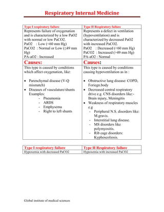
  • 1. Respiratory Internal Medicine Type I respiratory failure Type II Respiratory failure Represents failure of oxygenation Represents a defect in ventilation and is characterized by a low PaO2 (hypoventilation) and is with normal or low PaCO2. characterized by decreased PaO2 PaO2 : Low (<60 mm Hg) with increased PaCO2. PaCO2 : Normal or Low (≤49 mm PaO2 : Decreased (<60 mm Hg) Hg) PaCO2 : Increased (>49 mm Hg) PA-aO2 : Increased PA-aO2 : Normal Causes: Causes: This type is caused by conditions This type is caused by conditions which affect oxygenation, like: causing hypoventilation as in :  Parenchymal disease (V-Q  Obstructive lung disease: COPD, mismatch) Foriegn.body  Diseases of vasculature/shunts  Decreased central respiratory Examples: drive e.g. CNS disorders like:- - Pneumonia Brain injury, Meningitis - ARDS  Weakness of respiratory muscles - Emphysema e.g - Right to left shunts - Peripheral N.S. disorders like: M.gravis. - Interstitial lung disease. - MS disorders like polymyositis. - Rib cage disorders: Kyphoscoliosis. Type I respiratory failure Type II Respiratory failure Hypoxemia with decreased PaCO2 Hypoxemia with increased PaCO2 Global institute of medical sciences
  • 2. Respiratory Internal Medicine PRT Results Obstructive Pattern Restrictive Pattern FEV1 Decreased (<80% predicted) Decreased (May be preserved) (Decreased out of proportion to (Decreased in proportion to FVC) FVC) FVC Decreased (may by preserved) Decreased ((<80% predicted) FEV1/FVC Decreased (<0.7) Normal or increased (>0.7) (FEV1%) FEF25-75 <50% predicted Decreased in proportion to loss of lung volume TLC Normal or elevated Decreased DLCO Normal Decreased in intrinsic restrictive Decreased in Emphysema lung disease. Normal in neuromuscular or musculoskeletal restrictive disease. Respiratory disease Obstructive Restrictive 1. asthma 2. COPD – Chronic bronchitis Parenchymal Extra parenchymal Emphysema - sarcoidosis 1. Neuromuscular 3. Bronchiectasis - pneumoconiosis disease 4. Cystic fibrosis - idiopathic pulmonary - Diaphragmatic palsy fibrosis - GB syndrome 5. Bronchiolitis - drug/Radiation - Muscular dystrophy induced interstitial - Cervical spine injury lung disease. 2. Cest wall - Kphoscoliosis - Oesity - Akylosing spondylitis Global institute of medical sciences
  • 3. Respiratory Internal Medicine Obstructive lung disease Restrictive lung disease Total lung capacity Normal to increase Decrease Residual volume Increase Decrease Vital capacity Decrease Decrease FEV1/FVC Decrease (<0.7) Normal to increase (>0.7) FEF 25-75% Decrease Normal (forced expiratory flow rate) Diffusion capacity Normal (decrease in Decreased emphysema) FVC The forced vital capacity (FVC) represents the total volume of gas exhaled with maximum expiration following maximal inspiration. A reduced FVC suggests pulmonary restriction. FEV1% or FEV1/FVC Note that FEV1 expressed as a percentage is actually FEV1/FVC. The volume of gas exhaled during the first second while expiration from FVC is FEC1. FEV1 is often represented as a ration of the FVC (often referred to as FEV1% rather than FEV1/FVC) Normal to increased FEV1% (FEV1/FVC) suggest restrictive lung disease Decreased FEV1% (FEV1/FVC) suggests obstructive lung disease. DLCO : (Diffusion capacity for carbon monoxide) This reflects the ability of lungs to transfer gas across the alveolar – capillary interface. Decreased DLCO is consistent with a diagnosis of:  Interstitial lung disease  Emphysema  Pulmonary hypertension Global institute of medical sciences
  • 4. Respiratory Internal Medicine Increased PaO2-PaO2 (alveolar arterial difference in oxygen) Correctible with oxygen Not correctible with oxygen V/Q Mismatch shunt V/Q Mismatch: Shunt: - Airway disease (Asthma, - Alveolar collapse COPD) - Intra alveolar filling - Interstitial lung disease 1. pneumonia - Alveolar disease 2. pulm. Edema - Pulmonary vascular disease - intracardiac shunt - vascular shunt within lung Decreased D LCO (diffusion capacity) Increased DLCO (diffusion capacity) 1. interstitial lung disease:- scarring 1. alveolar haemorrhage:- as a Good of alveolar capillary units pasture‟s syndrome: haemoglobin diminishes area of alveolar capillary contained in erythrocytes in bed as well as pulmonary blood alveolar lumen binds Co so exhaled volume carbon monoxide concentration is 2. emphysema:- alveolar walls are diminished & DLCO is increased. destroyed so the surface area of 2. congestive heart failure:- may be alveolar capillary bed is diminished elevated if pulmonary blood 3. recurrent pulmonary embolism volume is increased. Once and primary pulmonary pulmonary edema ensues DLCO hypertension:- disease causes a may decrease as and the net DLCO decrease in cross sectional area and depends on the opposing influences. volume of pulmonary vasculature Global institute of medical sciences
  • 5. Respiratory Internal Medicine Asthma Extrinsic asthma /allergic asthma Intrinsic asthma/Idiosyncratic asthma - History of atopy is present - Attacks related to environmental - No history of atopy (concomitant exposures nasal polyps are usually present - IgE antibody levels are increased and patients may be aspirin - Early onset of disease (young) sensitive) - Usually is less severe, intermittent - Attacks not related to asthma that may however progress environmental exposures: occur to severe persistent asthma without provocation - IgE antibody levels are normal - Delayed onset of disease (adult onset asthma) - Usually have more severe persistent asthma. The microscopically identifiable features described in sputum are three ‘C’s Charcot Leyden crystals : derived from granules of eosnophils and found only in asthma Curshmann spirals : curiously twisted casts of airways : Whorls of shed epithelium Creola bodies : clumps of cells or isolated metaplastic cells. Hypoxia is the universal finding in Asthma Hypocapnia and respiratory alkalosis is seen in most asthmatic patients Hypercarbia and Respiratory acidosis are very late features of asthma and signify severe obstruction and respiratory failure. These are not universal findings in Asthma. Global institute of medical sciences
  • 6. Respiratory Internal Medicine Management of acute bronchial asthma: Sit patient up and give high dose O2 (100%) β2 agonist / Salbutamol (nebulized) Alternate β2 agonist Terbutaline Fenoterol etc. Steroids: hydrocortisone IV or Prednisolone orally Additive treatment if life threatening features are present 1. anticholinergic : iprotropium bromide 2. methylxanthines : aminophyllines Panacinar emphysema Centriacinar emphysema - acini are uniformly involved - central or proximal parts of from level of respiratory acini formed by respiratory bronchiole to terminal blind bronchioles are affected alveoli. whereas distal alveoli are - Lsions are more common in spared lower zone and bases - lesions are more severe and - Occurs in association with common in Upper lobes α1 antitrypsin deficiency - occurs predominantly in smokers - I the commonest pattern Global institute of medical sciences
  • 7. Respiratory Internal Medicine Asbestos related lung disease Pulmonary Pleural Non malignant Malignant Non malignant Malignant Diffuse Bronchogenic interstitial - Pleural plaques - Mesothelioma carcinoma - Diffuse pleural Both plerural fibrosis All histological thickening and peritoneal (asbestosis) subtypes of - Acute benign mesotheliomas Asbestosis is lung cancer pleural effusion develop in defined as a occur with - Rounded persons diffuse interstial increased atelectasis exposed to fibrosing disease frequency but asbestos of lung caused by adenocarcino exposure to ma has the asbestos fibres. highest incidence Silicosis has predilection for upper lobes Radiographs typically show fine nodularity in the upper zones of the lung Rounded opacities appear in the upper lobes on chest radiograph Silicosis is associated with calcific Hilar adenopathy Calcification of hilar lymph nodes may occur in as many as 20% oc cases and produce a charachteristic “egg shell” pattern. Silicosis is associated with tuberculosis Because silica is cytotoxic to alveolar macrophages, patients with silicosis are at greater risk of acquiring lung infections that involve these cells as a primary defense including myocobacterium tuberculosis, atypical mycobacteria and fungi Global institute of medical sciences
  • 8. Respiratory Internal Medicine Clinical spectrum of pulmonary aspergillosis saprophytic infection invasive aspergillosis Allergic aspergillosis aspergilloma (fungal - Invasive bronchial ball) - Allergic bronchial aspergillosis (IBA) asthma - Chronic pulmonary - Allergic aspergillosis (CPA) bronchopulmonary - Invasive pulmonary aspergillosis(ABPA) aspergillosis (IPA) - Extrinsic allergic alveolitis (type to hypersensitivity pneumonitis) kleibsella pneumonia kleibsella pneumonia presents as a typical ‘air space’ pneumonia with cough productive of purulent sputum. ‘purulent sputum production and „air space‟ diseases X ray are typical‟ causes of atypical pneumonias: 1. mycoplasma pneumonias 2. viral pneumonias - influenza - RSV - Adenovirus - Rhinovirus - Rubeola - Varicilla - Corona virus 3. Chlamydia pneumonia 4. coxiella burnetti 5. pneumocystis carinii Global institute of medical sciences
  • 9. Respiratory Internal Medicine Crona virus is an infrequent cause of pneumonia. Pneumocytic carinni pneumonia is characterized by a prominent eosinophilic exudates and mild interstitial pneumonitis. These is damage to type I pneumocytes and associated compensatory hypertrophy of type II pneumocytes. Classic histopathology in pneumocystis pneumonia Intra alveolar changes interstitial changes Mild interstitial pneumonitis Prominent eosinophilic foamy, vacuolated - Mononuclear interstitial cell intra alveolar exudeate (HE staining) infiltrate (mild) - Intra alveolar exudates contains the - Damaged type I pneumocytes organisms, which can be identified in - Hypertrophy / proliferation of type various stages of development II pneumocytis (reparative (throphozoite and cystic stage) by the response) use of special stains such as pneumocystis attaches to and 1. methamine silver / Toluidine stain damages type 1 pneumocytes. 2. wright giemsa stain There is associated 3. papanicolaou’s stain compensatory hypertrophy of type II pneumocytes. Global institute of medical sciences
  • 10. Respiratory Internal Medicine Montoux test Montoux test is carried out by injecting one tuberculin unit (1TU) of PPD in 0.1 ml on the flexor surgace of forearm (PPD RT 23 with Tween 80) Test is read after 72 hrs (not 48 hrs) look for erythema and induration <6 mm 6-9 mm >10 mm negative doubtful Positive - a positive test indicates that patient is infected with M. tuberculosis. It does not however prove that the person is ‘suffering’ from the disease - studies indicate that 92% of new cases occur in persons who already are tuberculin reactors - 6-9 mm induration does not indicate high chances of developin tuberculosis. Infact patients with 25 mm induration have more chances of developing tuberculosis than those with 6-9 mm induration. Global institute of medical sciences
  • 11. Respiratory Internal Medicine Sarcoidosis Sarcoidosis is a chronic multisystem disorder of unknown causes characterized by accumulation of T lymphocytes and mononuclear phagocytes in various tissues of body Non caseating SARcoid granulomas in affected organs Lung (90%) lymphnodes skin Others (75-90%) -Eye :- uveitis - interstitial - Erythema - B/L hilar - Kidney (rare):- lung disease. nodosum Lymphadenopathy renal . fibrosis of lung - Lupus hypercalcemia is the hallmark of perenium parenchyma sarcoidosis with or without - Pleura is (purple blue hypercalcuria - Parotid shiny involved in 5% of enlargement B/e - Skeletal :- cases u/e swollen arthritis involvement is the lesions on pleural effusion vuli - Nervous - Cavitation is nose, system:- rare cheeks, lips, peripheral ears). neuropathy - Heart :- cor pulmonle Inclusions seen in giant cells in sarcoidosis (remember as SARcoidosis) - Schaumann bodies - Asteroid bodies - Residual bodies Global institute of medical sciences
  • 12. Respiratory Internal Medicine Pneumothorax traumatic Spontaneous pneumothorax pneumothorax (non spontaneous pneumothorax) -These Primary spontaneous Secondary spontaneous penumothoraces occur pneumothorax pneumothorax as a result of : 1. blunt trauma or - Occur in persons with No • occurs in persons 2. penetrating trauma clinical evidence of lung with an underlying - Iatrogenic disease and without lung disease as a pneumothorax is precipitating event. complicationof the subcategory of - Most of these patients have underlying disease traumatic occult lung disease (sub process pneumothorax clinical) with subpleural blebs Causes of iatrogenic on CT scan Etiology of pneumothorax characterstics of affected secondary - Thoracocentesis people are :- - tall, thin, males pneumothorax - Insertion of central - smokers venous catheter - Mechanical - subpleural blebs on lungs ventilation (IPPV, assisted ventilation etc) - Surgery Obstructive lung disease Malignancy Interstitial lung disease Connective tissue Infection disease Global institute of medical sciences
  • 13. Respiratory Internal Medicine Facts to remember Most frequent histological type Adenocarcinoma Most frequent histological type in India Squamous cell carcinoma Most common histological varieny in life time non-smokers Adenocarcinoma Most common histological variety in young patients Adenocarcinoma Most common histological variety in females Adenocarcinoma Most common site for metastasis from Ca lung Liver Most common endocrine organ to be involved by metastasis Adrenals from Ca lung Ca lung which metastizes to opposite lung Adenocarcinoma Commonest tumor to metastise to heart Ca lung (Bronchognic Ca) Histological varieties that cavitate Squamous cell and large cell Histological varieties that are central in distribution Squamous cell and small cell Histological varieties that are peripheral in distribution Adenocarcinoma Pancoost tumor is histologically Squamous cell Most common variety associated with paraneoplatic syndrome Small cell variety Most common variety associated with hypokalemia Small cell (presumably d/t ACTH) Most common variety associated with hypercalcemia Squamous cell (presumably d/t PTH) Histological variety most responsive to chemotherapy Small cell Histological variety response to radiotherapy Small cell Global institute of medical sciences
  • 14. Respiratory Internal Medicine Histological variety associated with hest prognosis Squamous cell The most common malignancy of lung is adenocarcinoma (overall/world wide):- Most common lung cancer worldwide is Adenocarcinoma Most common lung cancer in India is swquammous cell carcinoma Most common lung cancer in women is adenocarcinoma Most common lung cancer in smokers is squammous cell carcinoma Most common lung cancer in nonsmokers is adenocarcinoma Most common lung cancer in young patients is adenocarcinoma Most common lung cancer to metastasize is small cell carcinoma. Ectopic hormones produced by small cell Paraneoplastic syndrome carcinomas - ACTH Cushings syndrome - SIADH/ANP Hyponatremia - Calcitonin Hypocalcemia - Gonadotropins gynaecomastia Global institute of medical sciences