SlideShare une entreprise Scribd logo
1  sur  37
2/10/2016 1bhargava
2/10/2016 bhargava 2
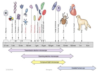
•Instrument that uses a lens or a combination of lenses
to magnify and resolve the fine details of an object.
•Early methods for examining physical evidence relied
solely on the microscope.
The Microscope
Vocabulary
• Magnification – larger
image
• Resolution – clearer image
• Numerical Aperture – light
gathering capacity of a lens
• Working Distance – the
distance from the bottom
of an objective to the in-
focus area of an object
(distance between
specimen and lens)
2/10/2016 3bhargava
Magnification
• Magnification is the enlargement of the image
• The magnification of a microscope is given by-
• Generally used class microscope has following
magnification-
Mmicroscope = Moccular X Meyepiece
2/10/2016 4bhargava
Resolution
• Resolution is defined as the
ability to distinguish two very
small and closely-spaced
objects as separate entities.
• Resolution is best when the
distance separating the two
tiny objects is small.
• Degree to which detail in
specimen is retained in
magnified image.
• Resolving power-
– Unaided eye – 0.1 mm apart
– Microscope - 0.2 µm apart
2/10/2016 5bhargava
Numerical Aperture
•NA is light gathering capacity of objective
•Limit of resolution = 0.61λ
NA
• NA (Numerical Aperture) = n sinα
Wavelength of
illumination
Aperture angle
Refractive index of air or liquid
between specimen and lens
•The N.A. of each objective lens is inscribed in the metal tube, and ranges from
0.25-1.4
•The higher the N.A., the better the light-gathering properties of the lens, and the
better the resolution.
2/10/2016 6bhargava
Simple Microscope
• Similar to a
magnifying glass
and has only one
lens.
2/10/2016 7bhargava
Compound Microscope
• Compound microscope
was constructed by
Robert Hooke (1665) & is
forerunner of present day
compound microscope.
– Most widely used
microscope
– Light passes through 2
lenses
– Can magnify up to 2000x
•Early Compound Microscopes
•Could magnify upto 30X2/10/2016 8bhargava
Compound Microscope
https://www.youtube.com/watch?v=RKA8_mif6-E
• A compound microscope consists of various components which gather
light and redirects the light path so that a magnified image of the
viewed object can be focused within a short distance.
I. Light source – source of illumination
II. Condenser – collimates the light
III. Sample Stage – specimen is placed over this
IV. Objective lens – produces a real intermediate image onto the ocular
front plane
V. Oculars – re-focus the intermediate image on the retina as a larger
virtual image
2/10/2016 9bhargava
Principle of Compound Microscope
2/10/2016 10bhargava
Original text
As seen through a compound
microscope
•This reversal is always seen
using a standard compound
microscope.
•It's the reason when we move
a slide right the image moves
left, and
•when we move a slide
downward the image moves
upward.
The specimen
Final image
Intermediate
image
Eye
2/10/2016 11bhargava
Modern Compound Microscope
The microscope is consists of:
•mechanical system which
supports the microscope,
•an optical system which
illuminates the object under
investigation
•light passes through a series of
lens to form an image of the
specimen.
2/10/2016 12bhargava
Comparison Microscope
2/10/2016 13bhargava
A - B - Two identical microscopes
C -C' - Specimens to compare
D - Comparison eyepiece (optical bridge)
2/10/2016 14bhargava
Unlike any other microscope,
it looks at two different
objects at the same time. As
its name implies, it is used to
compare objects
Youtube video:
https://www.youtube.com/watch?v=Ci1Qi3Ire_E
Comparison Microscope
• The comparison microscope consists of two
independent objective lenses joined together by
an optical bridge to a common eyepiece lens.
• When a viewer looks through the eyepiece lens of
the comparison microscope, the objects under
investigation are observed side-by-side in a
circular field that is equally divided into two parts.
2/10/2016 15bhargava
Bullet markings Photographed
using Comparison Microscope
• Modern firearms examination began with the
introduction of the comparison microscope, with its
ability to give the firearms examiner a side by side
magnified view of bullets.
2/10/2016 16bhargava
Forensic Applications
1. It enables side by side comparison of the rifling impressions on projectile found at the
crime scene with a test projectile fired in the laboratory.
2. Similar principle is used for comparison of cartridge cases, where we compare
a) firing pin marks (formed when a pin hits the primer and makes the cartridge
explode),
b) breech face marks (the impression made when the cartridge is pressed against the
end of the barrel during the explosion) and
c) ejector/extractor marks (caused when the cartridge is discharged or ejected from
the barrel).
3. The same principle is used to compare the tool marks such as- screwdriver, saw, saw
edged knife, axe, dagger etc.
4. Comparison microscope is used for looking at adhesive strips from a letter bomb parcel,
and comparing them with a roll of adhesive tape found at a potential perpetrator’s.
5. Another area of use of comparison microscopy is the examination of traces from cars
such as glass and artificial glass splinters, paint traces etc.
6. Used for comparison of hairs and fibres of different origins.
2/10/2016 bhargava 17
Stereoscopic Microscope
• The stereo or stereoscopic microscope is an optical microscope variant designed for low
magnification observation of a sample, typically using light reflected from the surface of an
object rather than transmitted through it.
• The instrument uses two separate optical paths with two objectives and eyepieces to
provide slightly different viewing angles to the left and right eyes.
• This arrangement produces a three-dimensional visualization of the sample being
examined.
• Reflected light illumination rather than transmitted illumination (Unlike a compound light
microscope).
• Light reflected from the surface of an object rather than light transmitted through an
object.
• Use of reflected light from the object allows examination of specimens that would be too
thick or otherwise opaque for compound microscopy.
• Stereomicroscopy overlaps macrophotography for recording and examining solid samples
with complex surface topography, where a three-dimensional view is needed for analyzing
the detail.
2/10/2016 bhargava 18
2/10/2016 bhargava 19
The stereoscopic microscope is actually
two monocular compound microscopes
properly spaced and aligned to present a
three dimensional image of a specimen to
the viewer, who looks through both
eyepiece lenses.
Advantage:
•Great working distance
•Enhanced depth of field
•Ease of sample manipulation on
stage
•Larger samples can be analyzed
•3-D view of image just like human
eyes
Disadvantage:
•Low magnification
•Low resolution
2/10/2016 bhargava 20
Fig: Details of a suspected document pen
over toner.
Fig: Detail of a bank note (embossed elements).
Forensic Applications
• Most important application of Stereoscopic Microscopy is in the
fields of Questioned Document Examination.
– It is especially adapted for the examination of inks, colors, erasures,
changes, interlineations, and overwriting.
– For the comparison of disturbed and undisturbed paper surfaces, pen,
and pencil points, the tint,
– Texture and condition of paper surfaces,
– The texture and quality of typewriter ribbons, written and printed
characters, and type faces.
2/10/2016 bhargava 21
Fluorescence Microscope
https://www.youtube.com/watch?v=PCJ13LjncMc
• Excites and observe fluorescent molecules.
• A fluorescence microscope is an optical microscope
that uses fluorescence and phosphorescence instead
of, or in addition to, reflection and absorption to study
properties of organic or inorganic substances.
• The "fluorescence microscope" refers to any
microscope that uses fluorescence to generate an
image.
2/10/2016 22bhargava
Fluorescence Microscopy
• When certain compounds are illuminated with high
energy light, they then emit light of a different, lower
frequency.
• This effect is known as fluorescence.
• Often specimens show their own characteristic auto-
fluorescence image, based on their chemical makeup.
• Specimens usually stained with fluorochromes.
2/10/2016 23bhargava
Excited state
Ground state
excitation
Shorter wavelength,
higher energy
emission
longer wavelength, less
energy
•Exposes specimen to ultraviolet,
violet, or blue light
•Shows a bright image of the object
resulting from the fluorescent light
emitted by the specimen
(fluorophores).
Fluorophores:
•Different fluorescent dyes can be used to stain
different structures or chemical compounds.
•Examples of commonly used fluorophores are
fluorescein or rhodamine.
•An ideal fluorescent image shows only the
structure of interest that was labelled with the
fluorescent dye.
2/10/2016 24bhargava
2/10/2016 25bhargava
lamp
sample
camera
Emission filter
Dichroic mirror
Excitation filter
Emission filter
Transmission
(%)
wave length (nm)
Excitation filter
2/10/2016 26bhargava
2/10/2016 27bhargava
Sample
Objective lens
Excitation light
Tube lens
Emission light
Pinhole
Detector
2/10/2016 28bhargava
Fluorescence
2/10/2016 29bhargava
Uses
1. To study the membrane dynamics (endocytosis, receptor bindings
etc.)
2. To measure the concentration of Ca+2 ions, pH changes and protein
interactions.
3. Determine the localisation of specific (multiple) proteins
4. Determine the shape of organs, cells, intracellular structures
5. Examine the dynamics of proteins
6. Study protein interactions or protein conformation
7. Examine the ion concetration etc.
2/10/2016 30bhargava
Polarized Light Microscope
• Polarized light microscopy is a techniques involving polarized light
for illumination of the sample, while blocking the directly
transmitted light with a polariser orientated at 90 degrees to the
illumination.
• Polarized light microscope is designed to observe specimens that
are visible primarily due to their optically anisotropic character
(birefringent).
• The microscope must be equipped with both a polarizer, positioned
in the light path somewhere before the specimen, and
• An analyzer (a second polarizer), placed in the optical pathway
between the objective rear aperture and the observation tubes or
camera port.
2/10/2016 31bhargava
When the electric field vectors of light are restricted to a single plane
by filtration, then the light is said to be polarized with respect to the
direction of propagation and all waves vibrate in the same plane.
2/10/2016 32bhargava
Fig- 1
Fig- 2
Fig- 3
2/10/2016 33bhargava
Polarizers
• Polarizers specifically transmit one polarization
angle of light
• Crossed polarizers transmit no light
X
2/10/2016 34bhargava
2/10/2016 35bhargava
Analyzer (upper polarizer) -- a polarizing prism
located above the microscope stage, between
the objective lens and the eyepiece.
This restricts the transmission of light vibrating
perpendicular to the polarizer. The analyzer
can be slipped in or out of the light path or
rotated for partially crossed polarized light.
Light passing through the polarizer will not
pass through the analyzer unless the vibration
direction of the light is changed between the
two prisms.
Polarizer (lower polarizer) -- a polarizing prism
located beneath the microscope stage
(between the light source and the object of
study).
This restricts transmission of light to that
vibrating in only one (N-S) direction.
Some microscopes have a different orientation
direction.
In effect, it plane polarizes the incident light
beam.
2/10/2016 36bhargava
Uses of Polarized Microscopy
• Polarizing microscopy has found wide
applications for the study of birefringent
materials; materials that split a beam of light
in two, each with its own refractive index
value.
• The determination of these refractive index
data provides information that helps to
identify minerals present in a soil sample or
the identity of a man-made fiber.
2/10/2016 37bhargava

Contenu connexe

Tendances

Tendances (20)

Different types of microscopes
Different types of microscopesDifferent types of microscopes
Different types of microscopes
 
Phase contrast microscope
Phase contrast microscopePhase contrast microscope
Phase contrast microscope
 
Fluorescence Microscopy
Fluorescence MicroscopyFluorescence Microscopy
Fluorescence Microscopy
 
Simple microscope
Simple microscopeSimple microscope
Simple microscope
 
MICROSCOPY.pptx
MICROSCOPY.pptxMICROSCOPY.pptx
MICROSCOPY.pptx
 
Microscopy
MicroscopyMicroscopy
Microscopy
 
Bright Field Microscopy.pptx
Bright Field Microscopy.pptxBright Field Microscopy.pptx
Bright Field Microscopy.pptx
 
phase contrast microscope
phase contrast microscopephase contrast microscope
phase contrast microscope
 
PHASE MICROSCOPY
PHASE MICROSCOPYPHASE MICROSCOPY
PHASE MICROSCOPY
 
Fluorescence microscope
Fluorescence microscopeFluorescence microscope
Fluorescence microscope
 
Fluorescence microscope
Fluorescence microscopeFluorescence microscope
Fluorescence microscope
 
Microscopy
MicroscopyMicroscopy
Microscopy
 
Compound microscope
Compound microscopeCompound microscope
Compound microscope
 
FLUORESCENCE MICROSCOPY .pptx
FLUORESCENCE MICROSCOPY .pptxFLUORESCENCE MICROSCOPY .pptx
FLUORESCENCE MICROSCOPY .pptx
 
Fluorescence microscope by Subhankar Das
Fluorescence microscope by Subhankar DasFluorescence microscope by Subhankar Das
Fluorescence microscope by Subhankar Das
 
Bright field microscopes
Bright field microscopesBright field microscopes
Bright field microscopes
 
Confocal microscope presentation pt
Confocal microscope presentation  ptConfocal microscope presentation  pt
Confocal microscope presentation pt
 
transmission Electron Microscopy (Tem)
transmission Electron Microscopy (Tem)transmission Electron Microscopy (Tem)
transmission Electron Microscopy (Tem)
 
Phase contrast & Flurosence microscope
Phase contrast & Flurosence microscopePhase contrast & Flurosence microscope
Phase contrast & Flurosence microscope
 
DARK FIELD MICROSCOPY
DARK FIELD MICROSCOPYDARK FIELD MICROSCOPY
DARK FIELD MICROSCOPY
 

Similaire à Light microscopy

To Study Principles of Microscopy: Light Microscope, Phase Contrast Microsco...
To Study Principles of Microscopy: Light Microscope, Phase Contrast  Microsco...To Study Principles of Microscopy: Light Microscope, Phase Contrast  Microsco...
To Study Principles of Microscopy: Light Microscope, Phase Contrast Microsco...Om Prakash
 
microscope- lecture 2.pptx
microscope- lecture 2.pptxmicroscope- lecture 2.pptx
microscope- lecture 2.pptxOsmanAli92
 
Microscope lecture notes for laboratory).ppt
Microscope lecture notes for laboratory).pptMicroscope lecture notes for laboratory).ppt
Microscope lecture notes for laboratory).pptSheikhAlshoteri2
 
Types of Light Microscopes used in Histological Studies.pptx
Types of Light Microscopes used in Histological Studies.pptxTypes of Light Microscopes used in Histological Studies.pptx
Types of Light Microscopes used in Histological Studies.pptxssuserab552f
 
2.0 - Types of Light Microscope.pptx ppt
2.0 - Types of Light Microscope.pptx ppt2.0 - Types of Light Microscope.pptx ppt
2.0 - Types of Light Microscope.pptx pptstephenopokuasante
 
2.0 - Types of Light Microscope wonder.pptx
2.0 - Types of Light Microscope wonder.pptx2.0 - Types of Light Microscope wonder.pptx
2.0 - Types of Light Microscope wonder.pptxstephenopokuasante
 
2.0 - Types of Light Microscope.ppt microscope
2.0 - Types of Light Microscope.ppt microscope2.0 - Types of Light Microscope.ppt microscope
2.0 - Types of Light Microscope.ppt microscopestephenopokuasante
 
Types of Microscopes with their applications
Types of Microscopes with their applicationsTypes of Microscopes with their applications
Types of Microscopes with their applicationsMicrobiology Note
 
Electron Microscope: Definition, Types, Parts, Application, Advantages, Disad...
Electron Microscope: Definition, Types, Parts, Application, Advantages, Disad...Electron Microscope: Definition, Types, Parts, Application, Advantages, Disad...
Electron Microscope: Definition, Types, Parts, Application, Advantages, Disad...Microbiology Note
 
light microscopy seminar.pptx
light microscopy seminar.pptxlight microscopy seminar.pptx
light microscopy seminar.pptxAhalya40
 
Microscopy - Magnification, Resolving power, Principles, Types and Applications
Microscopy - Magnification, Resolving power, Principles, Types and ApplicationsMicroscopy - Magnification, Resolving power, Principles, Types and Applications
Microscopy - Magnification, Resolving power, Principles, Types and ApplicationsNethravathi Siri
 

Similaire à Light microscopy (20)

Microscope PPT.pdf
Microscope PPT.pdfMicroscope PPT.pdf
Microscope PPT.pdf
 
Compound microscope
Compound microscopeCompound microscope
Compound microscope
 
Light microscope
Light microscopeLight microscope
Light microscope
 
Confocal microscope
Confocal microscopeConfocal microscope
Confocal microscope
 
To Study Principles of Microscopy: Light Microscope, Phase Contrast Microsco...
To Study Principles of Microscopy: Light Microscope, Phase Contrast  Microsco...To Study Principles of Microscopy: Light Microscope, Phase Contrast  Microsco...
To Study Principles of Microscopy: Light Microscope, Phase Contrast Microsco...
 
Microscopy
MicroscopyMicroscopy
Microscopy
 
microscope- lecture 2.pptx
microscope- lecture 2.pptxmicroscope- lecture 2.pptx
microscope- lecture 2.pptx
 
Microscope
MicroscopeMicroscope
Microscope
 
Microscope lecture notes for laboratory).ppt
Microscope lecture notes for laboratory).pptMicroscope lecture notes for laboratory).ppt
Microscope lecture notes for laboratory).ppt
 
Types of Light Microscopes used in Histological Studies.pptx
Types of Light Microscopes used in Histological Studies.pptxTypes of Light Microscopes used in Histological Studies.pptx
Types of Light Microscopes used in Histological Studies.pptx
 
Microscope
MicroscopeMicroscope
Microscope
 
2.0 - Types of Light Microscope.pptx ppt
2.0 - Types of Light Microscope.pptx ppt2.0 - Types of Light Microscope.pptx ppt
2.0 - Types of Light Microscope.pptx ppt
 
2.0 - Types of Light Microscope wonder.pptx
2.0 - Types of Light Microscope wonder.pptx2.0 - Types of Light Microscope wonder.pptx
2.0 - Types of Light Microscope wonder.pptx
 
2.0 - Types of Light Microscope.ppt microscope
2.0 - Types of Light Microscope.ppt microscope2.0 - Types of Light Microscope.ppt microscope
2.0 - Types of Light Microscope.ppt microscope
 
Types of Microscopes with their applications
Types of Microscopes with their applicationsTypes of Microscopes with their applications
Types of Microscopes with their applications
 
Electron Microscope: Definition, Types, Parts, Application, Advantages, Disad...
Electron Microscope: Definition, Types, Parts, Application, Advantages, Disad...Electron Microscope: Definition, Types, Parts, Application, Advantages, Disad...
Electron Microscope: Definition, Types, Parts, Application, Advantages, Disad...
 
light microscopy seminar.pptx
light microscopy seminar.pptxlight microscopy seminar.pptx
light microscopy seminar.pptx
 
Microscopy - Magnification, Resolving power, Principles, Types and Applications
Microscopy - Magnification, Resolving power, Principles, Types and ApplicationsMicroscopy - Magnification, Resolving power, Principles, Types and Applications
Microscopy - Magnification, Resolving power, Principles, Types and Applications
 
Presentation1 4
Presentation1 4Presentation1 4
Presentation1 4
 
Ppt f1
Ppt f1Ppt f1
Ppt f1
 

Plus de Saurabh Bhargava

Plus de Saurabh Bhargava (13)

Crime scene investigation lecture
Crime scene investigation  lectureCrime scene investigation  lecture
Crime scene investigation lecture
 
Scanning Electron Microscopy (SEM) lecture
Scanning Electron Microscopy (SEM) lectureScanning Electron Microscopy (SEM) lecture
Scanning Electron Microscopy (SEM) lecture
 
Introduction to Forensic science labs in India
Introduction to Forensic science labs in IndiaIntroduction to Forensic science labs in India
Introduction to Forensic science labs in India
 
Hyphenated techniques
Hyphenated techniquesHyphenated techniques
Hyphenated techniques
 
forensic facial reconstruction
forensic facial reconstructionforensic facial reconstruction
forensic facial reconstruction
 
Strychnine nux vomica
Strychnine nux vomicaStrychnine nux vomica
Strychnine nux vomica
 
aberrations
aberrations aberrations
aberrations
 
Plant poisons
Plant poisonsPlant poisons
Plant poisons
 
Forensic Odontology
Forensic OdontologyForensic Odontology
Forensic Odontology
 
Identification
IdentificationIdentification
Identification
 
Paternity index
Paternity indexPaternity index
Paternity index
 
Glass analysis
Glass analysisGlass analysis
Glass analysis
 
Narco & beos
Narco & beosNarco & beos
Narco & beos
 

Dernier

MULTIDISCIPLINRY NATURE OF THE ENVIRONMENTAL STUDIES.pptx
MULTIDISCIPLINRY NATURE OF THE ENVIRONMENTAL STUDIES.pptxMULTIDISCIPLINRY NATURE OF THE ENVIRONMENTAL STUDIES.pptx
MULTIDISCIPLINRY NATURE OF THE ENVIRONMENTAL STUDIES.pptxAnupkumar Sharma
 
Procuring digital preservation CAN be quick and painless with our new dynamic...
Procuring digital preservation CAN be quick and painless with our new dynamic...Procuring digital preservation CAN be quick and painless with our new dynamic...
Procuring digital preservation CAN be quick and painless with our new dynamic...Jisc
 
Incoming and Outgoing Shipments in 3 STEPS Using Odoo 17
Incoming and Outgoing Shipments in 3 STEPS Using Odoo 17Incoming and Outgoing Shipments in 3 STEPS Using Odoo 17
Incoming and Outgoing Shipments in 3 STEPS Using Odoo 17Celine George
 
AUDIENCE THEORY -CULTIVATION THEORY - GERBNER.pptx
AUDIENCE THEORY -CULTIVATION THEORY -  GERBNER.pptxAUDIENCE THEORY -CULTIVATION THEORY -  GERBNER.pptx
AUDIENCE THEORY -CULTIVATION THEORY - GERBNER.pptxiammrhaywood
 
Judging the Relevance and worth of ideas part 2.pptx
Judging the Relevance  and worth of ideas part 2.pptxJudging the Relevance  and worth of ideas part 2.pptx
Judging the Relevance and worth of ideas part 2.pptxSherlyMaeNeri
 
Virtual-Orientation-on-the-Administration-of-NATG12-NATG6-and-ELLNA.pdf
Virtual-Orientation-on-the-Administration-of-NATG12-NATG6-and-ELLNA.pdfVirtual-Orientation-on-the-Administration-of-NATG12-NATG6-and-ELLNA.pdf
Virtual-Orientation-on-the-Administration-of-NATG12-NATG6-and-ELLNA.pdfErwinPantujan2
 
FILIPINO PSYCHology sikolohiyang pilipino
FILIPINO PSYCHology sikolohiyang pilipinoFILIPINO PSYCHology sikolohiyang pilipino
FILIPINO PSYCHology sikolohiyang pilipinojohnmickonozaleda
 
Choosing the Right CBSE School A Comprehensive Guide for Parents
Choosing the Right CBSE School A Comprehensive Guide for ParentsChoosing the Right CBSE School A Comprehensive Guide for Parents
Choosing the Right CBSE School A Comprehensive Guide for Parentsnavabharathschool99
 
ECONOMIC CONTEXT - PAPER 1 Q3: NEWSPAPERS.pptx
ECONOMIC CONTEXT - PAPER 1 Q3: NEWSPAPERS.pptxECONOMIC CONTEXT - PAPER 1 Q3: NEWSPAPERS.pptx
ECONOMIC CONTEXT - PAPER 1 Q3: NEWSPAPERS.pptxiammrhaywood
 
Earth Day Presentation wow hello nice great
Earth Day Presentation wow hello nice greatEarth Day Presentation wow hello nice great
Earth Day Presentation wow hello nice greatYousafMalik24
 
INTRODUCTION TO CATHOLIC CHRISTOLOGY.pptx
INTRODUCTION TO CATHOLIC CHRISTOLOGY.pptxINTRODUCTION TO CATHOLIC CHRISTOLOGY.pptx
INTRODUCTION TO CATHOLIC CHRISTOLOGY.pptxHumphrey A Beña
 
Inclusivity Essentials_ Creating Accessible Websites for Nonprofits .pdf
Inclusivity Essentials_ Creating Accessible Websites for Nonprofits .pdfInclusivity Essentials_ Creating Accessible Websites for Nonprofits .pdf
Inclusivity Essentials_ Creating Accessible Websites for Nonprofits .pdfTechSoup
 
4.16.24 21st Century Movements for Black Lives.pptx
4.16.24 21st Century Movements for Black Lives.pptx4.16.24 21st Century Movements for Black Lives.pptx
4.16.24 21st Century Movements for Black Lives.pptxmary850239
 
What is Model Inheritance in Odoo 17 ERP
What is Model Inheritance in Odoo 17 ERPWhat is Model Inheritance in Odoo 17 ERP
What is Model Inheritance in Odoo 17 ERPCeline George
 
Field Attribute Index Feature in Odoo 17
Field Attribute Index Feature in Odoo 17Field Attribute Index Feature in Odoo 17
Field Attribute Index Feature in Odoo 17Celine George
 
Difference Between Search & Browse Methods in Odoo 17
Difference Between Search & Browse Methods in Odoo 17Difference Between Search & Browse Methods in Odoo 17
Difference Between Search & Browse Methods in Odoo 17Celine George
 
ACC 2024 Chronicles. Cardiology. Exam.pdf
ACC 2024 Chronicles. Cardiology. Exam.pdfACC 2024 Chronicles. Cardiology. Exam.pdf
ACC 2024 Chronicles. Cardiology. Exam.pdfSpandanaRallapalli
 
Grade 9 Quarter 4 Dll Grade 9 Quarter 4 DLL.pdf
Grade 9 Quarter 4 Dll Grade 9 Quarter 4 DLL.pdfGrade 9 Quarter 4 Dll Grade 9 Quarter 4 DLL.pdf
Grade 9 Quarter 4 Dll Grade 9 Quarter 4 DLL.pdfJemuel Francisco
 

Dernier (20)

MULTIDISCIPLINRY NATURE OF THE ENVIRONMENTAL STUDIES.pptx
MULTIDISCIPLINRY NATURE OF THE ENVIRONMENTAL STUDIES.pptxMULTIDISCIPLINRY NATURE OF THE ENVIRONMENTAL STUDIES.pptx
MULTIDISCIPLINRY NATURE OF THE ENVIRONMENTAL STUDIES.pptx
 
Procuring digital preservation CAN be quick and painless with our new dynamic...
Procuring digital preservation CAN be quick and painless with our new dynamic...Procuring digital preservation CAN be quick and painless with our new dynamic...
Procuring digital preservation CAN be quick and painless with our new dynamic...
 
Incoming and Outgoing Shipments in 3 STEPS Using Odoo 17
Incoming and Outgoing Shipments in 3 STEPS Using Odoo 17Incoming and Outgoing Shipments in 3 STEPS Using Odoo 17
Incoming and Outgoing Shipments in 3 STEPS Using Odoo 17
 
AUDIENCE THEORY -CULTIVATION THEORY - GERBNER.pptx
AUDIENCE THEORY -CULTIVATION THEORY -  GERBNER.pptxAUDIENCE THEORY -CULTIVATION THEORY -  GERBNER.pptx
AUDIENCE THEORY -CULTIVATION THEORY - GERBNER.pptx
 
Judging the Relevance and worth of ideas part 2.pptx
Judging the Relevance  and worth of ideas part 2.pptxJudging the Relevance  and worth of ideas part 2.pptx
Judging the Relevance and worth of ideas part 2.pptx
 
Raw materials used in Herbal Cosmetics.pptx
Raw materials used in Herbal Cosmetics.pptxRaw materials used in Herbal Cosmetics.pptx
Raw materials used in Herbal Cosmetics.pptx
 
Virtual-Orientation-on-the-Administration-of-NATG12-NATG6-and-ELLNA.pdf
Virtual-Orientation-on-the-Administration-of-NATG12-NATG6-and-ELLNA.pdfVirtual-Orientation-on-the-Administration-of-NATG12-NATG6-and-ELLNA.pdf
Virtual-Orientation-on-the-Administration-of-NATG12-NATG6-and-ELLNA.pdf
 
FILIPINO PSYCHology sikolohiyang pilipino
FILIPINO PSYCHology sikolohiyang pilipinoFILIPINO PSYCHology sikolohiyang pilipino
FILIPINO PSYCHology sikolohiyang pilipino
 
Choosing the Right CBSE School A Comprehensive Guide for Parents
Choosing the Right CBSE School A Comprehensive Guide for ParentsChoosing the Right CBSE School A Comprehensive Guide for Parents
Choosing the Right CBSE School A Comprehensive Guide for Parents
 
YOUVE_GOT_EMAIL_PRELIMS_EL_DORADO_2024.pptx
YOUVE_GOT_EMAIL_PRELIMS_EL_DORADO_2024.pptxYOUVE_GOT_EMAIL_PRELIMS_EL_DORADO_2024.pptx
YOUVE_GOT_EMAIL_PRELIMS_EL_DORADO_2024.pptx
 
ECONOMIC CONTEXT - PAPER 1 Q3: NEWSPAPERS.pptx
ECONOMIC CONTEXT - PAPER 1 Q3: NEWSPAPERS.pptxECONOMIC CONTEXT - PAPER 1 Q3: NEWSPAPERS.pptx
ECONOMIC CONTEXT - PAPER 1 Q3: NEWSPAPERS.pptx
 
Earth Day Presentation wow hello nice great
Earth Day Presentation wow hello nice greatEarth Day Presentation wow hello nice great
Earth Day Presentation wow hello nice great
 
INTRODUCTION TO CATHOLIC CHRISTOLOGY.pptx
INTRODUCTION TO CATHOLIC CHRISTOLOGY.pptxINTRODUCTION TO CATHOLIC CHRISTOLOGY.pptx
INTRODUCTION TO CATHOLIC CHRISTOLOGY.pptx
 
Inclusivity Essentials_ Creating Accessible Websites for Nonprofits .pdf
Inclusivity Essentials_ Creating Accessible Websites for Nonprofits .pdfInclusivity Essentials_ Creating Accessible Websites for Nonprofits .pdf
Inclusivity Essentials_ Creating Accessible Websites for Nonprofits .pdf
 
4.16.24 21st Century Movements for Black Lives.pptx
4.16.24 21st Century Movements for Black Lives.pptx4.16.24 21st Century Movements for Black Lives.pptx
4.16.24 21st Century Movements for Black Lives.pptx
 
What is Model Inheritance in Odoo 17 ERP
What is Model Inheritance in Odoo 17 ERPWhat is Model Inheritance in Odoo 17 ERP
What is Model Inheritance in Odoo 17 ERP
 
Field Attribute Index Feature in Odoo 17
Field Attribute Index Feature in Odoo 17Field Attribute Index Feature in Odoo 17
Field Attribute Index Feature in Odoo 17
 
Difference Between Search & Browse Methods in Odoo 17
Difference Between Search & Browse Methods in Odoo 17Difference Between Search & Browse Methods in Odoo 17
Difference Between Search & Browse Methods in Odoo 17
 
ACC 2024 Chronicles. Cardiology. Exam.pdf
ACC 2024 Chronicles. Cardiology. Exam.pdfACC 2024 Chronicles. Cardiology. Exam.pdf
ACC 2024 Chronicles. Cardiology. Exam.pdf
 
Grade 9 Quarter 4 Dll Grade 9 Quarter 4 DLL.pdf
Grade 9 Quarter 4 Dll Grade 9 Quarter 4 DLL.pdfGrade 9 Quarter 4 Dll Grade 9 Quarter 4 DLL.pdf
Grade 9 Quarter 4 Dll Grade 9 Quarter 4 DLL.pdf
 

Light microscopy

  • 2. 2/10/2016 bhargava 2 •Instrument that uses a lens or a combination of lenses to magnify and resolve the fine details of an object. •Early methods for examining physical evidence relied solely on the microscope. The Microscope
  • 3. Vocabulary • Magnification – larger image • Resolution – clearer image • Numerical Aperture – light gathering capacity of a lens • Working Distance – the distance from the bottom of an objective to the in- focus area of an object (distance between specimen and lens) 2/10/2016 3bhargava
  • 4. Magnification • Magnification is the enlargement of the image • The magnification of a microscope is given by- • Generally used class microscope has following magnification- Mmicroscope = Moccular X Meyepiece 2/10/2016 4bhargava
  • 5. Resolution • Resolution is defined as the ability to distinguish two very small and closely-spaced objects as separate entities. • Resolution is best when the distance separating the two tiny objects is small. • Degree to which detail in specimen is retained in magnified image. • Resolving power- – Unaided eye – 0.1 mm apart – Microscope - 0.2 µm apart 2/10/2016 5bhargava
  • 6. Numerical Aperture •NA is light gathering capacity of objective •Limit of resolution = 0.61λ NA • NA (Numerical Aperture) = n sinα Wavelength of illumination Aperture angle Refractive index of air or liquid between specimen and lens •The N.A. of each objective lens is inscribed in the metal tube, and ranges from 0.25-1.4 •The higher the N.A., the better the light-gathering properties of the lens, and the better the resolution. 2/10/2016 6bhargava
  • 7. Simple Microscope • Similar to a magnifying glass and has only one lens. 2/10/2016 7bhargava
  • 8. Compound Microscope • Compound microscope was constructed by Robert Hooke (1665) & is forerunner of present day compound microscope. – Most widely used microscope – Light passes through 2 lenses – Can magnify up to 2000x •Early Compound Microscopes •Could magnify upto 30X2/10/2016 8bhargava
  • 9. Compound Microscope https://www.youtube.com/watch?v=RKA8_mif6-E • A compound microscope consists of various components which gather light and redirects the light path so that a magnified image of the viewed object can be focused within a short distance. I. Light source – source of illumination II. Condenser – collimates the light III. Sample Stage – specimen is placed over this IV. Objective lens – produces a real intermediate image onto the ocular front plane V. Oculars – re-focus the intermediate image on the retina as a larger virtual image 2/10/2016 9bhargava
  • 10. Principle of Compound Microscope 2/10/2016 10bhargava Original text As seen through a compound microscope •This reversal is always seen using a standard compound microscope. •It's the reason when we move a slide right the image moves left, and •when we move a slide downward the image moves upward.
  • 12. Modern Compound Microscope The microscope is consists of: •mechanical system which supports the microscope, •an optical system which illuminates the object under investigation •light passes through a series of lens to form an image of the specimen. 2/10/2016 12bhargava
  • 14. A - B - Two identical microscopes C -C' - Specimens to compare D - Comparison eyepiece (optical bridge) 2/10/2016 14bhargava Unlike any other microscope, it looks at two different objects at the same time. As its name implies, it is used to compare objects Youtube video: https://www.youtube.com/watch?v=Ci1Qi3Ire_E
  • 15. Comparison Microscope • The comparison microscope consists of two independent objective lenses joined together by an optical bridge to a common eyepiece lens. • When a viewer looks through the eyepiece lens of the comparison microscope, the objects under investigation are observed side-by-side in a circular field that is equally divided into two parts. 2/10/2016 15bhargava
  • 16. Bullet markings Photographed using Comparison Microscope • Modern firearms examination began with the introduction of the comparison microscope, with its ability to give the firearms examiner a side by side magnified view of bullets. 2/10/2016 16bhargava
  • 17. Forensic Applications 1. It enables side by side comparison of the rifling impressions on projectile found at the crime scene with a test projectile fired in the laboratory. 2. Similar principle is used for comparison of cartridge cases, where we compare a) firing pin marks (formed when a pin hits the primer and makes the cartridge explode), b) breech face marks (the impression made when the cartridge is pressed against the end of the barrel during the explosion) and c) ejector/extractor marks (caused when the cartridge is discharged or ejected from the barrel). 3. The same principle is used to compare the tool marks such as- screwdriver, saw, saw edged knife, axe, dagger etc. 4. Comparison microscope is used for looking at adhesive strips from a letter bomb parcel, and comparing them with a roll of adhesive tape found at a potential perpetrator’s. 5. Another area of use of comparison microscopy is the examination of traces from cars such as glass and artificial glass splinters, paint traces etc. 6. Used for comparison of hairs and fibres of different origins. 2/10/2016 bhargava 17
  • 18. Stereoscopic Microscope • The stereo or stereoscopic microscope is an optical microscope variant designed for low magnification observation of a sample, typically using light reflected from the surface of an object rather than transmitted through it. • The instrument uses two separate optical paths with two objectives and eyepieces to provide slightly different viewing angles to the left and right eyes. • This arrangement produces a three-dimensional visualization of the sample being examined. • Reflected light illumination rather than transmitted illumination (Unlike a compound light microscope). • Light reflected from the surface of an object rather than light transmitted through an object. • Use of reflected light from the object allows examination of specimens that would be too thick or otherwise opaque for compound microscopy. • Stereomicroscopy overlaps macrophotography for recording and examining solid samples with complex surface topography, where a three-dimensional view is needed for analyzing the detail. 2/10/2016 bhargava 18
  • 19. 2/10/2016 bhargava 19 The stereoscopic microscope is actually two monocular compound microscopes properly spaced and aligned to present a three dimensional image of a specimen to the viewer, who looks through both eyepiece lenses. Advantage: •Great working distance •Enhanced depth of field •Ease of sample manipulation on stage •Larger samples can be analyzed •3-D view of image just like human eyes Disadvantage: •Low magnification •Low resolution
  • 20. 2/10/2016 bhargava 20 Fig: Details of a suspected document pen over toner. Fig: Detail of a bank note (embossed elements).
  • 21. Forensic Applications • Most important application of Stereoscopic Microscopy is in the fields of Questioned Document Examination. – It is especially adapted for the examination of inks, colors, erasures, changes, interlineations, and overwriting. – For the comparison of disturbed and undisturbed paper surfaces, pen, and pencil points, the tint, – Texture and condition of paper surfaces, – The texture and quality of typewriter ribbons, written and printed characters, and type faces. 2/10/2016 bhargava 21
  • 22. Fluorescence Microscope https://www.youtube.com/watch?v=PCJ13LjncMc • Excites and observe fluorescent molecules. • A fluorescence microscope is an optical microscope that uses fluorescence and phosphorescence instead of, or in addition to, reflection and absorption to study properties of organic or inorganic substances. • The "fluorescence microscope" refers to any microscope that uses fluorescence to generate an image. 2/10/2016 22bhargava
  • 23. Fluorescence Microscopy • When certain compounds are illuminated with high energy light, they then emit light of a different, lower frequency. • This effect is known as fluorescence. • Often specimens show their own characteristic auto- fluorescence image, based on their chemical makeup. • Specimens usually stained with fluorochromes. 2/10/2016 23bhargava
  • 24. Excited state Ground state excitation Shorter wavelength, higher energy emission longer wavelength, less energy •Exposes specimen to ultraviolet, violet, or blue light •Shows a bright image of the object resulting from the fluorescent light emitted by the specimen (fluorophores). Fluorophores: •Different fluorescent dyes can be used to stain different structures or chemical compounds. •Examples of commonly used fluorophores are fluorescein or rhodamine. •An ideal fluorescent image shows only the structure of interest that was labelled with the fluorescent dye. 2/10/2016 24bhargava
  • 26. lamp sample camera Emission filter Dichroic mirror Excitation filter Emission filter Transmission (%) wave length (nm) Excitation filter 2/10/2016 26bhargava
  • 28. Sample Objective lens Excitation light Tube lens Emission light Pinhole Detector 2/10/2016 28bhargava
  • 30. Uses 1. To study the membrane dynamics (endocytosis, receptor bindings etc.) 2. To measure the concentration of Ca+2 ions, pH changes and protein interactions. 3. Determine the localisation of specific (multiple) proteins 4. Determine the shape of organs, cells, intracellular structures 5. Examine the dynamics of proteins 6. Study protein interactions or protein conformation 7. Examine the ion concetration etc. 2/10/2016 30bhargava
  • 31. Polarized Light Microscope • Polarized light microscopy is a techniques involving polarized light for illumination of the sample, while blocking the directly transmitted light with a polariser orientated at 90 degrees to the illumination. • Polarized light microscope is designed to observe specimens that are visible primarily due to their optically anisotropic character (birefringent). • The microscope must be equipped with both a polarizer, positioned in the light path somewhere before the specimen, and • An analyzer (a second polarizer), placed in the optical pathway between the objective rear aperture and the observation tubes or camera port. 2/10/2016 31bhargava
  • 32. When the electric field vectors of light are restricted to a single plane by filtration, then the light is said to be polarized with respect to the direction of propagation and all waves vibrate in the same plane. 2/10/2016 32bhargava
  • 33. Fig- 1 Fig- 2 Fig- 3 2/10/2016 33bhargava
  • 34. Polarizers • Polarizers specifically transmit one polarization angle of light • Crossed polarizers transmit no light X 2/10/2016 34bhargava
  • 36. Analyzer (upper polarizer) -- a polarizing prism located above the microscope stage, between the objective lens and the eyepiece. This restricts the transmission of light vibrating perpendicular to the polarizer. The analyzer can be slipped in or out of the light path or rotated for partially crossed polarized light. Light passing through the polarizer will not pass through the analyzer unless the vibration direction of the light is changed between the two prisms. Polarizer (lower polarizer) -- a polarizing prism located beneath the microscope stage (between the light source and the object of study). This restricts transmission of light to that vibrating in only one (N-S) direction. Some microscopes have a different orientation direction. In effect, it plane polarizes the incident light beam. 2/10/2016 36bhargava
  • 37. Uses of Polarized Microscopy • Polarizing microscopy has found wide applications for the study of birefringent materials; materials that split a beam of light in two, each with its own refractive index value. • The determination of these refractive index data provides information that helps to identify minerals present in a soil sample or the identity of a man-made fiber. 2/10/2016 37bhargava