Contenu connexe
Similaire à 694.full (20)
694.full
- 1. Anatomic Pathology / Micropapillary Adenocarcinoma of Lung
Micropapillary Lung Adenocarcinoma
EGFR, K-ras, and BRAF Mutational Profile
Rosane De Oliveira Duarte Achcar, MD, Marina N. Nikiforova, MD, and Samuel A. Yousem, MD
Key Words: Micropapillary adenocarcinoma; Papillary adenocarcinoma; Bronchioloalveolar adenocarcinoma
DOI: 10.1309/AJCPBS85VJEOBPDO
Abstract Lung adenocarcinomas are histologically heterogeneous,
Micropapillary lung adenocarcinoma (MPA) which led the World Health Organization to highlight that
has been reported as an aggressive variant of most adenocarcinomas had a mixed growth pattern, with
adenocarcinoma, frequently manifesting at high characteristic patterns being solid, acinar, papillary, and le-
stage with a poor prognosis. We analyzed the clinical pidic.1 Lung adenocarcinomas with papillary growth show 2
and molecular profile of 15 primary MPAs for types of papillary architecture: true papillary structures with
K-ras, EGFR, and BRAF mutations and performed papillae containing a layered glandular epithelium surround-
fluorescence in situ hybridization for EGFR ing a fibrovascular core and micropapillary growth in which
amplification. In our study, 11 (73%) of 15 MPAs the papillary tufts lack a central fibrovascular core and exten-
harbored mutually exclusive mutations: 5 (33%) K-ras, sively shed within alveolar spaces.2,3 Micropapillary growth
3 (20%) EGFR, and 3 (20%) BRAF. Mutations in all 3 patterns have been associated with an aggressive clinical
genes occurred in patients with a smoking history and course compared with traditional papillary adenocarcinoma
tumors with mucinous differentiation and secondary and bronchioloalveolar carcinoma.2-10 Micropapillary adeno-
lepidic, acinar, and solid growth, suggesting that in carcinoma (MPA) often manifests at a high stage in nonsmok-
a Western population, cytomorphologic correlation ers, with intralobar satellites, and frequently metastasizes to
with genetic mutations is more unpredictable than the contralateral lung, mediastinal lymph nodes, bone, and
in Japanese cohorts. We conclude that K-ras, adrenal glands, with high mortality.4-12
EGFR, and BRAF mutations are disproportionately Because MPA represents a unique form of lung adeno-
seen in adenocarcinomas of lung with a dominant carcinoma, we analyzed 15 MPA cases for the common
micropapillary growth pattern compared with genetic mutations in lung adenocarcinoma to determine
conventional adenocarcinoma in our institutional whether a distinct genetic profile was associated with this
experience. histopathologic growth pattern. To our knowledge, no study
comparing K-ras, EGFR, and BRAF mutations in micropapil-
lary adenocarcinoma has been reported.
Materials and Methods
The 1997-2008 pathology files of the University of
Pittsburgh Medical Center, Pittsburgh, PA, were searched
for lung adenocarcinomas showing micropapillary growth.
The study was approved by the institutional review board
of the University of Pittsburgh Medical Center. Histologic
694 Am J Clin Pathol 2009;131:694-700 © American Society for Clinical Pathology
694 DOI: 10.1309/AJCPBS85VJEOBPDO
- 2. Anatomic Pathology / Original Article
classification was according to the revised World Health second, annealing at 55°C for 20 seconds, and extension at
Organization classification of lung malignancies of 2004, 72°C for 10 seconds. Postamplification fluorescence melting
with micropapillary adenocarcinoma defined according to the curve analysis was performed by gradual heating of samples
criteria of Silver and Askin13 for papillary adenocarcinoma, at a rate of 0.2°C/s from 45°C to 95°C. All PCR products that
in which greater than or equal to 75% of the adenocarcinoma showed deviation from the wild-type (placental DNA) melt-
manifested a micropapillary growth pattern with primary and ing peak were sequenced to verify the presence of mutation.
secondary budding from papillary stalks.1 Our internal study of more than 350 primary adenocar-
In this series, micropapillary growth was always inti- cinomas (exclusive of micropapillary adenocarcinoma) of the
mately admixed with a lesser component of papillary growth. lung revealed the following overall general rates of mutation
Because the two were nonseparable, we used dominant micro- in lung adenocarcinoma at our institution: EGFR (exons 19
papillary growth, in a background of simple papillae, as part and 21), 10%; K-ras, 23%; and BRAF, 5.5% (S. Dacic et al,
of the histologic definition of MPA. Of the 37 cases identified, 2009, unpublished data).
15 adenocarcinomas conformed to these criteria. There was The χ2 and Fisher exact tests were used for categorical
an average of 5 tumor sections (range, 2-11 sections) of each data. Two-tailed P values of less than .05 were considered
adenocarcinoma available for review. The adenocarcinomas significant.
were analyzed by patient age, sex, and smoking history and by
tumor location and stage. Tumors were also evaluated for the
following morphologic features: tumor size, grade, second-
Results
ary architectural growth patterns, and predominant cell type
(hobnail vs columnar); goblet, clear cell, and signet-ring cell The clinicohistopathologic and molecular profiles of the
differentiation; mucin production; psammoma bodies; host 15 cases of MPA are summarized in zTable 1z. The male/
inflammatory response; presence of necrosis; and visceral- female ratio was 8:7 (1.1), and ages ranged from 50 to 80
pleural and angiolymphatic invasion. years (mean and median, 68 years). Of the 15 patients, 13
Fluorescence in situ hybridization analysis for EGFR (87%) were 60 years or older at diagnosis. All patients cur-
gene amplification was performed with the dual-color EGFR rently smoked or had a history of smoking cigarettes. Tumor
SpectrumOrange/CEP7 SpectrumGreen probe and paraffin sizes ranged from 0.5 to 6.5 cm (mean, 3.4 cm; median, 3.0
pretreatment kit (Vysis, Downers Grove, IL) using previously cm), and 10 (67%) of the tumors were 3 cm or larger.
described methods.14 Amplification was determined by the Micropapillary growth constituted 75% to 100% of the
ratio of the number of EGFR signals per cell to the number of tumors zImage 1z and zImage 2z. In 13 cases, secondary
chromosome 7 centromere signals per cell. Amplification was minor growth patterns included a lepidic pattern in 7 (54%),
defined as a ratio of 2.0 or greater.
Direct DNA sequencing of codons 12 and 13 of exon
2 of the K-ras gene and exons 19 and 21 of the EGFR gene
was performed as previously described and according to the
manufacturer’s instructions using the BigDye Terminator v3.1
cycle sequencing kit on an ABI 3130 (Applied Biosystems,
Foster City, CA).14-16 All polymerase chain reaction (PCR)
products were sequenced in sense and antisense directions.
The sequences were analyzed by using Mutation Surveyor
software (SoftGenetics, State College, PA). Each case was
classified as positive or negative for the K-ras and EGFR
mutations based on the sequencing results.
Detection of the BRAF V600E mutation was performed
using real-time PCR and post-PCR fluorescence melting
curve analysis on a LightCycler (Roche Applied Science,
Indianapolis, IN), as previously reported.16 Briefly, a pair
of oligonucleotide primers flanking the mutation site was
designed, together with 2 fluorescent probes, with the sensor
probe spanning the nucleotide position 1799. Amplification
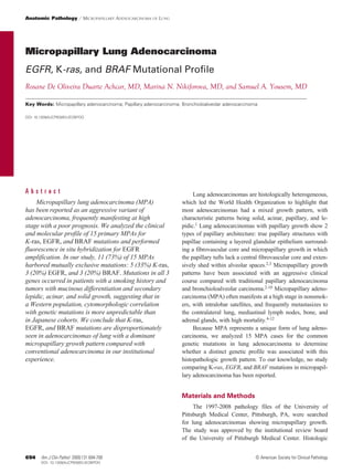
was performed in a glass capillary using 50 ng of DNA in zImage 1z Micropapillary lung adenocarcinoma. At low
a 20-µL volume. The reaction mixture was subjected to 40 magnification, a nodular growth pattern with prominent
cycles of rapid PCR consisting of denaturation at 94°C for 1 desmoplastic stroma is shown (H&E, ×40).
© American Society for Clinical Pathology Am J Clin Pathol 2009;131:694-700 695
695 DOI: 10.1309/AJCPBS85VJEOBPDO 695
- 3. Achcar et al / Micropapillary Adenocarcinoma of Lung
zImage 2z Micropapillary lung adenocarcinoma. Dyscohesive zImage 3z Micropapillary lung adenocarcinoma (MPA).
papillary clusters of cytologically malignant cells “float” within While MPA frequently shows “hobnail” cytologic and
air spaces and are focally associated with lepidic growth bronchioloalveolar features, acinar and tubular architectures
(H&E, ×120). are common, as are columnar and polygonal cell cytologic
features with abundant intracellular and extracellular mucin
production (H&E, ×100).
an acinar growth pattern in 5 (38%), and solid growth in 1 pure hobnail or terminal reserve differentiation, 2 had K-ras
(8%); 2 cases showed micropapillary growth exclusively. mutations, 1 an EGFR mutation, and 1 a BRAF mutation.
Cytologically, the tumors were extremely heterogeneous, Of the tumors with pure columnar or polygonal cell change,
although we attempted to separate out nonmucinous hobnail 2 had EGFR mutations, 1 a K-ras mutation, and 1 a BRAF
and terminal reserve unit–type micropapillary adenocarci- mutation. Of the 15 adenocarcinomas, 5 showed clear cell
noma (n = 4) from tumors having a predominant columnar or differentiation, whereas only 1 showed signet-ring change.
polygonal cell configuration (n = 8), with the remainder hav- Intracellular and extracellular mucin production with diastase-
ing mixed cell populations (n = 3) zImage 3z.7,17 Of the 4 with predigested periodic acid–Schiff and mucicarmine stains was
zTable 1z
Complete Clinicopathologic Data for 15 Cases of Micropapillary
Lung Adenocarcinoma*
Cytologic Features
Case No./ Tumor Tumor Mucin Psammoma Angiolymphatic
Sex/Age (y) Location Size (cm) Differentiation Hobnail Columnar Production Bodies Host Response Necrosis Invasion
1/M/67 RML 3.3 Moderate No Yes Yes No Moderate Yes Yes
2/M/74 LUL 4.0 Well Yes No No No Mild Yes Yes
3/F/80 LUL 1.3 Moderate Yes Yes No Yes Mild No No
4/M/64 LUL 6.0 Moderate Yes No No No Moderate Yes Yes
5/M/79 LLL 2.0 Moderate No Yes Yes No Mild Yes No
6/F/71 LUL 3.5 Moderate Yes No No No Mild Yes Yes
7/F/73 LUL 3.0 Moderate No Yes Yes No Moderate Yes Yes
8/F/68 RUL 2.0 Moderate No Yes No No Minimal Yes Yes
9/F/60 LUL 1.5 Well Yes Yes No No Mild Yes Yes
10/M/58 RLL 6.0 Moderate No Yes Yes No Mild Yes Yes
11/F/80 RUL 6.5 Moderate Yes Yes Yes No Mild Yes Yes
12/M/64 LUL 3.0 Moderate Yes No Yes Yes Mild Yes Yes
13/F/50 RLL 0.5 Moderate No Yes Yes Yes Minimal Yes No
14/M/68 LLL 4.0 Moderate No Yes Yes No Mild Yes Yes
15/F/70 LLL 4.5 Moderate No Yes No Yes Moderate Yes Yes
LLL, left lower lobe; LUL, left upper lobe; RLL, right lower lobe; RML, right middle lobe; RUL, right upper lobe.
* Hobnail, cells with Clara and type II pneumocyte differentiation, often called terminal reserved unit cells; columnar, columnar or polygonal cells resembling bronchiolar epithelium.
696 Am J Clin Pathol 2009;131:694-700 © American Society for Clinical Pathology
696 DOI: 10.1309/AJCPBS85VJEOBPDO
- 4. Anatomic Pathology / Original Article
seen in 8 (53%) of 15 cases. Psammoma bodies were noted in micropapillary adenocarcinoma; articles reporting on micro-
4 (27%) of 15 cases. papillary growth have indicated its presence in between 5%
Of 7 cases associated with secondary lepidic growth, 6 and 100% of their study populations.2-10 In our study of 15
demonstrated the following mutations: 3 (43%) of 7, K-ras; 2 cases of primary MPA, we used an original definition sug-
(29%) of 7, EGFR; and 1 (14%) of 7, BRAF V600E. Among the gested by Silver and Askin13 that a papillary adenocarcinoma
MPAs showing mucin production with histochemical stains, 4 be composed of greater than or equal to 75% papillary growth.
(50%) of 8 showed the following mutations: 2 (25%) of 8, BRAF By using this definition for MPA, we confirmed the general
V600E; 1 (13%) of 8, K-ras; and 1 (13%) of 8, EGFR. observations of other studies having a less stringent defini-
Molecular studies demonstrated that 11 (73%) of 15 tion. In particular, micropapillary adenocarcinoma is typically
cases had mutations involving the 3 genes investigated: 5 a malignancy of older people, with an equal sex distribution,
(33%) showed K-ras mutations, 3 (20%) demonstrated EGFR that manifests more often at a late stage with intrapulmonary
mutations, and 3 (20%) showed BRAF V600E mutations zFig- and extrapulmonary metastases. Our study is unique in focus-
ure 1z. Of the 3 EGFR mutations, 2 were deletions in exon 19 ing on the 3 major oncogenes reported in lung adenocarci-
and 1 was a point mutation in exon 21. EGFR amplification noma, although we recognize that between 30% and 70% of
was detected in 2 (13%) of 15 cases, with 1 of these 2 cases lung adenocarcinomas have complex genetic mutations not
associated with EGFR mutations. All K-ras mutations were at tied to EGFR, K-ras, and BRAF.18 Still, in our study group,
codon 12 leading to substitution of glycine with phenylalanine 73% of the cases demonstrated mutations of K-ras (33%),
(n = 1), cysteine (n = 3), or alanine (n = 1). Gly12Phe is a rare EGFR (20%), and BRAF (20%), making MPA unusual in its
type of K-ras mutation in which 2 nucleotides are substituted frequency of involvement of these 3 genes compared with
at codon 12 (GGT to TTT) instead of the usual 1-nucleotide our institutional percentages in lung adenocarcinoma: EGFR,
substitution. We attempted to correlate the mutations of these 10%; K-ras, 23%; and BRAF, 5.5% (see the “Materials and
3 genes with morphologic findings. No clear association of Methods” section). This percentage far outweighs the cumu-
mutations with the morphologic or clinical features shown in lative percentages of 40% usually associated with primary
Table 1 was noted (P > .05). conventional lung adenocarcinoma.18-20
Most studies on micropapillary growth and lung adeno-
carcinomas have focused on a higher than usual incidence of
Discussion EGFR mutations in papillary and micropapillary adenocarci-
Micropapillary growth in pulmonary adenocarcinomas nomas of lung.7,11 Our study, in fact, confirmed that EGFR
reflects an aggressive subset of lung adenocarcinomas with mutations are twice as common in MPA as reported in the
a poor prognosis.2,3,5,6,8-10 In analyzing the relevant stud- Western literature. However, our study also highlights that
ies, one problem is achieving agreement on a definition for EGFR is not the only gene associated with MPA, with K-ras
and BRAF mutations present in more than 50% of our study
population. Furthermore, while the association of EGFR
mutations with an absence of smoking has been emphasized
in Far Eastern and Western populations, our study also shows
that EGFR mutations in MPA also occur in cigarette smok-
Mutation ers.11,17,21,22 In our study, we defined a history of cigarette
Visceral-Pleural Pathologic EGFR EGFR K-ras BRAF
smoking in absolute terms—any patient with a history of
Invasion Stage Exon 19 Exon 21 Exon 2 V600E cigarette smoking was considered as having a risk of develop-
ing adenocarcinoma, in contrast with other studies, such as a
Yes T2 N0 M0 No No No No
Yes T2 N0 M0 Yes No No No study by Motoi et al,11 who defined nonsmokers as patients
No T1 NX M0 No No Yes No having less than a 15-pack-year history of smoking.
Yes T2 N1 M0 No No Yes No
No T1 NX M0 No No No No EGFR mutations have also been reported primarily in
No T2 N0 M0 No No Yes No adenocarcinomas having type II alveolar pneumocyte dif-
Yes T2 N1 M1 Yes No No No
ferentiation, variously reported as “hobnail configuration”
No T1 N0 M0 No Yes No No
Yes T4 N2 M0 No No Yes No or “terminal reserve unit” cytologic features.7,11,17 Although
Yes T2 N2 M0 No No Yes No
EGFR mutations were certainly seen in cells with these
Yes T4 N0 M0 No No No Yes
Yes T4 N2 M0 No No No Yes differentiation characteristics, EGFR mutations were also
No T4 N0 M0 No No No No observed in MPAs with columnar and polygonal cell dif-
Yes T2 N0 M0 No No No No
Yes T2 N0 M0 No No No Yes ferentiation, whereas K-ras and BRAF mutations were seen
in adenocarcinomas with hobnail or terminal reserve unit cell
differentiation. Furthermore, although it has been emphasized
© American Society for Clinical Pathology Am J Clin Pathol 2009;131:694-700 697
697 DOI: 10.1309/AJCPBS85VJEOBPDO 697
- 5. Achcar et al / Micropapillary Adenocarcinoma of Lung
A B EGFR exon 21
EGFR exon 19
L858R mutation
15 bp deletion
G G G C T G G C C A A A
T C G C T A T C A A GG A A T T AA G
G
C D
0.078
–(d/dt) Fluorescence (705/530)
K-ras codon 12 BRAF V600E
Gly12Cys mutation 0.073
0.069 WT
G G A G C TG G T G G C G T A
0.065
0.063
T
0.058
0.053
0.048
40 45 50 55 60 65 70 75 80
Temperature (°C)
zFigure 1z Examples of mutations found in micropapillary lung adenocarcinoma. A, Direct nucleotide sequencing shows a
15-base-pair (bp) deletion in exon 19 of the EGFR gene. B, Point mutation (CTG to CGG) at codon 858 exon 21 of the EGFR
gene detected by sequencing. C, K-ras codon 12 mutation (GGT to TGT) detected by sequencing leads to glycine to cysteine
amino acid change. D, BRAF V600E mutation detected by real-time polymerase chain reaction (PCR) and post-PCR melting
curve analysis demonstrates the mutant peak at a melting temperature of 58ºC in addition to the wild-type (WT) peak.
that mucin-producing, goblet cell–type bronchioloalveolar Japanese cohort: EGFR mutations in 4 exons occurred in 59%
adenocarcinomas are unassociated with EGFR mutations, our of cases; patients were usually nonsmoking women, and there
micropapillary adenocarcinomas produced mucin and had was a strong association with a bronchioloalveolar and papil-
EGFR mutations, as well as K-ras and BRAF mutations. lary growth pattern. Our study in a Western population, using
The majority of our MPA cases demonstrated secondary more rigorous criteria for the definition of MPA, suggests that
lepidic and acinar growth patterns ranging from 1% to 25% of this tumor has a wider demographic and molecular spectrum.
the tumor. There was no strong correlation between second- Motoi et al11 did not define MPA by absolute percentages of
ary growth pattern and molecular profile, although overall, micropapillary growth, the result being the inclusion of con-
K-ras mutations were more frequent in tumors with lepidic ventional adenocarcinomas and papillary adenocarcinomas in
and papillary growth, whereas BRAF mutations were slightly the analysis, in which micropapillary architecture represented
more frequent in tumors with acinar growth. Nevertheless, in only the major component in relative terms but not necessarily
this group of MPAs, it is important to emphasize that in con- in percentages greater than 75%. We confirmed their observa-
trast with the findings of other studies, lepidic growth in these tion that EGFR mutations are more common in MPA than in
adenocarcinomas did not exclude EGFR mutations.11 conventional adenocarcinoma, but we also noted that MPA
To date, only 2 studies7,11 have looked specifically at is present in smokers, displays mucinous differentiation, and
the molecular alterations in MPAs, and neither used the is associated with K-ras and BRAF mutations, as noted by
definitions of Silver and Askin.13 In the study by Ninomiya others.23,24
et al,7 MPA was defined as an adenocarcinoma with 25% In the literature, EGFR mutations have largely been
of the tumor having micropapillary growth. By using this associated with bronchioloalveolar adenocarcinoma, inva-
definition, the authors showed that MPA closely resembled sive adenocarcinomas with prominent lepidic growth,
their EGFR+ adenocarcinomas in their demographics in this and papillary adenocarcinoma.7,11,20,23,24 Micropapillary
698 Am J Clin Pathol 2009;131:694-700 © American Society for Clinical Pathology
698 DOI: 10.1309/AJCPBS85VJEOBPDO
- 6. Anatomic Pathology / Original Article
adenocarcinoma of the lung needs to be added to this group, 5. Kamiya K, Hayashi Y, Douguchi J, et al. Histopathological
although as emphasized before, mutations in this group are features and prognostic significance of the micropapillary
pattern in lung adenocarcinoma. Mod Pathol.
not restricted to EGFR, thus warranting a comprehensive 2008;21:992-1001.
molecular analysis should personalized therapies be con- 6. Makimoto Y, Nabeshima K, Iwasaki H, et al. Micropapillary
templated. For example, BRAF mutations occur in approxi- pattern: a distinct pathological marker to subclassify tumours
mately 5% of pulmonary adenocarcinomas, often with a with a significantly poor prognosis within small peripheral lung
adenocarcinoma (≤20 mm) with mixed bronchioloalveolar and
papillary architecture, but in the micropapillary subgroup, invasive subtypes (Noguchi’s type C tumours). Histopathology.
the incidence rises to approximately 20%, similar to that 2005;46:677-684.
seen in thyroid and ovarian papillary adenocarcinoma.16 It 7. Ninomiya H, Hiramatsu M, Inamura K, et al. Correlation
may be worthwhile to add this gene to a molecular panel between morphology and EGFR mutations in lung
adenocarcinomas: significance of the micropapillary pattern
when one sees this architectural pattern given that some
and the hobnail cell type [published online ahead of print June
personalized therapies for BRAF mutation (akin to tyrosine 20, 2008]. Lung Cancer. 2009;63:235-240.
kinase inhibitor therapy in EGFR-mutated adenocarcinoma) 8. Roh MS, Lee JI, Choi PJ, et al. Relationship between
have been identified.25-30 micropapillary component and micrometastasis in the regional
This study represents the first attempt to rigorously lymph nodes of patients with stage I lung adenocarcinoma.
Histopathology. 2004;45:580-586.
evaluate the molecular alterations in MPAs by using a
9. Sánchez-Mora N, Presmanes MC, Monroy V, et al.
uniform definition and to correlate this growth pattern with Micropapillary lung adenocarcinoma: a distinctive histologic
K-ras, EGFR, and BRAF mutations. We conclude that subtype with prognostic significance: case series. Hum Pathol.
K-ras, EGFR, and BRAF mutations occur at an increased 2008;39:324-330.
frequency in lung adenocarcinomas showing greater than 10. Yokose T, Suzuki K, Nagai K, et al. Favorable and
unfavorable morphological prognostic factors in peripheral
75% micropapillary growth and that these mutations are adenocarcinoma of the lung 3 cm or less in diameter. Lung
seen in smokers and in adenocarcinomas with mucin produc- Cancer. 2000;29:179-188.
tion. K-ras mutations certainly predominate, but BRAF and 11. Motoi N, Szoke J, Riely GJ, et al. Lung adenocarcinoma:
EGFR mutations occur at a higher incidence than in conven- modification of the 2004 WHO mixed subtype to include
the major histologic subtype suggests correlations between
tional lung adenocarcinomas reported in the literature and in papillary and micropapillary adenocarcinoma subtypes, EGFR
our institutional experience. mutations and gene expression analysis. Am J Surg Pathol.
2008;32:810-827.
From the Department of Pathology, University of Pittsburgh 12. Noguchi M, Morikawa A, Kawasaki M, et al. Small
Medical Center, Pittsburgh, PA. adenocarcinoma of the lung: histologic characteristics and
prognosis. Cancer. 1995;75:2844-2852.
Address reprint requests to Dr Yousem: Dept of Pathology, 13. Silver SA, Askin FB. True papillary carcinoma of the
University of Pittsburgh Medical Center, Presbyterian Campus, lung: a distinct clinicopathologic entity. Am J Surg Pathol.
Room A610, 200 Lothrop St, Pittsburgh, PA 15213-2582. 1997;21:43-51.
Acknowledgments: We thank Kathy Cieply for fluorescence 14. Striebel JM, Dacic S, Yousem SA. Gross cystic disease
in situ hybridization studies and Linda Shab and Tom Bauer for fluid protein-(GCDFP-15): expression in primary lung
photographic aid. adenocarcinoma. Am J Surg Pathol. 2008;32:426-432.
15. Naoki K, Chen TH, Richards WG, et al. Missense mutations
of the BRAF gene in human lung adenocarcinoma. Cancer
Res. 2002;62:7001-7003.
References 16. Yousem SA, Nikiforova M, Nikiforov Y. The histopathology
of BRAF-V600E-mutated lung adenocarcinoma. Am J Surg
1. Travis WD, Brambilla E, Müller-Hermelink HK, et al, eds. Pathol. 2008;32:1317-1321.
Pathology and Genetics of Tumours of the Lung, Pleura, 17. Yatabe Y, Kosaka T, Takahashi T, et al. EGFR mutation is
Thymus and Heart. Lyon, France: IARC Press; 2004. WHO specific for terminal respiratory unit type adenocarcinoma.
Classification of Tumours; vol 10. Am J Surg Pathol. 2005;29:633-639.
2. Amin MB, Tamboli P, Merchant SH, et al. Micropapillary 18. Eberhard DA, Johnson BE, Amler LC, et al. Mutations in the
component in lung adenocarcinoma: a distinctive histologic epidermal growth factor receptor and in KRAS are predictive
feature with possible prognostic significance. Am J Surg Pathol. and prognostic indicators in patients with non–small-cell lung
2002;26:358-364. cancer treated with chemotherapy alone and in combination
3. Miyoshi T, Satoh Y, Okumura S, et al. Early-stage lung with erlotinib. J Clin Oncol. 2005;23:5900-5909.
adenocarcinomas with a micropapillary pattern, a distinct 19. Lynch TJ, Bell DW, Sordella R, et al. Activating mutations in
pathologic marker for a significantly poor prognosis. Am J Surg the epidermal growth factor receptor underlying responsiveness
Pathol. 2003;27:101-109. of non–small-cell lung cancer to gefitinib. N Engl J Med.
4. Aida S, Shimazaki H, Sato K, et al. Prognostic analysis of 2004;350:2129-2139.
pulmonary adenocarcinoma subclassification with special 20. Shigematsu H, Gazdar AF. Somatic mutations of epidermal
consideration of papillary and bronchioloalveolar types. growth factor receptor signaling pathway in lung cancers. Int J
Histopathology. 2004;45:468-476. Cancer. 2006;118:257-262.
© American Society for Clinical Pathology Am J Clin Pathol 2009;131:694-700 699
699 DOI: 10.1309/AJCPBS85VJEOBPDO 699
- 7. Achcar et al / Micropapillary Adenocarcinoma of Lung
21. Jackman DM, Yeap BY, Sequist LV, et al. Exon 19 deletion 26. Chou TY, Chiu CH, Li LH, et al. Mutation in the tyrosine
mutations of epidermal growth factor receptor are associated kinase domain of epidermal growth factor receptor is a
with prolonged survival in non–small cell lung cancer predictive and prognostic factor for gefitinib treatment in
patients treated with gefitinib or erlotinib. Clin Cancer Res. patients with non–small cell lung cancer. Clin Cancer Res.
2006;12:3908-3914. 2005;11:3750-3757.
22. Kawakami T, Nabeshima K, Makimoto Y, et al. Micropapillary 27. Dhomen N, Marais R. New insight into BRAF mutations in
pattern and grade of stromal invasion in pT1 adenocarcinoma cancer. Curr Opin Genet Dev. 2007;17:31-39.
of the lung: usefulness as prognostic factors. Mod Pathol. 28. Miller VA, Riely GJ, Zakowski MF, et al. Molecular
2007;20:514-521. characteristics of bronchioloalveolar carcinoma and
23. Miller VA, Kris MG, Shah N, et al. Bronchioloalveolar adenocarcinoma, bronchioloalveolar carcinoma subtype,
pathologic subtype and smoking history predict sensitivity to predict response to erlotinib. J Clin Oncol. 2008;26:1472-1478.
gefitinib in advanced non–small-cell lung cancer. J Clin Oncol. 29. Solit DB, Garraway LA, Pratilas CA, et al. BRAF
2004;22:1103-1109. mutation predicts sensitivity to MEK inhibition. Nature.
24. Tsuchiya E, Furuta R, Wada N, et al. High K-ras mutation rates 2006;439:358-362.
in goblet-cell-type adenocarcinomas of the lungs. J Cancer Res 30. Trovisco V, Soares P, Sobrinho-Simoes M. B-RAF mutations
Clin Oncol. 1995;121:577-581. in the etiopathogenesis, diagnosis, and prognosis of thyroid
25. Cappuzzo F, Hirsch FR, Rossi E, et al. Epidermal growth carcinomas. Hum Pathol. 2006;37:781-786.
factor receptor gene and protein and gefitinib sensitivity
in non–small-cell lung cancer. J Natl Cancer Inst.
2005;97:643-655.
700 Am J Clin Pathol 2009;131:694-700 © American Society for Clinical Pathology
700 DOI: 10.1309/AJCPBS85VJEOBPDO