Renal stones
•Télécharger en tant que PPT, PDF•
140 j'aime•44,134 vues

Pathologic & other aspects

Recommandé
Contenu connexe
Tendances
Tendances (20)
En vedette
En vedette (20)
Similaire à Renal stones
Similaire à Renal stones (20)
Plus de Mohammad Manzoor
Plus de Mohammad Manzoor (20)
Dernier
Dernier (20)
Renal stones
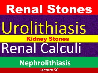
- 1. Renal Stones Lecture 50 Urolithiasis Renal Calculi Nephrolithiasis Kidney Stones
- 2. Renal Stones A kidney stone, is a solid concretion or crystal aggregation formed in the kidneys from dietary minerals in the urine.
- 3. Classification- by location • Urinary stones are typically classified by their location in the • kidney (nephrolithiasis), •ureter (ureterolithiasis), •bladder (cystolithiasis),
- 4. Classification- by chemical composition 1.1. Calcium saltsCalcium salts 2.2. Uric acidUric acid 3.3. Mg ammonium POMg ammonium PO44 4.4. CystineCystine 5.5. Other (xanthine, etc.Other (xanthine, etc.))
- 5. Causes
- 6. Dietary Causes • LOW FLUID INTAKE • HIGH DIETARY INTAKE OF ANIMAL PROTEIN, • SODIUM, • REFINED SUGARS, FRUCTOSE • HIGH FRUCTOSE CORN SYRUP, • OXALATE, • GRAPEFRUIT JUICE • APPLE JUICE higher Vitamin C
- 7. Risk Factors
- 8. Risk Factors
- 9. Risk Factors • Male sex • Obesity • Family History • H/o stone disease (1/2 will have recurrence)
- 10. Risk Factors •Occupation • Small bowel disease (i.b.d.) • Medical conditions causing hypercalcuria • Medical conditions causing aciduria
- 11. Pathogenesis- MucoProtein Matrix • An organic mucoprotein matrix, making up 1% to 5% of the stone by weight, is present in all calculi.
- 12. Pathogenesis- suPersaturation The most important determinant(Renal stones) is an increased urinary concentration of the stones' constituents, such that it exceeds their solubility (supersaturation). A low urine volume in some metabolically normal patients may also favor supersaturation.
- 13. Supersaturation- Slow urine flow
- 15. SOLUBILITY • Solubility is affected by •Urine pH, •Volume and •Total excretion
- 16. Pathogenesis- Calcium Oxalate Stones Hypercalcemia & Hypercalciuria • Calcium oxalate stones are associated in about 5%of patients with hypercalcemia and hypercalciuria, such as occurs with hyperparathyroidism, diffuse bone disease, sarcoidosis, and other hypercalcemic states.
- 17. Pathogenesis 55% Hypercalciuria without hypercalcemia • This is caused by several factors, including: • Hyperabsorption of calcium from the intestine (Absorptive hypercalciuria), • Intrinsic impairment in renal tubular reabsorption of calcium (Renal hypercalciuria), • Idiopathic fasting hypercalciuria with normal parathyroid function.
- 18. Pathogenesis- 20% Hyperuricosuric calcium nephrolithiasis • 20% of calcium oxalate stones are associated with • increased uric acid secretion (hyperuricosuric calcium nephrolithiasis), with or without hypercalciuria. • The mechanism of stone formation in this setting involves • “nucleation” of calcium oxalate by uric acid crystals in the collecting ducts.
- 19. Pathogenesis- Hyperoxaluria • 5% are associated with hyperoxaluria, either HEREDITARY (primary oxaluria) or, more commonly, ACQUIRED by intestinal overabsorption in patients with enteric diseases. Enteric hyperoxaluria, also occurs in vegetarians, because much of their diet is rich in oxalates. 5%
- 20. Pathogenesis- HYPOCITRATURIA • Hypocitraturia, associated with acidosis and chronic diarrhea of unknown cause, may produce calcium stones. •
- 21. Pathogenesis Idiopathic calcium stone disease • In a variable proportion of individuals with calcium stones, no cause can be found (idiopathic calcium stone disease).
- 22. Pathogenesis Magnesium ammonium phosphate stones • Formed largely after infections by bacteria (e.g., Proteus and some staphylococci) that convert urea to ammonia. • The resultant alkaline urine causes the precipitation of magnesium ammonium phosphate salts.
- 23. Pathogenesis Magnesium ammonium phosphate stones • These form some of the largest stones, • as the amounts of urea excreted normally are huge. • Indeed, so-called staghorn calculi occupying large portions of the renal pelvis are almost always a consequence of infection.
- 24. Pathogenesis- URIC ACID stones • Uric acid stones are common in individuals with hyperuricemia, such as gout, and diseases involving rapid cell turnover, such as the leukemias.
- 25. However, more than half of all patients with uric acid calculi have neither hyperuricemia nor increased urinary excretion of uric acid. • In this group, it is thought that an unexplained tendency to excrete urine of pH below 5.5 may predispose to uric acid stones, because uric acid is insoluble in acidic urine. In contrast to the radiopaque calcium stones, uric acid stones are radiolucent.
- 26. Pathogenesis- CYSTINE STONES • Cystine stones are caused by • genetic defects in the renal reabsorption of amino acids, including cystine, leading to cystinuria. • Stones form at low urinary pH.
- 27. Pathogenesis • It can therefore be appreciated that 1. increased concentration of stone constituents, 2.changes in urinary pH, 3. decreased urine volume, and 4.the presence of bacteria influence the formation of calculi.
- 28. Pathogenesis • However, many calculi occur in the absence of these factors; • conversely, individuals with hypercalciuria, hyperoxaluria, and hyperuricosuria often do not form stones.
- 29. • It has therefore been postulated that stone formation is enhanced by a deficiency in inhibitors of crystal
- 30. Inhibitors of Crystal Formation • Pyrophosphate, • Diphosphonate, • Citrate, • Glycosaminoglycans, • Osteopontin, and •Nephrocalcin (Glycoprotein).
- 31. Morphology- Gross• Unilateral 80% Favored sites: •Renal calyces •Pelves •Bladder.
- 32. Morphology- in Renal Mass •Small-2 to 3 mm. • May have smooth contours or • May take the form of an irregular, jagged mass of spicules.
- 33. Morphology • Often many stones are found within one kidney. • On occasion, progressive accretion of salts leads to the development of branching structures known as staghorn calculi, which create a castof the pelvic and calyceal system.
- 36. Smaller Stones • In general, smaller stones are most hazardous, • because they may pass into the ureters, producing colic, one of the most intense forms of pain, and • ureteral obstruction.
- 37. If stones grow to sufficient size (usually at least 3 millimeters (0.12 in)) they can cause obstruction of the ureter. • Ureteral obstruction causes • Postrenal azotemia and • Hydronephrosis (distension and dilation of the renal pelvis and calyces), as well as • Spasm of the ureter.
- 38. Ureteral Obstruction leads to pain, most commonly felt in the flank, lower abdomen, and groin (a condition called renal colic). • Renal colic can be associated with nausea, vomiting, fever, blood in the urine, pus in the urine, and painful urination. Renal colic typically comes in waves lasting 20 to 60 minutes, beginning in the flank or lower back and often radiating to the groin or genitals (The Hallmark of obstructive ureteral stones).
- 39. Larger Stones • Larger stones cannot enter the ureters and are more likely to remain silent within the renal pelvis. • Commonly, these larger stones first manifest themselves by hematuria.
- 40. Clinical features cont… • Stones also predispose to superimposed infection, both by their obstructive nature and by the traumathey produce.
- 42. Renal colic caused by kidney stones is commonly accompanied by: • Urinary urgency, • Rrestlessness, • Hematuria, Sweating, Nausea, Vomiting.
- 43. Calcium saltCalcium salt stonesstones 80% of kidney stones contain calcium80% of kidney stones contain calcium The type of salt depends onThe type of salt depends on – Urine pHUrine pH – Availability of oxalateAvailability of oxalate General appearance:General appearance: – White, hard, radioopaqueWhite, hard, radioopaque – Calcium POCalcium PO44: staghorn in renal pelvis: staghorn in renal pelvis (large)(large) – Calcium oxalate: present in ureter (small)Calcium oxalate: present in ureter (small)
- 44. Calcium salt stonesCalcium salt stones Causes of calcium salt stones:Causes of calcium salt stones: Hypercalciuria:Hypercalciuria: – Increased urinary calcium excretionIncreased urinary calcium excretion – Men: > 7.5 mmols/dayMen: > 7.5 mmols/day – Women > 6.2 mmols/dayWomen > 6.2 mmols/day – May or may not be due to hypercalcemiaMay or may not be due to hypercalcemia
- 45. Calcium salt stonesCalcium salt stones • Hyperoxaluria:Hyperoxaluria: – Causes the formation of calcium oxalatesCauses the formation of calcium oxalates without hypercalciuriawithout hypercalciuria – Diet rich in oxalatesDiet rich in oxalates – Increased oxalate absorption in fatIncreased oxalate absorption in fat malabsorptionmalabsorption • Primary hyperoxaluria:Primary hyperoxaluria: – Due to inborn errorsDue to inborn errors – Urinary oxalate excretion: > 400Urinary oxalate excretion: > 400 mmols/daymmols/day
- 47. Calcium salt stonesCalcium salt stones • Treatment:Treatment: – Treatment of primary causes such asTreatment of primary causes such as infection, hypercalcemia, hyperoxaluriainfection, hypercalcemia, hyperoxaluria – Oxalate-restricted dietOxalate-restricted diet – Increased fluid intakeIncreased fluid intake – Acidification of urine (by dietaryAcidification of urine (by dietary changes)changes) • Calcium salt stones are formed inCalcium salt stones are formed in alkaline urinealkaline urine
- 48. Uric acid stonesUric acid stones• About 8% of renal stones contain uric acidAbout 8% of renal stones contain uric acid • May be associated with hyperuricemia (with orMay be associated with hyperuricemia (with or without gout)without gout) • Form in acidic urineForm in acidic urine • General appearance:General appearance: – Small, friable, yellowishSmall, friable, yellowish – May form staghornMay form staghorn – Radiolucent (plain x-rays cannot detect)Radiolucent (plain x-rays cannot detect) – Visualized by ultrasound or i.v. pyelogramVisualized by ultrasound or i.v. pyelogram
- 49. Uric acid stonesUric acid stones • Treatment:Treatment: –Purine-restricted dietPurine-restricted diet –Alkalinization of urineAlkalinization of urine (by dietary changes)(by dietary changes) –Increased fluid intakeIncreased fluid intake
- 50. Uric acid stones
- 51. Mg ammonium POMg ammonium PO44 stonesstones About 10% of all renal stones contain Mg amm.About 10% of all renal stones contain Mg amm. POPO44 Also calledAlso called struvitestruvitekidney stoneskidney stones Associated with chronic urinary tract infectionAssociated with chronic urinary tract infection – Microorganisms (such as fromMicroorganisms (such as from ProteusProteus genus)genus) that metabolizethat metabolize urea into ammoniaurea into ammonia – Causing urine pH to become alkaline and stoneCausing urine pH to become alkaline and stone formationformation STRUVITE: A crystalline mineral found in guano. It is a hydrous phosphate of magnesia and ammonia. Guano is the excrement of seabirds, cave-dwelling bats, or birds more generally.
- 52. Mg ammonium POMg ammonium PO44 stonesstones • Commonly associated with staghorn calculiCommonly associated with staghorn calculi • 75% of staghorn stones are of struvite type75% of staghorn stones are of struvite type • Treatment:Treatment: – Treatment of infectionTreatment of infection – Urine acidificationUrine acidification – Increased fluid intakeIncreased fluid intake
- 53. Mg ammonium phosphate (struvite) stone Staghorn Stone ( Branching Stone)
- 54. Cystine stonesCystine stones • A rare type of kidney stoneA rare type of kidney stone • Due to homozygous cystinuriaDue to homozygous cystinuria • Form in acidic urineForm in acidic urine • Soluble in alkaline urineSoluble in alkaline urine • Faint radio-opaqueFaint radio-opaque Treatment:Treatment: – Increased fluid intakeIncreased fluid intake – Alkalinization of urine (by dietary changes)Alkalinization of urine (by dietary changes) – Penicillamine (binds to cysteine to form aPenicillamine (binds to cysteine to form a compound more soluble than cystine)compound more soluble than cystine)
- 55. Cystine stone
- 57. Kidney Stones Form in acidic urine • Uric acid Stones • Cystine Stones Form in alkaline urine • Calcium Salt Stones • Struvite Stones
- 58. Kidney Stones Radioopaque • Calcium Salt Stones • Cystine stones (Faint) • Struvite Radiolucent • Uric Acid Stones
Notes de l'éditeur
- Pre-disposing medical factors, such as hyperparathyroidism, are a complex list, and will not be addressed here
- From what is now known about stone disease it is understandable that there are some factors which will predictable lead to an increased incidence of stone disease. Occupation has been shown to be related to stone disease when it prevents a person from maintaining adequate degree of hydration and when it leads to a large amount of extra renal fluid loss. Such occupations would include construction work, farming and other activities that keep people out in hot weather and increased sweating. Family history is a factor in certain forms of stone disease such as cystinuria which is an inherited metabolic disorder. In general family history may be more of a factor in that habits of diet and poor hydration are probably adapted by other members of the family. Small bowel disease i.e... Krohn’s disease are associated with an increased incidence of stone disease because of the associated steattorhea. The fatty acids in the gut bind with intraluminal calcium leaving the oxalate in a more easily absorbed state. The net effect is hyperoxaluria. IBD may also result in aciduria since there can be considerable amount of basic fluid lost via the gi tract. There are a number of diseases that result in hypercalcuria such as hyperparathyroidism etc. that lead to increased calcium stone formation. Similarly cliical disease leading to hyperuricouria i.e... gout, can produce uric acid stones.
- The picture of acute renal colic is well described in textbooks of physical diagnosis. The mechanism for pain production is through the sudden obstruction of the kidney with stretching of the collecting system and renal capsule. It is the stretching of these structures that causes the pain. The pain is not the result of a foreign body on the mucosa of the ureter. In most cases sudden renal obstruction produces flank pain. It is not possible from the pain to predict what level the stone is at. The only time one can predict the stone’s location is when the patient begins to complain of symptoms of bladder irritation. When this occurs the stone is in the most distal portion of the ureter (intra-mural ureter) which produces some inflammation of the bladder neck and hence the bladder irritative symptoms. The associated gi symptoms of nausea and vomiting may be pronounced and mislead you into thinking that the patient has a gi problem. Look for hematuria, most patients passing a stone will have at least microscopic hematuria. Chronic stone disease may have little associated pain. Although there may be renal and ureteral obstruction present often this has been long standing and no longer produces the dramatic pain of acute obstruction. Often there will be chronic infection which resist attempts at cure.