SlideShare une entreprise Scribd logo
1  sur  46
The Role of Incretins in Glucose Homeostasis and Diabetes
                         Treatment
Abstract

Incretins are gut hormones that are secreted from enteroendocrine cells into the blood within minutes
after eating. One of their many physiological roles is to regulate the amount of insulin that is secreted
after eating. In this manner, as well as others to be described in his review, their final common raison
d'être is to aid in disposal of the products of digestion. There are two incretins, known as glucose-
dependent insulinotropic peptide (GIP) and glucagon-like peptide-1 (GLP-1), that share many
common actions in the pancreas but have distinct actions outside of the pancreas. Both incretins are
rapidly deactivated by an enzyme called dipeptidyl peptidase 4 (DPP4). A lack of secretion of
incretins or an increase in their clearance are not pathogenic factors in diabetes. However, in type 2
diabetes (T2DM), GIP no longer modulates glucose-dependent insulin secretion, even at
supraphysiological (pharmacological) plasma levels, and therefore GIP incompetence is detrimental
to β-cell function, especially after eating. GLP-1, on the other hand, is still insulinotropic in T2DM, and
this has led to the development of compounds that activate the GLP-1 receptor with a view to
improving insulin secretion. Since 2005, two new classes of drugs based on incretin action have been
approved for lowering blood glucose levels in T2DM: an incretin mimetic (exenatide, which is a potent
long-acting agonist of the GLP-1 receptor) and an incretin enhancer (sitagliptin, which is a DPP4
inhibitor). Exenatide is injected subcutaneously twice daily and its use leads to lower blood glucose
and higher insulin levels, especially in the fed state. There is glucose-dependency to its insulin
secretory capacity, making it unlikely to cause low blood sugars (hypoglycemia). DPP4 inhibitors are
orally active and they increase endogenous blood levels of active incretins, thus leading to prolonged
incretin action. The elevated levels of GLP-1 are thought to be the mechanism underlying their blood
glucose-lowering effects.

I. Background and Introduction

Incretins are hormones that are released from the gut into the bloodstream in response to ingestion of
food, and they then modulate the insulin secretory response to the products within the nutrients in the
food. The insulin secretory response of incretins, called the incretin effect, accounts for at least 50%
of the total insulin secreted after oral glucose. Therefore, by definition, incretin hormones are
insulinotropic (i.e., they induce insulin secretion) at usual physiological concentrations seen in the
plasma after ingestion. The concept of incretins is at least a century old (Table 1). In 1902, Bayliss
and Starling published their landmark manuscript, “The Mechanism of Pancreatic Secretion.” The
authors found that acid infused into the digestive system caused pancreatic secretion of juices
through the pancreatic duct from the pancreas, even after they cut the ennervation to the intestine.
Until that time, it was thought that nervous system signals controlled secretion of pancreatic juices.
They carried out ground-breaking studies that led them to conclude that the nature of the signal to the
pancreas was most likely a chemical stimulus: they removed extracts from the intestinal wall after it
had been stimulated by acid, injected the extracts into the bloodstream, and once again they could
see juices coming from the pancreatic duct of the animal that had been injected. Therefore, they
proved that the extracts must have contained a substance that must normally be secreted from the
intestinal wall into the bloodstream to stimulate the flow of pancreatic juice. They called the substance
“secretin.” In his “Cronian Lectures,” Starling introduced the word “hormone” (derived from the Greek
word meaning “impetus”) for clinical factors that are released from one site and act on another
(Starling, 1905. The example of this was that the intestinal extracts contained secretin, which induced
obvious “exocrine” secretion of pancreatic juices. Moore wrote in 1906 that Bayliss and Starling
considered the possibility that the duodenum also supplied a chemical excitant for the “internal”
secretion of the pancreas. They wrote: “This line of argument seems to have occurred to the
discoverers of secretin themselves, for Starling mentions a case of diabetes which was tested by
Spriggs by injections of secretin solutions but with negative results.” Moore (1906) also described
experiments carried out on individual young diabetic patients to whom he gave, by mouth, extracts of
intestinal mucosa. This is therefore the first attempt at “incretin-based” therapies for treating diabetes,
although, of course, the investigator did not call it that. He reported achieving some success, but his
experiments were essentially doomed, because we now know that the chemical excitants are
peptides that would have been degraded when given orally. After World War I ended and insulin was
extracted from pancreatic islets by Banting and Best in 1921, there was further work on the possibility
of food entering the gut leading to secretion of an excitant into the bloodstream that would ultimately
lead to insulin secretion and lowering of blood glucose. Different groups published various and often
contradictory results from experiments on the effects of extracts of duodenal mucosa on fasting blood
glucose and/or on hyperglycemia induced by injection or ingestion of glucose. In 1932, La Barre used
the word “incretin” to refer to an extract from upper gut mucosa that produces hypoglycemia but does
not induce exocrine secretion, although he did not prove incontrovertibly that incretins existed.
Progress on the incretin concept was rapidly made once radioimmunoassays for insulin became
available. Between 1964 and 1967, at least three groups showed independently that glucose, given
orally, induced a greater insulin response (by radioimmunoassay) than i.v. glucose injection even if
the blood glucose levels attained were higher because of the i.v. glucose (Elrick et al., 1964; McIntyre
et al., 1964; Perley and Kipnis, 1967). The three groups therefore knew that the oral glucose was
indeed inducing release of “incretins” into the bloodstream that subsequently increased insulin
secretion, more than did glucose itself. In 1971, John C. Brown isolated and deduced the amino acid
structure of a peptide he had isolated from intestinal mucosa (Brown and Dryburgh, 1971).
Exogenous administration of the peptide inhibited gastric acid secretion in dogs, so he called it gastric
inhibitory polypeptide (GIP)1 (Brown and Dryburgh, 1971). Brown and colleagues subsequently found
that it had insulinotropic properties and suggested that it be called glucose-dependent insulinotropic
peptide, retaining the acronym GIP (Dupre et al., 1973). They not only demonstrated GIP to be
insulinotropic but also demonstrated the glucose dependence of the insulinotropic activity; i.e.,
plasma glucose must be elevated in order for GIP to induce insulin secretion. GIP was therefore the
first incretin to be isolated and its properties characterized.

Time-line to use of incretin-based therapies in the treatment of diabetes

Year  The Development of Incretin-Based Therapies
1869  Islets of Langerhans (nests of cells that appeared different from the surrounding pancreatic
      tissue) in the pancreas were described.
1901  The role of islets of Langerhans (what ultimately became known as an endocrine function) in
      diabetes was described.
1902  The role of a substance (called secretin) secreted by gut cells that stimulates the digestive
      juices for the pancreas (what ultimately became known as exocrine function) was described.
1905  This type of substance, a presumed “chemical messenger,” was now called a “hormone.”
1906  The role of a gut-derived hormone to treat diabetes was first alluded to.
1921– Extraction of insulin from pancreas and its potential to treat type 1 diabetes was shown.
1922
1932  The term “incretin” was used for the first time to refer to a substance derived from the gut,
      presumably a hormone, that regulates insulin secretion after eating.
1960  Radioimmunoassay was developed for measurement of plasma insulin levels.
1964– Clinical proof that a gut-derived factor positively modulated insulin secretion.
1967
1971  The first incretin, GIP, was isolated and sequenced.
1985  The second incretin, GLP-1, was described.
Year     The Development of Incretin-Based Therapies
1992–    Studies show that exogenous GIP does not lower blood glucose in T2DM, but exogenous
1994     GLP-1 does so.
2002     Exendin-4, a GLP-1 receptor agonist extracted from Gila monster lizard saliva, was shown to
         powerfully stimulate insulin secretion in a glucose-dependent manner in subjects with and
         without T2DM.
2005     Exenatide (synthetic exendin-4) came into clinical use for T2DM.
2006     Sitagliptin, an orally active dipeptidyl peptidase 4 inhibitor, came into use in T2DM.

 As late as 1985, another peptide produced in gut, glucagon-like peptide-1 (GLP-1) (7-36), was found
to be a fragment of the recently sequenced proglucagon molecule and was shown to be a potent
insulinotropic factor (Schmidt et al., 1985). It was therefore the second and final incretin to be
characterized. As with GIP, glucose dependence of its insulin-secreting capacity also applies to
GLP-1. GIP and GLP-1 together account for the full incretin effect (Fig. 1). Subsequently, clinical
investigators demonstrated that GLP-1 could increase insulin secretion and normalize blood glucose
in subjects with type 2 diabetes mellitus (T2DM) when given intravenously so as to raise plasma
GLP-1 to supraphysiological levels: supraphysiological levels of GIP did not do so, and thus were
sown the seeds of new therapies based on the actions of GLP-1 for treating T2DM.

II. General Description of Incretins
A. Glucose-Dependent Insulinotropic Peptide

The first incretin hormone described, GIP, is a single 42-amino acid peptide derived from the post-
translational processing of a 153-amino acid precursor encoded by the gip gene (Takeda et al., 1987
and a member of a family of structurally related hormones that includes secretin, glucagon, and
vasoactive intestinal peptide. GIP was originally observed to inhibit gastric acid secretion and
gastrointestinal (GI) motility in dogs, predominantly at supraphysiological dosages (Brown et al.,
1975). Other exciting studies uncovered that GIP exerts glucose-dependent stimulatory effects on
insulin secretion, thereby ensuring prompt insulin-mediated uptake of glucose into tissues.
Furthermore, this action occurred at physiological plasma levels of GIP (Dupre et al., 1973). It is
synthesized in and released in response to nutrients from enteroendocrine cells (called K cells)
primarily in the proximal small intestine (duodenum and jejunum). In the fasted state, circulating levels
of GIP are low relative to levels attained after eating, and GIP release into the bloodstream is then
stimulated by food ingestion containing glucose or fat (Dupre et al., 1973; Pederson et al., 1975;
Elliott et al., 1993). Oral fat alone (e.g., oral corn oil), without any carbohydrate being present, induces
GIP secretion, but this is not sufficient to stimulate insulin secretion at fasting glucose concentrations,
indicating that the effects of GIP on insulin release do not occur if plasma levels of glucose are also
not concurrently increasing; i.e., GIP-mediated insulin secretion is glucose-dependent (Ross and
Dupre, 1978). GIP achieves its insulinotropic effects by binding to its specific receptor (GIPR), which
is positively coupled to increases in intracellular cAMP and Ca2+ levels in β cells. In addition to being
insulinotropic, GIP is involved in fat metabolism in adipocytes: it enhances insulin-stimulated
incorporation of fatty acids into triglycerides, stimulates lipoprotein lipase activity, modulates fatty acid
synthesis (Yip and Wolfe, 2000), and promotes β-cell proliferation and cell survival (Trümper et al.,
2001, 2002). GIP is degraded very quickly by dipeptidyl peptidase 4 (DPP4). In T2DM, plasma
concentrations are reported to be normal or increased (Ross et al., 1977; Vilsbøll et al., 2001), but the
insulinotropic effect is deficient. Although the mechanisms underlying the reduced β-cell response to
GIP remain unclear, more recent studies suggest that hyperglycemia alters the physiological
response as a result of down-regulation of GIPR expression/activity (Lynn et al., 2001; Zhou et al.,
2007). Therefore, GIP/GIPRs have not become targets for treating T2DM.
View larger version:

Schematic representation of incretin secretion and action. GIP and GLP-1 are secreted after food
ingestion, and they then stimulate glucose-dependent insulin secretion. Once released, GIP and
GLP-1 are subject to degradation by DPP4 on lymphocytes and on endothelial cells of blood vessels.
The red cells in the islets are insulin-containing (β) cells and the green cells are glucagon-containing
(α) cells.

B. Glucagon-Like Peptide-1

GLP-1, deduced during cloning and characterization of the proglucagon gene (Bell et al., 1983), is a
post-translational cleavage product of the proglucagon gene by the prohormone convertase PC1/3
(Zhu et al., 2002; Ugleholdt et al., 2004) (Fig. 2) and is a second peptide with incretin activity that
potently stimulates glucose-dependent insulin secretion (Schmidt et al., 1985). In addition to encoding
glucagon, the proglucagon gene encodes two glucagon-like peptides that have approximately 50%
amino acid homology to glucagon; these are designated GLP-1 and glucagon-like peptide-2 (GLP-2,
which is not insulinotropic, has no glucose-lowering properties, and is therefore not an incretin).
These two peptides are mainly produced in enteroendocrine L cells that are scattered among the
enterocytes throughout the small bowel and ascending colon, where they are secreted into the
bloodstream in response to nutrient ingestion (Doyle and Egan, 2007; Jang et al., 2007). In humans,
GLP-1 exists in multiple forms. The majority (at least 80%) of circulating biologically active GLP-1 in
humans is the COOH-terminally amidated form, GLP-1 (7-36) amide, with lesser amounts of the
minor glycine extended form, GLP-1 (7-37), also detectable (Orskov et al., 1986). Like GIP, GLP-1
achieves its insulinotropic effects by binding to its specific receptor (GLP-1R) that is positively
coupled to increases in intracellular cAMP and Ca2+ levels in β cells. In addition to its insulinotropic
effects, it inhibits gastric emptying, decreases food intake (Willms et al., 1996), inhibits glucagon
secretion (Komatsu et al., 1989), and slows the rate of endogenous glucose production (Prigeon et
al., 2003), all of which should help to lower blood glucose in T2DM. It has also been shown to protect
β cells from apoptosis (Farilla et al., 2002) and to stimulate β-cell proliferation by up-regulation of the
β-cell transcription factor pancreatic duodenal homeobox-1 protein (PDX-1) (Perfetti et al., 2000;
Stoffers et al., 2000), which is known to augment insulin gene transcription and up-regulate
glucokinase and glucose transporter2 (GLUT2) (Wang et al., 1999). There is no evidence that
defective secretion of GLP-1 is a cause of T2DM (further discussed in depth in section III.C).
Continuous GLP-1 treatment in T2DM can normalize blood glucose, improve β-cell function, and
restore first-phase insulin secretion and “glucose competence” to β cells (Holz et al., 1993; Zander et
al., 2002); hence, GLP-1/GLP-1Rs are therapeutic targets for treating T2DM. Continuous GLP-1
administration is required for maintenance of glucose homeostasis because of its short half-life (1.5–2
min); similar to GIP, it is degraded by DPP4. The following sections provide our current understanding
of the physiology, the therapeutic potential, and the targets of incretin-based therapies.


Schematic representation of proGIP and proglucagon. GIP is a single 42-amino acid peptide derived
from the post-translational processing of proGIP by PC1/3 in enteroendocrine K cells. It is the only
functional proGIP product in all species examined to date, and there is a greater than 90% amino acid
identity between human, rat, murine, porcine, and bovine sequences. GLP-1 is a post-translational
cleavage product of the proglucagon gene by PC1/3 in enteroendocrine L cells and GLP-1 (7-36)
amide is a major form of circulating biologically active GLP-1 in humans. In mammals, proglucagon is
expressed in pancreas, enteroendocrine L cells, brain, and taste cells with an identical mRNA
transcript in each tissue. Fish and bird proglucagon mRNA in pancreas and liver, however, have
different 3′-ends because of differential splicing upon pancreatic expression. The Xenopus laevis
proglucagon gene encodes three unique GLP-1-like peptides, each with insulinotropic properties that
are capable of activating the human GLP-1R, and one of which seems more potent than human
GLP-1 (Irwin et al., 1997).

III. Synthesis, Secretion, and Degradation of Incretins
A. Synthesis, Secretion, and Degradation of Glucose-Dependent Insulinotropic Peptide
1. Pro-Glucose-Dependent Insulinotropic Peptide Gene Structure, Expression and Post-
Translational Processing.

As already stated, GIP is synthesized within and secreted from K cells in the small intestine, the
highest density of K cells being in duodenum and jejunum; few, if any, K cells are present in distal
ileum (Buffa et al., 1975; Buchan et al., 1978). Human GIP is a single 42-amino acid peptide derived
from the processing by PC1/3 of proGIP, a 153-amino acid precursor (Fig. 2) that is encoded by a
459-bp open reading frame and whose gene is localized to chromosome 17q. It is composed of six
exons, and the majority of GIP-encoding sequences are in exon 3 (reviewed extensively in Fehmann
et al., 1995). This sequence includes a 51-amino acid N-terminal peptide containing a signal peptide
with a cleavage site at glycine and a 60-amino acid C-terminal peptide flanking the 42-amino acid GIP
hormone, which presently seems to be the only biologically active peptide derived from proGIP (Fig.
2). The rat GIP cDNA has a 432-bp open reading frame encoding a 144-amino acid polypeptide, and
the figure is similar but not identical to the human gene (Higashimoto et al., 1992). There is a >90%
amino acid homology of GIP between human, porcine, bovine, mouse, and rat. Expression of the gip
gene in the gut is regulated by nutrients (Tseng et al., 1994; Kieffer et al., 1995). Glucose and lipid
administrations into the rat GI tract increase GIP mRNA levels (Higashimoto et al., 1995). Likewise,
Northern blot analysis and radioimmunoassays have shown that duodenal GIP mRNA, as well as
serum and tissue peptide levels, increase in streptozotocin (STZ)-induced diabetic rats, suggesting
that hyperglycemia can up-regulate gip gene expression; STZ, which is a β-cell toxin often used to
cause insulin-dependent diabetes for research purposes and to screen for compounds that might
have protective effects on β-cell apoptosis, produces a progressive increase in blood glucose levels
over time as β cells die. Studies of the rodent gip promoter show that the first 193 bp upstream of the
transcription initiation site are sufficient to direct specific expression through the binding of
transcription factors that include GATA-4, Isl-1, and PDX-1 (Boylan et al., 1997; Jepeal et al., 2003,
2005, 2008). GATA-4 binds to a consensus motif for the GATA family, located between bp -193 and
-182, that is highly conserved across rat, mouse, and human species and Isl-1 binds to a second cis-
regulatory region (between bp -156 and -151) downstream of the GATA-4 binding site. Isl-l seems to
function in conjunction with GATA-4 to promote gip gene expression (Jepeal et al., 2003). The
essential β-cell transcription factor, PDX-1, also is essential to direct cell-specific expression of GIP
within K cells. In the embryo, PDX-1 is required for pancreatic development and ultimately for
development of the β cells within it. The pdx1 gene is first expressed in a well delineated domain in
the foregut that gives rise to the pancreas; this is the same area that contains the GIP-expressing
cells (Offield et al., 1996). PDX-1 protein is expressed in the nucleus of GIP-expressing K cells in
both embryos and adult mice, and PDX-1(-/-) mice have a greater than 90% reduction in GIP-
expressing cells compared with wild-type mice in the foregut mucosa at embryonal day 18.5 (Jepeal
et al., 2005). The number of some types of enteroendocrine cells, such as gastrin-expressing cells,
was also reported reduced in PDX-1(-/-) newborn mice; however, GIP-expressing cells were not
specifically quantified, but presumably because they were so severely reduced in the embryo, they
would also be reduced in the newborn (Larsson et al., 1996; Offield et al., 1996). Other experiments
performed on transgenic mice also point to the necessity of PDX-1 for the full complement of GIP-
expressing cells (Boyer et al., 2006), illustrating the importance of PDX-1 not only for its well known
effects on pancreatic and β-cell development, but also for K-cell development. PDX-1 is capable of
binding to the second cis-regulatory element located between bp -156 and -151 of the gip promoter,
and overexpression of PDX-1 in transient transfection assays leads to a specific increase in the
activity of gip/Luc reporter constructs (Jepeal et al., 2005). In addition, the human gip gene promoter
contains potential binding sites for a number of transcription factors: these include Sp1, activator
proteins-1 and -2, and DNase I footprinting with the recombinant cAMP responsive element (CRE)
binding proteins. Mutation analyses actually show that this promoter contains two CREs that are
required for basal promoter activity (Someya et al., 1993). Pax6 is another transcription factor that is
essential for GIP expession as shown by the fact that its deletion prevented K cell development as
well as many other classic gut hormones (Larsson et al., 1998). There are reports in the literature that
some enteroendocrine cells contain both GIP and proglucagon gene products (so called K/L cells) in
rodents and humans (Mortensen et al., 2003; Theodorakis et al., 2006) and in those cells PDX-1 is
also expressed. In a report by Fujita et al. (2008), the investigators found that Pax6 and PDX-1 are
both expressed in K/L cells and they bind to the proximal sequence (-184 to -145 bp) of the human
gip promoter. gip promoter activity was enhanced by exogeneous Pax6 or PDX-1 and diminished by
inhibition of endogenous Pax6 or PDX-1 by dominant-negative forms.

It is noteworthy that the rat salivary gland also expresses the gip gene (Tseng et al., 1993); other
species have not been reported on. However, based on feeding experiments in humans, there is
indirect evidence that GIP is produced in salivary glands and its secretion into saliva is reduced after
feeding, as opposed to small bowel-derived GIP, whose secretion increases after feeding (Messenger
et al., 2003) (there is no evidence that proglucagon or any of its usual products are produced in
salivary glands). Lens epithelial cells, again in rats, are reported to contain the gip gene (Nakajima et
al., 2002); finally, a subpopulation of adult rat hippocampal cells express both the gip gene and
protein, where it may have a role in proliferation of progenitor cell turnover (Nyberg et al., 2005).

Proconvertases (PCs) are endoproteinases that cleave prohormones to biologically active ones and
studies using specific PC-null mice and cell lines using adenovirus-mediated overexpression of PC
enzymes indicate that PC1/3 is both essential and sufficient for the cleavage of proGIP to the
biologically active 42-amino acid peptide; it obviously colocalizes with GIP in K cells (Ugleholdt et al.,
2006).

2. Glucose-Dependent Insulinotropic Peptide Secretion and Degradation.

GIP secretion from the gut occurs in a regulated manner; circulating levels of GIP are low in the
fasted state and rise within minutes of food ingestion. Amino acids are weak stimuli. Oral or
intraduodenal infusion of glucose and/or fat increases GIP secretion in a dose-dependent manner
(Schirra et al., 1996). The degree to which nutrients regulate GIP secretion is species-dependent
because fat is a more potent stimulator of GIP secretion than carbohydrates in humans, whereas, in
the rodent and pig, carbohydrates are more potent than fat (Baggio and Drucker, 2007). Sucrose,
galactose, and fructose also stimulate GIP secretion, although mannose does not (Flatt et al., 1989).
The postprandial level of circulating GIP is dependent on meal size (Hampton et al., 1986; Vilsbøll et
al., 2003c). GIP secretion is reduced in patients with intestinal malabsorption (Besterman et al., 1978,
1979), presumably as a result of insufficient K cells, as well as in chronic pancreatitis (Ebert et al.,
1976). GIP secretion coupling pathways are poorly understood and have not been studied to the
same extent as has secretion from L cells, because GIP has not become a therapeutic target for
T2DM, but presumably elevation of intracellular Ca2+ levels is required. In addition, some K cells
contain sweet receptors, which are G protein-coupled (GPCRs), and their activation by sugars and
sweeteners may lead to GIP secretion (Jang et al., 2007; Egan and Margolskee, 2008). Other
GPCRs that are activated by fats and may be responsible for initiating GIP secretion are being
described as mechanisms by which GLP-1 is secreted are being studied, and these will be discussed
below in section III.B.2.

GIP secretion in response to oral glucose ingestion or different test meals has been quantified in
numerous studies. In humans, fasting plasma GIP levels, assayed from peripheral veins, are
approximately 9 to 11 pM (total GIP) and peak plasma concentrations of 50 to 120 pM are achieved
after eating, depending on health status of the subject and the amount and quality of the food
consumed (Vilsbøll et al., 2001).

Once released, GIP is subject to degradation by DPP4, which is bound to lymphocytes (where it is
called CD-26) and endothelial cells of blood vessels of gut and liver as well as being present in
soluble form in the circulation. The first two amino acids (Tyr Ala) at the N terminus of full-length GIP
(1-42) are cleaved in 1 to 2 min in rodents and 5 to 7 min in humans by DPP4 and converted to GIP
(3-42), which has insignificant, if any, insulinotropic activity (Kieffer et al., 1995; Deacon et al., 2000).
It is then excreted by the kidney. The elimination rates of GIP (1-42) and GIP (3-42) are similar in
subjects with T2DM and those without (Vilsbøll et al., 2006); hence, more rapid
degradation/elimination of GIP is unlikely to be a factor in defective insulinotropic effects seen in
T2DM.

B. Synthesis, Secretion and Degradation of Glucagon-Like Peptide-1
1. Proglucagon Gene Structure and Expression.

The human proglucagon gene located on the long arm of chromosome 2 has six exons, of which
exons 2 to 5 encode distinct functional domains (Bell, 1986), and five introns. It spans approximately
9.4 kb (White and Saunders, 1986). Just a single gene encodes the proglucagon sequence in
mammals and proglucagon, 180 amino acids long, is very similar in all mammalian species (greater
than 90% amino acid sequence homology). Glucagon is encoded in exon 3, and GLP-1 and -2 are
encoded in exons 4 and 5, respectively (White and Saunders, 1986). A single size structurally
identical mRNA transcript is produced in all tissues that contain proglucagon (Novak et al., 1987;
Drucker and Asa, 1988). These tissues are the L cells of the intestine, the α cells of islets of
Langerhans, some taste cells in the tongue, and some neurons in the brainstem and hypothalamus
(Drucker and Asa, 1988; Eissele et al., 1992; Fehmann et al., 1995; Shin et al., 2008).

It seems that the 5′-flanking regions of human and rat proglucagon genes are essential for their
tissue-specific expression in the pancreas, brain, and intestine (White and Saunders, 1986; Lee et al.,
1992; Jin and Drucker, 1995; Nian et al., 1999). Indeed, the DNA sequence located between -1.3 and
-2.3 kb of rat proglucagon promoter, the proglucagon gene upstream enhancer element (GUE),
contains multiple cis-acting positive and negative transcriptional elements contributing to the
transcriptional control of proglucagon gene expression in intestinal L cells (Jin and Drucker, 1995),
and initial studies of proglucagon gene expression using transgenie mice have shown that -1.3 kb of
5′-flanking sequences are responsible for the pancreatic α cell- and brain-specific expression of rat
proglucagon gene (Efrat et al., 1988). Further studies identified the first 300 bp of the 5′-flanking
region of the proglucagon gene as containing a minimum promoter region (G1) and four enhancer
elements (G2–G5) contributing to the pancreatic α cell specificity of glucagon gene expression
(Philippe et al., 1988; Herzig et al., 2000), via the binding to these elements to a cocktail of
transcription factors, including Isl-1, Pax6, Cdx2/3, Brn4, Pbx, Foxa1, and c-Maf. Study of the human
proglucagon gene transcription using transfected reporter genes and cell lines in vitro and transgenic
mice in vivo has indicated that -1.6 kb of the 5′-flanking region of human proglucagon promoter is
required for proglucagon gene transcription in the brain and intestine, but not in pancreatic islets,
whereas -6 kb of human proglucagon promoter are required for expression in islet cell lines (Nian et
al., 1999).

A typical CRE also exists in the 5′-flanking region of the rat proglucagon gene (Philippe et al., 1988),
and increased levels of cAMP stimulate proglucagon gene expression in both pancreatic α cells and
intestinal L cells via protein kinase A (PKA)- or Epac/MAPK-dependent pathway (Knepel et al., 1990;
Drucker et al., 1994; Brubaker et al., 1998; Lotfi et al., 2006; Jin, 2008). In humans, however, whether
cAMP signaling is capable of stimulating human proglucagon gene expression is unknown. Pax6, a
critical determinant of islet cell development (Habener et al., 2005), as well as K cell development, as
described in section III.A.1, also activates proglucagon gene transcription in the intestine and
pancreas, via binding to the G1, G3, and G5 elements in the proglucagon promoter (Jin, 2008). This
is supported by the following two observations: 1) the dominant-negative (SEYNeu) form of Pax6 in
mice leads to marked reduction of proglucagon mRNA transcripts in the intestine and pancreatic α
cells (Sander et al., 1997), and neither GLP-1 nor GLP-2-immunopositive enteroendocrine cells are
detected in the intestinal mucosa (Hill et al., 1999); 2) adenoviral-expressed Pax6 activated both
proglucagon promoter-luciferase activity and expression of the endogenous proglucagon gene in
enteroendocrine cell lines, and furthermore, increased levels of endogenous proglucagon gene
expression were observed in primary rat intestinal cell cultures in vitro, and in rat colonic epithelium in
vivo, after adenoviral-mediated overexpression of Pax-6 (Trinh et al., 2003). Similar to α cells in islets,
L cells do not express PDX-1: only the K/L cells require it, as well as Pax6 (Fujita et al., 2008).
Recent studies have indicated that the Wnt signaling pathway via β-catenin/T-cell factor-4, the major
effector of the Wnt signaling pathway, is a potent mediator of proglucagon gene expression and
GLP-1 production in L cells but not in islets (Ni et al., 2003; Yi et al., 2005). In these studies,
proglucagon gene expression and GLP-1 production were activated by inhibition of glycogen
synthase kinase-3β, a major negative modulator of the Wnt pathway, and by β-catenin
overexpression in L cells but not in islets, and this effect was mediated by binding of the transcription
factor TCF-4 to the proglucagon gene promoter G2 enhancer that is abundantly expressed in the gut
but not in pancreatic islets. More recently, Yi et al. (2008) suggested the existence of cross-talk
between the insulin and Wnt signaling pathways and the regulation of proglucagon gene expression
and GLP-1 production by this cross-talk in the L cells. Insulin has been shown to repress proglucagon
gene transcription in the pancreatic islets by regulation of binding of FoxO1 to the G3 element of the
proglucagon gene promoter (Philippe, 1989, 1991; McKinnon et al., 2006). It is noteworthy that the
opposite may be true in enteroendocrine cells of the gut. Insulin was shown to actually activate
proglucagon gene expression and GLP-1 production in a murine L cell line and in primary rat L cells
as well as in the mouse intestine in vivo, and this effect of insulin was thought to be mediated by
nuclear β-catenin accumulation and binding of β-catenin/T-cell factor-4 to the proglucagon gene
promoter, suggesting a potential novel function for insulin—namely up-regulation of GLP-1 (and
GLP-2) production, via cross-talk with Wnt signaling pathway (Yi et al., 2008). This dichotomy of
insulin effect has not been looked for in humans. It also seems that different downstream signaling
pathways are in action in the L cells and pancreatic α cells, because protein kinase B (PKB) activity is
involved in proglucagon gene expression in the α cells (Schinner et al., 2005), but not in the L cell
lines (Yi et al., 2008). In an α cell line, orexin-A, a neuropeptide hormone that regulates food intake
and energy homeostasis, inhibits proglucagon gene expression via activation of the
PKB/PI-3K/FoxO1-dependent pathway, and also inhibits glucagon secretion via decreases of
intracellular α cell cAMP and Ca2+ concentration (Göncz et al., 2008). However, the effect of orexin-A,
if any, in the L cells has not been determined. (The players in the regulation of proglucagon
expression in brain and taste cells are also virtually unknown.)

2. Tissue-Specific Post-Translational Processing of Proglucagon, Product Secretion, and
Degradation.

Peptide products with different physiological properties in tissues arise as a consequence of tissue-
specific, differential, post-translational processing of proglucagon, and this allows one protein to serve
a variety of functions (Mojsov et al., 1986; Orskov et al., 1986). Seven PCs have been identified (von
Eggelkraut-Gottanka and Beck-Sickinger, 2004); two, PC1/3 and PC2, seem localized to only
neuroendocrine cells, whereas a third, furin, is found in both neuroendocrine and nonendocrine cells.
Recent findings have shown that the expression level of PC2 is high in the pancreatic α cells and
absent in the gut, whereas PC1/3 is definitely expressed in gut endocrine cells (Rouillé et al., 1994;
Scopsi et al., 1995). The activity of the specific PC ultimately determines entirely different peptide
products. It seems that of all the PCs, only PC1/3 and PC2 are essential for proglucagon processing
(Rothenberg et al., 1995; Rouillé et al., 1995). PC2, in conjunction with 7B2, a chaperone protein that
is responsible for maturation of PC2 as well as its enzymatic activity, is responsible for the pancreatic
α cell-specific processing of proglucagon to glucagon (Rouillé et al., 1995). Consistent with this, mice
lacking active PC2 exhibit multiple endocrine disorders, have severely impaired processing of
proglucagon to glucagon, and consequently become severely hypoglycemic (Furuta et al., 1997). The
proglucagon-derived peptides from the pancreatic α cells include glucagon, glicentin-related
pancreatic peptide, intervening peptide-1, and the major proglucagon fragment (Fig. 3). In addition to
being present in K cells, where, as stated above, it is required for the cleavage of proGIP, PC1/3 is
also present in intestinal L cells and is responsible for the processing of proglucagon to GLP-1 (and
GLP-2) (Rothenberg et al., 1995; Rouillé et al., 1995) (Fig. 2). In agreement with this, mice with a
targeted deletion of the PC1/3 gene cannot process proglucagon to GLP-1 and GLP-2 (Zhu et al.,
2002; Ugleholdt et al., 2004). In a human case of PC1/3 deficiency, however (of which there are just
three cases described in the literature), the patient suffered from a plethora of endocrinopathies and
had some mature GLP-1 in her plasma, indicating a lack of absolute dependence for PC1/3 in
humans (Jackson et al., 2003). It is noteworthy that Wideman et al. (2006) recently demonstrated that
up-regulation of PC1/3 expression using adenovirus system in pancreatic α cells leads to increase of
islet GLP-1 secretion, resulting in improved glucose-stimulated insulin secretion and enhanced
survival of islets in response to cytokine treatment. Similar effects have been observed also in PC1/3-
expressing α cells derived from mice lacking active PC2 (Wideman et al., 2007). Moreover, this ability
of PC1/3-expressing α cells was attenuated in GLP-1R(-/-) mice and transplantation of PC1/3-
expressing α cells prevented STZ-induced hyperglycemia by preserving β-cell area and islet
morphology via increased β-cell proliferation (Wideman et al., 2007). Besides GLP-1 and GLP-2, the
other proglucagon-derived peptides from the L cells include glicentin, oxyntomodulin, and intervening
peptide-2 (Fig. 3). Although proglucagon exists in the brain and processing of proglucagon also
occurs (Drucker and Asa, 1988), the PC enzymes have not been specifically studied in brain. In taste
cells of the tongue, however, both PC1/3 and PC2 (as well as 7B2) are present; consequently,
GLP-1, GLP-2, and glucagon are all present (Shin et al., 2008).


Schematic representation of tissue-specific post-translational processing of proglucagon in the
intestinal L cell and the pancreatic α cell. PC1/3 is responsible for the processing of proglucagon in
the L cell to release GLP-1 (and GLP-2), and PC2, in conjunction with 7B2, is responsible for the
pancreatic α cell-specific processing of proglucagon. Processing of proglucagon also occurs in brain.
In taste cells of the tongue, both PC1/3 and PC2 (as well as 7B2) are present, and consequently
GLP-1, GLP-2, and glucagon are all present.
The primary physiological stimuli for the secretion of GLP-1 are fat- and carbohydrate-rich meals, but
mixed meals or individual nutrients, including glucose and other sugars, sweeteners, fatty acids,
amino acids, and dietary fiber, also can stimulate GLP-1 secretion (Baggio and Drucker, 2007).
Although functional GLP-1 has been found in taste buds and brain, the majority if not all of the GLP-1
measured in peripheral blood is synthesized in L cells, stored in granules, and released after eating
from the L cells that are distributed throughout the small and large intestine (Eissele et al., 1992).
More L cells are located in the distal ileum and colon than in the duodenum and jejunum, in contrast
to GIP-secreting K cells. K/L cells seem most abundant in terminal ileum (Fujita et al., 2008). Plasma
levels of GLP-1 increase rapidly within just a few minutes after oral glucose in rodents and humans.
Meal ingestion results in a biphasic pattern of GLP-1 secretion, with an early phase beginning within 5
to 15 min and a prolonged second phase following within 30 to 60 min (Herrmann et al., 1995). Thus,
the early phase and the prolonged second phase of GLP-1 secretion may be due to both direct
nutrient contact with the L cells and K/L cells in upper small intestine and by nutrient (most likely fat,
as nutrient-derived carbohydrate should have been absorbed before arriving in the ileum), neural, and
other gut-derived (and even non–gut-derived) endocrine factors activating L cells in the distal bowel
(Roberge and Brubaker, 1991; Rocca and Brubaker, 1999; De León et al., 2006). It now seems
probable that the mechanism by which the early phase of GLP-1 secretion occurs is that sugars
activate sweet taste receptors on L and or K/L cells (Theodorakis et al., 2006; Jang et al., 2007). The
L cell (as well as the K cell) is an open-type intestinal epithelial endocrine cell that is in direct contact
with nutrients (Eissele et al., 1992) through its microvilli on the luminal surface, in contact with the
enteric nervous system as well as the central nervous system (CNS) via the vagus, and in contact
with the microvasculature through its basolateral membrane (Hansen et al., 1999; Anini et al., 2002).
This allows GLP-1 secretion from L cells, as well as GIP from K cells, to be regulated by a variety of
nutrient, neural, and endocrine signals. Indeed, several studies have postulated that GLP-1 secretion
from the L cells is regulated by a complex proximal-distal loop that involves both endocrine and
neural factors with the vagus nerves having an essential role in this loop (Balks et al., 1997; Rocca
and Brubaker, 1999; Anini and Brubaker, 2003a). In this loop also, GIP, acetylcholine, and gastrin-
releasing peptide act as mediators: the afferent vagus nerve is activated by GIP, which subsequently
stimulates GLP-1 secretion through the efferent vagus nerve and enteric neurons that release
acetylcholine and gastrin-releasing peptide (Roberge et al., 1996; Rocca and Brubaker, 1999;
Persson et al., 2000; Anini et al., 2002; Anini and Brubaker, 2003a). It seems that GLP-1 secretion is
also affected by other neurotransmitters and peptides, including GABA and calcitonin gene-related
peptide (Brubaker, 1991; Gameiro et al., 2005). Furthermore, non-nutrient factors, including leptin
(Anini and Brubaker, 2003b) and insulin (Lim and Brubaker, 2006), have also been identified as
stimulators of GLP-1 secretion. Conversely, somatostatin, which is produced from the intestinal
enteroendocrine D cells (as well as from δ cells in islets of Langerhans) and whose secretion is
increased by GLP-1, has been identified as the inhibitor of GLP-1 secretion, implicating the existence
of a negative local feedback loop in the gut (Brubaker, 1991; Hansen et al., 2000; Chisholm and
Greenberg, 2002).

Recent studies have uncovered some of the intracellular signaling pathways that mediate nutrient-
induced GLP-1 secretion from L cells, which is then most likely modified by the neural and endocrine
factors mentioned above. Several studies show that specific GPCRs present on L cells are necessary
for GLP-1 secretion. In particular, long-chain free fatty acids and lipids stimulate GLP-1 secretion
through interaction with GPCRs, including GPR120 (Hirasawa et al., 2005), GPR119 (Chu et al.,
2008), and GPR40 (Edfalk et al., 2008). GPR120 is highly expressed in the intestine and the
stimulation of GPR120 by free fatty acids promotes the secretion of GLP-1 via increase of intracellular
Ca2+ levels and activation of p42/44 MAPK (Hirasawa et al., 2005). GPR119 mRNA is found in
intestinal subregions of humans and mouse and in cultured L cell lines, and in situ hybridization
studies also show that most GLP-1-producing cells express GPR119 mRNA. The engagement of
GPR119 by phospholipids and fatty acid amides stimulates GLP-1 secretion (Chu et al., 2008).
AR231453, a potent GPR119 agonist, stimulated intracellular cAMP accumulation and GLP-1 release
from the GLUTag L cell line and, when given by oral gavage before oral glucose, plasma levels of
GLP-1 were higher than in those animals not given AR231453. It also increased plasma levels of GIP
even though GPR119 was undetectable by in situ hybridization in K cells. Furthermore, the effects of
AR231453 on incretin secretion were absent in GPR119-deficient mice (Chu et al., 2008). More
recently, Edfalk et al. (2008) suggested that GPR40 modulated FFA-stimulated insulin secretion from
β cells not only directly but also indirectly via regulation of incretin secretion. In that study, GPR40
expression was uncovered in endocrine cells of the GI tract as well as in β cells, and PDX-1 was
required for its expression in β cells, stomach, and duodenum. However, in distal GI, such as ileum,
where PDX-1 is not expressed and where the greatest number of L cells reside, GPR40 expression
was also present, indicating that PDX-1 expression is not absolutely necessary for its expression.
Secretion of GLP-1 and GIP were both diminished, though not absent, in GPR40-null mutant mice in
response to a fat diet (Edfalk et al., 2008). Therefore, it seems likely that many different GPRs
present on both K and L cells complement one another to bring about fatty acid-mediated incretin
secretion. It is also likely that different GPRs reside in the various parts of the bowel so that fat, in
various stages of digestion as it moves though the bowel, can continue to bring about
enteroendocrine hormone secretion.

With regard to glucose stimulation of GLP-1 secretion, and analogous to β cells, expression of
glucokinase, the glucose sensor in β cells, has been found in the mouse intestinal L cells (Jetton et
al., 1994), and GLUT2, the most abundant glucose transporter in β cells, seems to be required for
GLP-1 secretion (Cani et al., 2007). In addition, it is likely that both GIPR and GLP-1R are required
for the full regulation of oral glucose-induced GLP-1 and GIP secretion (Cani et al., 2007). The
regulation of glucose-induced GLP-1 secretion by taste transduction elements was identified by our
recent studies. Jang et al. (2007) demonstrated that glucose (and the sucrose analog, sucralose,
commonly used as a noncaloric sweetener) led to secretion from L cells via a signaling pathway quite
similar to that used by taste cells in the tongue. The human L cell line NCI-H716 and duodenal L cells
expressed taste transduction elements, including sweet taste receptors, the taste G protein
gustducin, and several other signaling elements. Ingestion of glucose by α-gustducin-null mice
revealed deficiencies in secretion of GLP-1 and isolated small bowel and intestinal villi from these
mice showed markedly defective GLP-1 secretion in response to glucose. Furthermore, GLP-1
release from NCI-H716 cells was promoted by the sugar sucralose and blocked by the sweet receptor
antagonist lactisole. Down-regulation of α-gustducin expression by small interfering RNA also
lessened glucose-induced GLP-1 secretion. In conclusion, the L cells of the gut “taste” glucose
through the same mechanisms used by taste cells of the tongue, and this has an essential role in
glucose-induced GLP-1 secretion (Jang et al., 2007).

Typical basal (fasting) levels of bioactive GLP-1, measured from peripheral veins, are in the range of
5 to 10 pM and increase by 2- to 3-fold after meal ingestion, depending on the size and composition
of meal (Elliott et al., 1993). The first two N-terminal amino acids (His Ala) of native GLP-1 are rapidly
cleaved by DPP4, and the resulting GLP-1 (9-36) fragment is not insulinotropic (Hansen et al., 1999).
GLP-1 is also degraded by neutral endopeptidase 24.11 (NEP-24.11) (Plamboeck et al., 2005), which
is a membrane-bound zinc metallopeptidase (Turner et al., 2001), and six potential cleavage sites in
GLP-1 have been identified (Hupe-Sodmann et al., 1995). High levels of this enzyme are found in the
kidney and GLP-1, and its metabolites are rapidly cleared through the kidneys (Ruiz-Grande et al.,
1993), implying the involvement of NEP-24.11 in renal clearance of GLP-1. Therefore, many
modifications have been made to synthetic GLP-1 so as to increase its biological half-life and
consequently its efficacy in vivo, and therapeutic strategies based on modulating GLP-1 levels and
GLP-1 activity through administration of GLP-1 and its analogs or by inhibiting its degradation have
been tested and/or are under development for treating T2DM. The elimination rates of GLP-1 are
similar in T2DM and nondiabetic subjects, and, as with GIP, more rapid degradation of GLP-1 is
unlikely to be a contributory cause for glucose-responsiveness in T2DM (Vilsbøll et al., 2003a).
Exenatide, a synthetic form of exendin-4 (Ex-4, a GLP-1R agonist that is naturally synthesized in the
salivary glands of the Gila monster lizard and does not possess a DPP4 recognition site), and
sitagliptin, a small, orally active DPP4 inhibitor, are now FDA-approved and are being used for
lowering blood glucose in T2DM. For example, in the ACCORD study, 12.1% of the intensively
treated subjects with diabetes received exenatide (Gerstein et al., 2008).

C. Incretin Secretion in Type 2 Diabetes

T2DM is characterized by a severely impaired or absent GIP insulinotropic effect (Nauck et al., 1986)
(to be further discussed in section V) that most likely results in worsening insulin secretion. However,
T2DM seems unlikely to result from deficient incretin secretion. One of the reasons frequently given
for using exenatide or DPP4 inhibitors is that they lead to “normalization” of incretin levels that are
supposedly reduced compared with nondiabetic subjects (Drucker and Nauck, 2006). However, on
closer analysis of all the data in print, it is far from certain that incretin secretion is reduced in this
condition. For GIP, the data seem to actually favor the opposite conclusion. Based on results
obtained in the course of oral glucose tolerance testing and during meal testing, GIP secretion and
fasting levels seem to be actually increased, in both the impaired and diabetic state (Theodorakis et
al., 2004; Vollmer et al., 2008) (Fig. 4), whereas the insulinotropic effect is almost totally lost in T2DM.
Although different radioimmunoassays have been used over the years, most studies seem to agree
that the secretion of GIP is normal or even higher in patients with T2DM compared with healthy
control subjects (Crockett et al., 1976; Ross et al., 1977; Ebert and Creutzfeldt, 1980; Jones et al.,
1989; Vilsbøll et al., 2001).

For GLP-1 secretion in T2DM, the data has been confusing, especially those from earlier studies,
although a consensus is finally emerging thanks to studies performed in newly diagnosed subjects
and subjects with impaired glucose tolerance before receiving any treatment for glucose control
(Vollmer et al., 2008) (Fig. 4). In addition, better assays are now available and better methodologies,
such as proper storage of samples and not repeated assaying of plasma samples that have been
thawed and refrozen (this leads to degradation of incretins), have lead to more reliable quantification.
In general, GLP-1 levels reach maximum secretion 17 to 20 min after oral glucose administration,
followed by a slow decline toward fasting levels; unfortunately, many older studies began sampling 30
min after oral glucose, thereby missing peak secretion. In contrast, peak secretion occurs 60 to 90
min after a mixed meal. Data from the Baltimore Longitudinal Study of Aging shows that GLP-1
secretion is not deficient in either the fasting state or after oral glucose in glucose-impaired or diabetic
subjects not taking any drugs affecting glucose homeostasis (Fig. 4). Vollmer et al. (2008) actually
found a trend toward higher plasma GLP-1 levels in 17 well controlled subjects with T2DM [mean
hemoglobin A1c (HbA1c) level of 6.8%] after a mixed meal (Vollmer et al., 2008). HbA1c is a measure
used in clinical practice to monitor long-term blood glucose control in diabetes management. Levels
of <7% indicate good control. Therefore, it is evident that T2DM develops in the setting of normal
incretin secretion and reduced secretion cannot be evoked as causing the disease. Older studies,
using patients with worse metabolic control, on multiple drugs and suffering from diabetes for longer
times, have found impairments in GLP-1 secretion (Toft-Nielsen et al., 2001; Vilsbøll et al., 2001) and
so chronic hyperglycemia and its metabolic consequences may be the cause of the slightly impaired
GLP-1 secretion seen in those earlier studies. In addition, in T2DM, insulin secretion is typically
reduced just after ingestion of glucose or a meal (this is called defective early-phase insulin
secretion), but studies showing deficient GLP-1 secretion found reductions in secretion at 60 to 150
min, which is well after early-phase insulin secretion has occurred and at a time where there is
actually exaggerated insulin secretion; therefore, defective insulin secretion did not coincide in time
with defective GLP-1 secretion, so one cannot postulate cause and effect (Vilsbøll et al., 2001). It
seems fair therefore to conclude that abnormalities of incretin secretion are unlikely to be a primary
pathogenic factor in the development of T2DM and are instead a consequence of the diabetic state
(Vaag et al., 1996; Meier et al., 2005; Knop et al., 2007). In addition, it is clear that the incretin effect
of GLP-1 in T2DM is better preserved, in contrast to that of GIP (Nauck et al., 1993a), because an
infusion of GLP-1 in T2DM so as to reach pharmacologic concentrations in plasma can normalize
fasting (Nauck et al., 1993b; Gutniak et al., 1997; Rachman et al., 1997) and postprandial (Gutniak et
al., 1997; Rachman et al., 1997; Meier et al., 2003a) glucose concentrations, resulting from increase
of glucose-stimulated insulin secretion, decrease of glucagon secretion and slowing of gastric
emptying (Gutniak et al., 1992; Nauck et al., 1993b; Holst and Gromada, 2004). Continuous i.v.
infusion of GLP-1 also lowers postprandial plasma glucose (PPG) levels in subjects with type 1
diabetes by delaying gastric emptying (Gutniak et al., 1992). These effects of GLP-1 have been
consistently shown in a number of human studies (Nathan et al., 1992; Nauck et al., 1993a,b; Elahi et
al., 1994). In particular, continuous subcutaneous infusion (4.8 pmol/kg/min) of GLP-1 for 6 weeks in
T2DM subjects was associated with significant reductions in both fasting plasma glucose (FPG) and
PPG as well as HbA1c with a slight decrease of body weight (Zander et al., 2002). Prolonging the
GLP-1 infusion (3.2 pmol/kg/min) for 3 months in patients with T2DM resulted in a restoration of first-
phase insulin secretion as well as an improvement of late-phase secretion during a glucose clamp,
but no significant changes in body weight and plasma glucagon levels were noted (Meneilly et al.,
2003). In another study, repeated i.v. infusion (1∼1.2 pmol/kg/min) of GLP-1 also normalized FPG in
patients with T2DM (Nauck et al., 1997). Thus, there has been considerable interest in an incretin-
based therapeutic approach for treating T2DM. However, continuous GLP-1 infusion or repeated
GLP-1 injections are impractical and expensive ways to lower blood glucose and so the strategies
mentioned above have been developed.


FIG. 4.

Hormone responses to oral glucose (75 g) in nondiabetic (green), glucose intolerant (blue), and newly
diagnosed type 2 diabetic (red, T2DM) subjects within Baltimore Longitudinal Study of Aging: the
impaired and diabetes subjects were matched for body mass index. These subjects were not taking
any glucose-lowering medications. It is evident that glucose-mediated incretin secretion (GIP and
GLP-1) is not deficient in newly diagnosed T2DM.

IV. Incretin Receptors
A. Glucose-Dependent Insulinotropic Peptide Receptor

The presence of GIPRs was first demonstrated in a transplantable hamster insulinoma (Maletti et al.,
1984) and an insulin-secreting hamster β-cell line In111 (Amiranoff et al., 1984, 1985), followed by
cloning from rat cerebral cortex cDNA library (Usdin et al., 1993), and subsequently hamster (Yasuda
et al., 1994) and human GIPRs (Yamada et al., 1995) were cloned. The gene encoding rat and
mouse GIPRs contains 15 exons (Boylan et al., 1999). The human gipr gene comprises 14 exons that
span approximately 14 kb (Yamada et al., 1995) and is localized to chromosome 19, band q13.3. Its
protein exists as two isoforms of 466 and 493 amino acids. Northern blot, reverse
transcription/polymerase chain reaction, and in situ hybridization studies have shown that the gipr
gene is expressed in both α and β cells in pancreatic islets (Moens et al., 1996), GI tract, adipose
tissue, adrenal cortex, pituitary, heart, testis, endothelium of major blood vessels, bone, trachea,
spleen, thymus, lung, kidney, thyroid, and several brain areas (Usdin et al., 1993; Yasuda et al.,
1994; McIntosh et al., 1996; Yip et al., 1998). Both GIP (1-42) and several GIP fragments, truncated
at the N and C termini, bind to the GIPR with high affinity, but none of the related peptides from the
glucagon family do so (Wheeler et al., 1995). The GIPR is a glycoprotein belonging to the class II G
protein-coupled receptor superfamily that includes receptors for glucagon, GLP-1, secretin,
vasoactive intestinal polypeptide, and pituitary adenylyl cyclase-activating protein. As with other
GPCRs of this class, GIPR comprises an N-terminal extracellular domain that is essential for high-
affinity GIP binding and receptor activation; a central transmembrane domain (the first
transmembrane domain of which is important for receptor activation and cAMP coupling); and a C-
terminal cytoplasmic domain that mediates intracellular signaling by physical association with Gs
protein (Usdin et al., 1993; Gelling et al., 1997; Wheeler et al., 1999). Although the majority of the
cytoplasmic domain of the GIPR mediates intracellular signal transduction, a minimum length of
approximately 405 amino acids is required for efficient transport and plasma membrane insertion
(Baggio and Drucker, 2007). Ligand binding to the GIPR activates a heterotrimeric Gs protein that in
turn activates adenylate cyclase, elevates intracellular cAMP and Ca2+ levels, and activates PKA, as
well as a host of other signaling pathways, including PI-3K, PKB, MAPK, and phospholipase A2,
which result in a cascade of intracellular events that mediate the potentiation of glucose- and
depolarization-stimulated exocytosis of insulin-containing granules.

The regulators of GIPR expression are not all known. Rat gipr gene contains a TATA-less promoter,
and the first 100 bp of this promoter directed high levels of gene expression (Boylan et al., 1999). The
5′-flanking region of the gipr gene is sufficient to direct transcription in a rat insulinoma cell line 2
(RIN38), and this region contains negative regulatory sequences distal to the transcription start site
controlling the cell-specific expression of the gipr gene (Boylan et al., 1999). In addition, the 5′-
flanking region of gipr gene contains the binding sites for Oct-1, Sp1, and Sp3 transcription factors;
binding of these transcription factors is critical not only for high levels of gipr transcription but also for
cell-specific expression of the GIPR (Boylan et al., 2006).

B. Glucagon-Like Peptide-1 Receptor

GLP-1R is in the same class of receptors as GIPR. Although, as stated above, GLP-1, GLP-2, and
glucagon result from post-translational modifications of the proglucagon molecule encoded by one
gene, ligand binding of the three hormones to their unique receptors is highly specific with no relevant
cross-reactivity to receptors for the other two peptides. Glucagon, for example, binds GLP-1R with
100- to 1000-fold less affinity than does GLP-1 (Thorens, 1992; Fehmann et al., 1994; Doyle and
Egan, 2007). Ex-4 and its N-terminally truncated peptide exendin (9-39) (Ex-9-39) bind GLP-1R and
act as potent and specific GLP-1R agonists and antagonists, respectively (Göke et al., 1993). Thus
Ex-9-39 is also used as an investigational tool to uncover the physiological effects of GLP-1 signaling.
The glp-1r gene was first cloned from rat pancreatic islets cDNA library in 1992 (Thorens, 1992), and
the highly homologous human receptor was next cloned from a human pancreatic insulinoma (Dillon
et al., 1993; Thorens et al., 1993) and a gut tumor cell line (Graziano et al., 1993). The human glp-1r
gene is located on chromosome 6p21.1 (Stoffel et al., 1993). The rat and human GLP-1Rs are 463
amino acids in length and are 90% identical (Thorens, 1992; Thorens et al., 1993), differing at 42
amino acid positions (Tibaduiza et al., 2001). Although alternate splicing results in two different
transcripts for GLP-1R in rat and human (Thorens, 1992; Dillon et al., 1993), and numerous attempts
have been made to identify alternative GLP-1R, only one structurally and functionally identical
GLP-1R has been described. Ex-4 is also a ligand for the known GLP-1R and yet the lizard also
synthesizes GLP-1 in its gut, but it does not have a unique Ex-4 receptor. GLP-1R has, over the
years, been reported to be expressed in a variety of tissues: pancreatic ducts, many cell types within
pancreatic islets, thyroid C cells, kidney, lung, heart, gastrointestinal track, skin, pituitary, and multiple
regions of the peripheral and central nervous system, including hypothalamus, hippocampus and
cortex. In islets, it is clear that GLP-1Rs are expressed in all β cells, whereas much contradictory
information has been published with regard to their presence in the other islet cell types (Fehmann
and Habener, 1991; Heller and Aponte, 1995; Heller et al., 1997). A recent study using in situ
hybridization and double and triple fluorescence microscopy in mouse, rat, and human pancreas
seems to have cleared the waters regarding which islet cell types express GLP-1Rs (Tornehave et
al., 2008). Judging from those data, GLP-1R may be almost exclusively restricted to the β cells,
because it was not observed in α cells and was rarely observed in δ (somatostatin-secreting) cells,
and is present in cells lining the pancreatic ducts. Therefore, additional studies are required to
determine whether GLP-1R actually exists in δ cells [The presence of GLP-1Rs (or GIPRs for that
matter) on pancreatic polypeptide- or ghrelin-expressing cells in islets was not investigated.] It is
noteworthy that functional GLP-1Rs are also detected on intragemmal nerve fibers of taste buds of
rodent and monkey (Shin et al., 2008). There is considerable controversy with respect to the
presence of functional GLP-1Rs in human and rodent muscle, adipose tissue, and liver, although
GLP-1R expression was detected in muscle and adipose tissue of dog (Sandhu et al., 1999) and in
muscle, liver, and fat of rodents (Campos et al., 1994; Egan et al., 1994). GLP-1R mRNA transcripts
have also been detected in spleen, thymus, and lymph nodes from nondiabetic and diabetic mice
(Hadjiyanni et al., 2008). In brain, activated GLP-1Rs are involved in regulation of satiety and food
intake, memory and learning, and, similar to GIP, hippocampal cell turnover.

Like other GIPRs in its superfamily, GLP-1R comprises an N-terminal extracellular region that is
important for GLP-1 recognition and binding, seven α-helical transmembrane domains (the second
and fourth transmembrane domains is also important for GLP-1 binding), and a C-terminal,
cytoplasmic region that contains the major determinants required for specific G protein coupling.
GLP-1R is capable of signaling through Gαs subunit as well as additional G protein subunits such as
Gαq, Gαo, and Gαi (Montrose-Rafizadeh et al., 1999; Hällbrink et al., 2001). Under physiological
conditions studied to date, however, it seems that GLP-1R activation leads to increased intracellular
cAMP and Ca2+ concentrations and activation of downstream pathways, including PKA, PKC, PI-3K,
Epac2, and MAPK signaling pathway (Drucker et al., 1987; Thorens, 1992; Wheeler et al., 1993; Holz
et al., 1995; Montrose-Rafizadeh et al., 1999). Distinct domains within the third intracellular loop of
GLP-1R are responsible for the activation of the different G proteins (Hällbrink et al., 2001). However,
the coupling of additional G proteins to GLP-1R in vivo remains to be clarified, although the coupling
of the receptor to specific G proteins could be explained by the existence of subtypes of GLP-1R (to
be discussed in section VII.B.3) (which have not been shown to be present in humans), or could
result from a consequence of tissue-specific distribution of G proteins (Hällbrink et al., 2001). It is
likely that glycosylation of GLP-1R also regulates and optimizes its function by facilitating its correct
insertion into the cell membrane. Treatment with tunicamycin, which prevents glycosylation, resulted
in a decrease in the number of GLP-1 binding sites in the membrane of rat insulinoma RINm5F cells
without an inhibition of transcription of mRNA but a reduction in cAMP production occurred in
response to GLP-1 stimulation (Göke et al., 1994). The signal transduction activity of GLP-1R is also
regulated by palmitoylation at the cysteine 438 residue (Vázquez et al., 2005b). However, the
significance of these effects in vivo is unknown.

V. The Incretin Effect
A. The Incretin Effect of Glucose-Dependent Insulinotropic Peptide and Its Impact in Type 2
Diabetes
The incretin function of GIP has been identified in studies using GIP antagonists (Tseng et al., 1996b;
Lewis et al., 2000) and GIPR antisera (“immunoneutralization studies”) (Ebert and Creutzfeldt, 1982).
Treatment of animals with these compounds caused a reduction in the insulin response to oral
glucose that resulted in impaired glucose tolerance. In vitro studies of treatment of isolated islets or
perfused pancreas with GIP indicate that it increases insulin secretion (Dupre et al., 1973; Schauder
et al., 1975). Other studies demonstrate that the elevated GIP concentrations elicited by oral glucose
can almost completely account for the added insulin secretory response seen in oral versus
intravenous glucose administration (Nauck et al., 1989). Studies using the specific GIPR antagonist
(Pro3)GIP in Lepob/Lepob mice (mice with defective leptin that become obese) show that daily
administration of (Pro3)GIP reduced pancreatic insulin content (Gault et al., 2005) and that GIP might
be responsible for as much as 80% of the incretin effect seen after oral ingestion (Gault et al., 2002,
2003). This seems to confirm the pre-eminence of GIP as an incretin. GIPR(-/-) mice have fasting
blood glucose levels comparable with those of wild-type mice, and after intraperitoneal glucose
challenge, glucose-stimulated insulin secretion also seems comparable with that of wild-type mice. In
addition, glucose-stimulated insulin secretion from isolated islets is preserved in GIPR(-/-) mice
(Miyawaki et al., 1999). However, in response to oral glucose administration, GIPR(-/-) mice have
impaired glucose tolerance as a result of a 50% reduction in insulin secretion compared with wild-type
animals. These results demonstrate that GIP's primary role is that of an incretin and that insulin
secretion from β cells is regulated not only by glucose but also by GIP, especially in the postprandial
state (Miyawaki et al., 1999). It is noteworthy that isolated islets from GIPR(-/-) mice show increased
responsivity to GLP-1, although serum GLP-1 levels in GIPR(-/-) mice are unaltered (Pamir et al.,
2003). Furthermore, these mice exhibit a decrease in intrapancreatic insulin content and insulin gene
expression (Pamir et al., 2003). Although it has been reported that intracerebroventricular
administration of GIP does not affect food intake (Woods et al., 1981) and there was no significant
difference in food intake between WT and GIPR(-/-) mice, GIPR(-/-) mice fed a high-fat diet do not
become obese or insulin-resistant. Moreover, GIPR(-/-) mice with defective leptin [GIPR(-/-),
Lepob/Lepob] are protected from diet-induced obesity and its associated complications that occur with
defective leptin signaling, including T2DM and fatty liver (Miyawaki et al., 2002).

The incretin effect of GIP in stimulating insulin secretion is almost lost in T2DM (Nauck et al., 1993a;
Vilsbøll et al., 2002), and many studies indicate the existence of a specific defect in GIP action in
these patients. It has been speculated that this loss of insulinotropic action of GIP may occur as a
result of either chronic desensitization of GIPRs (Tseng et al., 1996a) or a reduction in the expression
of GIPRs on pancreatic β cells (Holst et al., 1997; Lynn et al., 2001; Xu et al., 2007; Zhou et al.,
2007). Several studies have examined the expression of GIPR in experimental models of diabetes.
Indeed, it has been shown that the expressions of both GIPR mRNA and protein are decreased in β
cells of diabetic-fatty Zucker (ZDF) rats containing a leptin receptor missense mutation (Lynn et al.,
2001; Piteau et al., 2007). Another study suggested a role for circulating free fatty acids in down-
regulation of GIPR expression (Lynn et al., 2003). Zhou et al. (2007) suggested that ubiquitination
may also be critical for the hyperglycemia-associated down-regulation of GIPR expression.
Hyperglycemia triggered the association of GIPR and ubiquitin ligase complexes, resulting in
reduction of GIPR protein levels on β-cell membranes (but not GLP-1R) and down-regulation of GIP
action.

Receptor desensitization has been described for several receptors, including glucagon, somatostatin,
β-adrenergic receptor, and GLP-1. The GIPR is susceptible to very rapid and reversible homologous
desensitization in vitro (Jones et al., 1989; Tseng et al., 1996a; Wheeler et al., 1999). This
desensitization process has been shown to be mediated by a number of separate mechanisms,
including receptor internalization, down-regulation, and uncoupling from G proteins. Upon agonist
stimulation, GIPRs are phosphorylated by protein kinase, resulting in their uncoupling from interaction
with G protein (Premont et al., 1995), and regulators of G protein signaling and G protein receptor
kinase 2 proteins have been demonstrated to mediate this desensitization mechanism (Druey et al.,
1996; Koelle and Horvitz, 1996; Neill et al., 1997; Tseng and Zhang, 1998). Functional studies on the
role of the COOH-terminal tail (CT) of GPCRs have been implicated in receptor desensitization and
endocytosis (Reneke et al., 1988; Hausdorff et al., 1990), and the CTs of glucagon receptor (Buggy et
al., 1997) and GLP-1R (Widmann et al., 1997) have been shown to be required for receptor
desensitization. In the case of GIPRs, CT deletion analyses demonstrate that the majority of the CT
of GIPR is important for regulating the rate of receptor internalization but not for GIPR expression and
signaling (Wheeler et al., 1999). Specific serine residues within the CT, particularly serines 426 and
427, play an important role in regulating the rate of receptor internalization, whereas serines 406 and
411 are important for receptor desensitization (Wheeler et al., 1999; Baggio and Drucker, 2007). In
summary, the incretin effect of GIP is deficient in T2DM; a unifying consensus has not yet emerged,
but increasing evidence suggests that persistent hyperglycemia leads to reduced GIPR protein levels.
A recent study in ZDF animals supports the role of hyperglycemia in GIPR down-regulation. Piteau et
al. (2007) treated ZDF rats with phlorizin, bringing blood glucose levels down from 28 to 10 mM.
Nontreated ZDF rats had GIPR mRNA levels that were 94% less than those of lean rats, and insulin
secretion did not increase in response to exogenous GIP in the ZDF animals. But after phlorizin
treatment, mRNA levels returned to those of lean rats. In addition, insulinotropic responsiveness to
GIP was restored in the treated rats. GIP also promotes insulin biosynthesis as well as β-cell
proliferation and survival, and whether these pleiotropic effects (to be discussed in section VI.A and
Table 2) as well as the insulinotropic effects of GIP are also deficient in humans with T2DM is not
known.

Actions of GIP and GLP-1 that affect blood glucose levels

                                     GIP      GLP-1


Islets
   Insulin secretion                 ↑        ↑
   Insulin synthesis                 ↑        ↑
   Insulin, glucokinase and GLUT2    ↑        ↑
expression
   Glucagon secretion                ↑        ↓
                                              (indirect)
   Somatostatin secretion            ↑        ↑
                                              (indirect)
   β cell proliferation              ↑        ↑
   β cell apoptosis                  ↓        ↓
Sweet taste modulator                —        Yes
Gastrointestinal tract
   Gastric emptying                  —        ↓
   Gastric acid secretion            ↓        ↓
   Motility                          —        ↓
Central nervous system
   Food intake                       —        ↓
   Satiety                           —        ↑
Muscle
GIP      GLP-1


   Glucose uptake                         —        ↑
Liver
   Glucose production                     ↓        ↓
                                          (indirect (indirect
                                          )         )

  •     ↑, increase; ↓, decrease; —, no effect or not reported

B. The Incretin Effect of Glucagon-Like Peptide-1 and Its Impact in Type 2 Diabetes

Blockade of endogenous GIP action by injection of its antiserum resulted in partial preservation of
incretin activity. This observation led to suspicion of the existence of additional incretin hormones,
and insulinotropic effects of GLP-1 were first described in rodent in 1985 (Schmidt et al., 1985). In
humans, insulinotropic effects of GLP-1 were first described in 1987 (Kreymann et al., 1987). In that
study, the ability of GLP-1 to enhance glucose-dependent insulin secretion was of such magnitude
that the authors believed that its effects, in conjunction with those of GIP, were sufficient to account
for the total incretin effect. GLP-1 (7-37) and GLP-1 (7-36) amide seem to be equipotent insulinotropic
fectors (Orskov et al., 1986). Although GLP-1 is circulating in much lower concentrations than GIP,
GLP-1 is one of the most potent insulinotropic factors that stimulate insulin secretion in a glucose-
dependent manner, far exceeding GIP's effects on a molar basis. In animals and humans, it is
essential for normal postprandial glucose homeostasis and the complete early/first-phase insulin
response (Otonkoski and Hayek, 1995). Secretion of GLP-1 highly correlates with glucose level and
insulin secretion, and its insulinotropic effects are observed at increased circulating glucose
concentrations, but disappear once plasma glucose returns to normal (Nauck et al., 2002).

Evidence for the functional importance of GLP-1 as a physiologically relevant incretin derives from
studies that have inactivated GLP-1 signaling using immunoneutralizing antisera, or GLP-1R
antagonists or knockout mice. A specific antibody to immunoneutralize circulating GLP-1 and the
GLP-1R antagonist Ex-9-39 have been used to demonstrate the essential physiological role of
endogenous GLP-1 in the control of glucose-dependent insulin secretion and glucose homeostasis in
rodent and human subjects. Removal of GLP-1 action, whether by Ex-9-39 or by its
immunoneutralization, causes increases in both fasting and postprandial glycemia and reduces
glucose-stimulated insulin release in both animals and humans (Kolligs et al., 1995; Wang et al.,
1995; D'Alessio et al., 1996; Edwards et al., 1999; Baggio et al., 2000). Administration of Ex-9-39 to
rodents or humans results in a suppression of the incretin effect by 50 to 70% (Kolligs et al., 1995;
Flamez et al., 1999; Schirra et al., 2006). In agreement with these results, GLP-1R(-/-) mice with a
targeted deletion of all GLP-1Rs have mild fasting hyperglycemia and abnormally high blood glucose
levels after oral and intraperitoneal glucose challenges that are associated with reduced circulating
levels of glucose-stimulated insulin secretion, whereas GLP-1R(-/-) mice have normal body weight,
food intake, and fasting and postprandial plasma levels of glucagon (Scrocchi et al., 1996; Scrocchi et
al., 1998). However, GLP-1R signaling is not required for actual glucose responsiveness of
pancreatic β cells, as demonstrated by the fact that isolated islets from GLP-1R(-/-) mice still have a
well preserved glucose response in terms of insulin secretion, and both islet and total pancreatic
insulin content are not significantly lower than in wild-type mice (Flamez et al., 1998). Pancreatic
insulin mRNA transcripts are also similar in wild-type and GLP-1R(-/-) mice (Scrocchi et al., 1998).
Moreover, GLP-1R(-/-) islets exhibit reduced basal but enhanced GIP-stimulated cAMP production
and abnormalities in basal and glucose-stimulated intracellular Ca2+ concentration (Flamez et al.,
1998). In these mice, the lack of GLP-1R signaling is compensated for, at least in part, via increased
GIP secretion and up-regulation of the GIP-insulin axis on pancreatic islets (Pederson et al., 1998). In
addition, GIPR(-/-) mice exhibit an enhanced GLP-1-mediated insulin secretory response in
pancreatic islets (Pamir et al., 2003). Thus, disruption of single incretin receptor genes in mice causes
only modest glucose intolerance, with a well preserved insulin-secretory response to glucose that is
partially modified as a result of complementary up-regulation of the remaining intact incretin hormone.
However, double incretin receptor knockout mice, which have disruption of both glp-1r and gipr,
surprisingly show only a modest and obviously incomplete perturbation in glucose homeostasis,
suggesting that other mechanisms are capable of compensating for the absence of the actions of GIP
and GLP-1 (Hansotia et al., 2004; Preitner et al., 2004).

The GLP-1R is no exception to the susceptibility of members of its subfamily to undergo
desensitization, at least in vitro. It undergoes rapid internalization, primarily via mechanisms
dependent on clathrin-coated pits (Widmann et al., 1995, 1997; Vázquez et al., 2005a) and caveolin
(Syme et al., 2006). This internalization is mediated by phosphorylation of specific residues in the
GLP-1R C-terminal tail (Widmann et al., 1997) that regulates receptor subcellular localization,
trafficking, and signaling. In addition, GLP-1R also undergoes homologous and heterologous
desensitization in vitro, which correlates with phosphorylation of GLP-1R C-terminal tail (Widmann et
al., 1996a,b, 1997; Baggio et al., 2004c). Xu et al. (2007) suggested that protein levels of GLP-1R are
significantly decreased in β cells exposed to high blood glucose concentrations in vivo and in vitro,
and insulin response to GLP-1 is reduced in this system. But research in our laboratory (Zhou et al.,
2007) did not find this to be the case in human islets. Even after long-term GLP-1R agonist
administration, such as with exenatide treatment, there does not seem to be any attenuation of
GLP-1R-dependent effects on glucose homeostasis in vivo (Klonoff et al., 2008), and an elevated
steady state of plasma GLP-1 for 6 to 12 weeks in clinical trials has resulted in effective reduction in
blood glucose levels without any loss of potency (Zander et al., 2002; Meneilly et al., 2003).
Therefore, no physiological significance for either the homologous or heterologous in vitro
desensitization has been established in vivo. In T2DM, the insulinotropic properties of GLP-1 seem to
be largely intact (Nauck et al., 1993a). In addition, in support of fully functional GLP-1Rs in T2DM, an
acute infusion of exenatide perfectly restores first-phase insulin secretion in response to i.v. glucose
in subjects with T2DM (Fehse et al., 2005).

In addition to stimulating insulin secretion, GLP-1 also promotes insulin biosynthesis as well as β-cell
proliferation and survival, thereby augmenting supplies of insulin for secretion. It should be noted that
GLP-1, and not GIP, also suppresses glucagon secretion in a glucose-dependent manner,
contributing to the glucose-lowering effect of GLP-1. Deceleration of gastric emptying by GLP-1, but
not GIP (Meier et al., 2004), also adds to the glucose-lowering effect of incretins after meal ingestion
in humans. In addition, GLP-1 may enhance peripheral glucose disposal and insulin sensitivity (Todd
and Bloom, 2007).

GLP-1 and GIP in β-cell lines have additive effects on insulin secretion but only at concentration that
have submaximal insulinotropic effects (Montrose-Rafizadeh et al., 1994). Therefore, the final
pathway to insulin secretion is most likely a common one. But under usual postprandial physiological
conditions, they are most likely each acting at submaximal concentrations. GLP-1 is at least 2 orders
of magnitude more potent than GIP as an insulinotropic agent. There is some evidence that GLP-1
can recruit cells into a secretory mode in response to glucose (i.e., make the cells glucose-
competent), whereas there is no such evidence for GIP (Holz et al., 1993; Montrose-Rafizadeh et al.,
1994). It is conceivable, however, GLP-1 might prime the cells for GIP actions, although this was
found not to be the case in elderly patients with T2DM (Meneilly and Elahi, 2006).

            Incretin effect on insulin
          secretion-GIP + GLP-1 People with Type 2 diabetes
    80                 Control subjects

    60

    40
                                  Incretin
                                    effect
    20
  m


                                             m
  U


                                             U
  u
  n


                                             u
                                             n
  s


                                             s
  )
  (


                                             )
                                             (
  /
  I


                                             /
                                             I
  i
  l


                                             i
                                             l
      0
                  60        120      180         0   60     120   180
                 Time (min)                          Time (min)

            Oral glucose load
            Intravenous glucose infusion


 Nauck et al. Diabetologia. 1986


VI. The Pleiotropic Effect of Incretin in Pancreas
A. The Pleiotropic Effects of Glucose-Dependent Insulinotropic Peptide in Pancreas
1. Effects on β Cells.

GIP binding to its specific receptor on pancreatic β cells facilitates the secretion of insulin in the
presence of PPG concentration via activation of proximal signal transduction pathways, and the ability
of GIP to promote glucose-stimulated insulin secretion in pancreatic β cells is attributed to GIPR
activation leading to membrane depolarization and increases in the intracellular Ca 2+ concentration as
well as direct effects on insulin exocytosis (Fig. 5). Ligand binding to GIPR activates several
mechanisms, including stimulation of the adenylyl cyclase/cAMP/PKA), inhibition of KATP channels,
increases in intracellular Ca2+, activation of phospholipase A2 (PLA2) (Ehses et al., 2001), specific
protein kinase signaling pathways, including PKB (Trümper et al., 2001), mitogen-activated protein
kinases (Ehses et al., 2002), and stimulation of exocytosis (Ding and Gromada, 1997). The rise in
cAMP significantly up-regulates activity of PKA, which leads to a cascade in which phosphorylation of
regulatory proteins, including GLUT2 (a low Km glucose transporter), SUR1 (subunits of the
pancreatic β-cell KATP channel), α-SNAP (a vesicle-associated protein), and other ion channels (Ca2+
channels, for example), induces membrane depolarization and subsequent exocytosis of insulin, as
well as phosphorylation of critical transcription factors, which results in significant up-regulation of
insulin gene promoter activity (Doyle and Egan, 2003). A PKA-independent mechanism is also
involved in GIP-mediated insulin secretion. The cAMP-binding protein cAMP-GEFII (Epac2) mediates
cAMP-dependent, PKA-independent insulin secretion, and this effect of Epac2 on insulin secretion is
mediated by Rim2 and depends on intracellular Ca2+ as well as on cAMP (Kashima et al., 2001). GIP
stimulation of insulin secretion from pancreatic β cells is also mediated by PLA2 (Ehses et al., 2001).
Metabolism of glucose has been shown to induce hydrolysis of membrane phospholipids, leading to
the accumulation of arachidonic acid (AA), which amplifies insulin secretion (Turk et al., 1987; Turk et
al., 1993). GIP stimulates AA release from clonal β cells (βTC-3), and coupling of GIPR to AA
production in β cells is mediated by cAMP and a Ca 2+-independent PLA2 (Ehses et al., 2001).
Incretin- and glucose-stimulated insulin secretion is also mediated via opening of voltage-dependent
Ca2+ channels as well as activation of several species of K+ channels, including KATP channels, Ca2+-
activated K+ (KCA) channels, and voltage-gated K+ (KV) channels (for review, see Doyle and Egan,
2003). Although all K+ channels are present in pancreatic β cells, most of the studies have
concentrated on studying KATP and KCA channels, and less is known about the mechanisms involved
in insulin modulation by KV channels. However, one study identified a potential role for GIP in the
regulation of KV1.4 endocytosis and GIP-induced down-regulation of KV1.4 via PKA-dependent
phosphorylation and endocytosis of the channel protein, resulting in the modulation of insulin
secretion (Kim et al., 2005b).


A schematic representation of the main molecular events during incretin-induced insulin secretion
from a β-cell. The binding of incretin ligands or agonists to the incretin receptors results in production
of cAMP via adenylyl cyclase (AC) activation and subsequent activation of PKA and the Epac family
of cAMP-regulated guanine nucleotide exchange factors (cAMP-GEFs), which leads to elevation of
intracellular Ca2+ levels via a depolarization of plasma membrane by inhibition of KATP and KV
channels after ATP generation from glucose and consequent opening of voltage-gated L-type Ca2+
channels. Intracellular Ca2+ levels are further increased via stimulation of IP3R and RyR on the ER.
Long-term GLP-1 treatment also stimulates the expression of GLUT2 transporter and glucokinase
(the β-cell glucose sensor), which lead to increased mitochondrial ATP synthesis. In addition, L-type
Ca2+ channels are phosphorylated by PKA, resulting in an increase of their open probability and thus
facilitation of enhanced Ca2+ influx. The changes in intracellular Ca2+ concentrations lead to fusion of
insulin-containing vesicles to the plasma membrane and subsequent rapid exocytosis of insulin from
β cells. In addition, the exocytosis of insulin-containing vesicles is directly regulated by PKA and
Epac2 via interaction with the regulators of exocytosis and ATP.

In addition to potentiating the release of insulin from β cells, GIP replenishes insulin in β cells by
increasing insulin gene transcription and biosynthesis, and enhances the glucose-sensing system by
increasing the expression of components of β-cell glucose sensors. Acute treatment of GIP
synergizes with glucose, causing more insulin to be released from β cells both in vivo and in vitro, and
the amount of insulin released from β cells is proportional to the rate of glucose metabolism
(Newgard, 1994), which is determined by the plasma membrane glucose transporters, the enzymes
responsible for glucose metabolism and their activity (Matschinsky et al., 1993). In the RIN 1046-38
rat insulinoma cell line, extended exposure to GIP up-regulates the expression of both GLUT1-
mediated glucose uptake and hexokinases catalyzing the initial phosphorylation of glucose, allowing
glycolysis to proceed (Wang et al., 1996). In addition, GIPR(-/-) mice exhibit decreased insulin gene
transcription and protein biosynthesis (Pamir et al., 2003), confirming the role of GIP in insulin
biosynthesis. Together, these results suggest that GIP confers long-term beneficial effects on insulin
secretion and cellular health by replenishing insulin stores.
Some evidence indicates that GIP acts synergistically with glucose as a growth- and antiapoptotic
factor for β cells, providing another mechanism by which GIP promotes long-term accommodation of
β-cell function to changing nutritional status. GIP potentiates glucose-induced β-cell proliferation and
protects β cells from cell death induced by various stimuli, including exposure to wortmannin, STZ,
glucolipotoxicity, or serum or glucose deprivation (Ehses et al., 2003; Kim et al., 2005c). Moreover,
overexpression of a dominant-negative human GIPR in murine β cells results in diminished islet size
(Herbach et al., 2005), and GIP reduces glucolipotoxicity-induced cell death in islets from wild-type
but not GIPR(-/-) mice (Kim et al., 2005c). Recent studies have begun to unravel the molecular
signaling pathways that mediate the proliferative and antiapoptotic actions of GIP in the β-cell, and it
is clear that growth and antiapoptotic pathways rely on similar pathways, including activation of
cAMP/PKA, PKA/CREB, MAPK, and PI-3K-dependent activation of PKB (Trümper et al., 2001, 2002;
Ehses et al., 2002, 2003; Kim et al., 2005c) (Fig. 6). These signaling modules are dependent on
glucose metabolism and Ca2+ influx, and cAMP-PKA-Rab1-extracellular signal-regulated kinase 1/2
pathway may also be one way in which GIP regulates β-cell proliferation (Ehses et al., 2002). In
particular, GIP phosphorylates several transcription factors such as FoxO1 and p70 S6K, crucial
regulators of translation known to be involved in glucose-induced β-cell mitogenesis, suggesting
possible mediators of GIP-mediated β-cell growth (Ehses et al., 2002). FoxO1 has been shown to
play a critical role in GIP-mediated β-cell survival and apoptosis. GIP improved cell survival in INS-1
(clone 832/13) cells and murine islets via GIP-induced phosphorylation of FoxO1, resulting in
inactivation of the pro-apoptotic bax gene (Kim et al., 2005c). In addition, a 2-week infusion of GIP
into diabetic (ZDF) rats was shown to significantly reduce β-cell apoptosis because of down-
regulation of the bax gene and up-regulation of the antiapoptotic bcl-2 gene: these effects were
mediated by activation of PI-3K/PKB, and subsequent phosphorylation and nuclear exclusion of
FoxO1 (Kim et al., 2005c). The protective effects of GIP on β-cell apoptosis in the diabetic state are
very intriguing to us. It is clear from many studies that GIP is not insulinotropic in T2DM and that in
animal models, such as the ZDF rat discussed above (section V.A), GIP does not stimulate insulin
secretion, and its receptor is severely down-regulated (94% reduction compared with lean
nondiabetic rats) (Piteau et al., 2007). So, if the GIP receptor is so severely down-regulated in the
ZDF rats, how did 2 weeks of GIP infusion have its antiapoptotic effects? Unlike the more recent
work, in which the authors found that reduction in hyperglyemia by phlorizin treatment reversed GIPR
down-regulation (Piteau et al., 2007), Kim et al. (2005c) did not first lower hyperglycemia before
giving exogenous GIP. Is there a case to be made that the antiapoptotic effect, in vivo, is not through
the known GIPR on β cells? Could this mean that by not developing GIPR agonists to treat T2DM, we
are missing the other potential effect of GIPR stimulation? Is there a case to be made that GLP-1R
and GIPR stimulation, together, might be beneficial in T2DM? In β-cell lines at least, a direct
antiapoptotic effect of GIP seems to hold up, because another study from the same research group
corroborated that GIP has antiapoptotic properties and that the antiapoptotic effect of GIP is, at the
very least, partially due to GIP-stimulated increases in bcl-2 gene expression (Kim et al., 2008b). In
that study, stimulation of INS-1 (clone 832/13) cells with GIP resulted in enhanced expression of
bcl-2, and this was mediated by a pathway involving AMP-activated protein kinase, cAMP-responsive
CREB activator 2, and CREB. GIP treatment also reduces caspase 3 activation via cAMP and MAPK
regulation (Ehses et al., 2003; Kim et al., 2008b), and there is a possibility that GIP improves β-cell
survival by reducing ER stress (Yusta et al., 2006).
Schema outlining the incretin downstream signal transduction pathways in a β cell. GLP-1R activation
(and GIPR activation, to some extent, but this has not been as well studied as GLP-1R activation)
recruits signaling mechanisms that considerably overlap, leading to promotion of β-cell proliferation
and prevention of β-cell apoptosis. Dashed line indicates mechanism that is not fully delineated.

2. Effects on Glucagon Secretion.

Several studies have suggested that GIPR seems to be also present in pancreatic α cells (Moens et
al., 1996) and compatible with this observation, exposure of α cells to GIP increases cAMP content to
levels that are required for nutrient-induced glucagon secretion (Pipeleers et al., 1985; Moens et al.,
1996), and GIP stimulates Ca2+-induced exocytosis in isolated rat α cells by a PKA-mediated
mechanism (Ding and Gromada, 1997). However, several lines of evidence indicate that GIP
stimulates glucagon secretion in human and animal models but only under certain circumstances. For
example, a glucagonostatic GIP effect has been observed in patients with liver cirrhosis and basal
hyperglucagonemia (Dupre et al., 1991) and in human and animal models during euglycemic
conditions (Pederson and Brown, 1978; Meier et al., 2003b), but in healthy persons under
hyperglycemic conditions or in T2DM, no GIP effects on glucagon secretion were reported (Nauck et
al., 1993a; Meier et al., 2001; Vilsbøll et al., 2003b). Most experiments in the literature were carried
out under hyperglycemic conditions to increase GIP-mediated insulin secretion, suggesting that an
absence of GIP effect on glucagon secretion in those studies may be due to increased glucose
concentrations and insulin-mediated counteraction of effects to stimulate glucagon secretion.

B. Pleiotropic Effects of Glucagon-Like Peptide-1 in Pancreas
1. Effects on β Cells.
GLP-1 is one of the most potent substances known to stimulate glucose-dependent insulin secretion
from islet β cells and, like GIP, its stimulatory activity is exerted via binding to its receptor on β cells.
This binding results in activation of adenylyl cyclase with consequent production of cAMP and
subsequent activation of PKA and the Epac family, which led to inhibition of K ATP channels, elevation
of intracellular Ca2+ levels, increases in mitochondrial ATP synthesis, and enhanced exocytosis of
insulin from insulin-secretory vesicles (Fig. 5).

PKA, a key component in the regulation of insulin secretion by cAMP, mediates many of the
phosphorylation reactions required for insulin secretion including KATP channels, the L-type Ca2+
channels on the ER and GLUT2 glucose transporters. GLP-1-mediated activation of PKA results in
phosphorylation of S1448 on the SUR1 KATP channel subunit via an ADP-dependent mechanism,
facilitating its closure (Light et al., 2002). This is followed by membrane depolarization and triggering
of the insulin secretory pathway. Treatment with the PKA inhibitor 8-bromoadenosine-3′,5′-cyclic
monophosphorothioate, Rp-isomer (Gromada et al., 1998) or H89 (Light et al., 2002) abolishes
GLP-1-induced inhibition of the KATP channels and SUR1(-/-) islets lack an insulin secretory response
but exhibit a normal rise in cAMP to GLP-1 and GIP, implicating cAMP-dependent PKA-independent
signal transduction pathway by which incretins exert their effects (Nakazaki et al., 2002; Shiota et al.,
2002). It is now evident that the action of cAMP produced by GLP-1 signaling is mediated not just by
PKA, but also by Epac2 (de Rooij et al., 1998; Kawasaki et al., 1998), and Epac2 also inhibits the
function of KATP channels in rodent and human β cells via interaction with SUR1 (Shibasaki et al.,
2004; Kang et al., 2006a). A recent study has also demonstrated that the scaffold protein β-arrestin-1
facilitates GLP-1-stimulated cAMP production via interaction with GLP-1Rs (Sonoda et al., 2008).

GLP-1 signaling also antagonizes voltage-dependent K+ (Kv) channels via cAMP/PKA-dependent
pathway in β cells, which prevents β-cell repolarization by reducing KV currents (MacDonald et al.,
2002, 2003). However, Kv currents are not antagonized by treatment with cAMP analog or the
constitutively active PKA catalytic subunit, and the Epac-selective activator 8-pT-2′-O-Me-cAMP,
implying a role for additional signaling pathways required for antagonism of the K v current
(MacDonald et al., 2003). This study has identified a role of PI-3K with subsequent activation of PKCζ
in the antagonism of the Kv current by GLP-1 and that this occurs via epidermal growth factor receptor
(EGFR) transactivation, not via the G protein-regulated isoform p110γ (MacDonald et al., 2003).

L-type Ca2+ channels are also phosphorylated by PKA, leading to increase of their open probability
and enhancement of Ca2+ influx (Lester et al., 1997; Fraser et al., 1998; Hulme et al., 2003). As well
as effects on Ca2+ influx, activation of GLP-1R increases intracellular Ca2+ through two intracellular
Ca2+-releasing receptors in the endoplasmic reticulum (ER): the inositol 1,4,5 triphosphate receptors
(IP3R) activated by PKA and the ryanodine receptors (RyR) activated by Epac2 (Kang et al., 2003;
Tsuboi et al., 2003; Doyle and Egan, 2007). Phosphorylation of IP3R by PKA alters Ca2+ release
properties of IP3Rs (Bugrim, 1999), contributing to increasing intracellular Ca2+ concentrations.
Pretreatment of β cells with ryanodine, and not with Xestospongin C, an IP3R inhibitor (Kang et al.,
2001), blocked Ca2+ release from ER in response to 8-pT-2′-O-Me-cAMP (Kang et al., 2003),
suggesting that Epac2 increases the release of Ca2+ from ER into cytosol through RyR. A recent
study has indicated that GLP-1 elevates intracellular Ca2+ concentration via stimulation of the nicotinic
acid adenine dinucleotide phosphate and cyclic ADP-ribose production (Kim et al., 2008a), catalyzed
by cyclic ADP-ribose cyclases, that stimulates glucose-induced Ca2+ mobilization followed by insulin
secretion (Kato et al., 1995, 1999). GLP-1-induced increase in intracellular Ca2+ concentrations is a
key trigger for fusion of insulin-containing vesicles to the plasma membrane, and exocytosis of insulin
from β cells rapidly follows.

Direct effects of GLP-1 signaling on insulin-laden vesicles have also been described. PKA and Epac2
directly regulate exocytosis of insulin-containing vesicles via interaction with the regulators of
exocytosis. Lester et al. (2001) proposed a mechanism by which changes in insulin secretion are
GENE THERAPY
GENE THERAPY
GENE THERAPY
GENE THERAPY
GENE THERAPY
GENE THERAPY
GENE THERAPY
GENE THERAPY
GENE THERAPY
GENE THERAPY
GENE THERAPY
GENE THERAPY
GENE THERAPY
GENE THERAPY
GENE THERAPY
GENE THERAPY
GENE THERAPY
GENE THERAPY
GENE THERAPY
GENE THERAPY
GENE THERAPY

Contenu connexe

Tendances

Newer insulins in clinical practice
Newer insulins in clinical practiceNewer insulins in clinical practice
Newer insulins in clinical practiceDr. Arun Sharma, MD
 
Human insulin
Human insulinHuman insulin
Human insulinDiya Khan
 
NOVEL APPROACHES TO INSULIN DELIVERY.
NOVEL APPROACHES TO INSULIN DELIVERY.NOVEL APPROACHES TO INSULIN DELIVERY.
NOVEL APPROACHES TO INSULIN DELIVERY.manish kumar
 
Novel approaches to deliver Insulin
Novel approaches to deliver InsulinNovel approaches to deliver Insulin
Novel approaches to deliver InsulinSidharth Mehta
 
Insulin through inhalation ppt
Insulin through inhalation pptInsulin through inhalation ppt
Insulin through inhalation pptDeepak Sarangi
 
Insulin degludec n Insulin peglispro
Insulin degludec n Insulin peglisproInsulin degludec n Insulin peglispro
Insulin degludec n Insulin peglisproArsalan Masoud
 
NOVEL APPROACHES TO DELIVER INSULIN
NOVEL APPROACHES TO DELIVER INSULINNOVEL APPROACHES TO DELIVER INSULIN
NOVEL APPROACHES TO DELIVER INSULINSHASHANK SONI
 
Entero Insular Axis , Dr sherif W. Mansour.
Entero Insular Axis   ,   Dr sherif W. Mansour.Entero Insular Axis   ,   Dr sherif W. Mansour.
Entero Insular Axis , Dr sherif W. Mansour.drsherif36
 
Insulin Ce
Insulin CeInsulin Ce
Insulin CeJaymax13
 
Newly Approved Insulin Inhalers
Newly Approved Insulin InhalersNewly Approved Insulin Inhalers
Newly Approved Insulin InhalersNIJEESH CH
 
Lessons from the Failed Inhaled Insulins Dec 2011 at World Congress on Diabet...
Lessons from the Failed Inhaled Insulins Dec 2011 at World Congress on Diabet...Lessons from the Failed Inhaled Insulins Dec 2011 at World Congress on Diabet...
Lessons from the Failed Inhaled Insulins Dec 2011 at World Congress on Diabet...Vinod Nikhra
 
Insulin presentation
Insulin presentationInsulin presentation
Insulin presentationAmmar Akhtar
 
INSULIN DELIVERY METHODS _ For Medical/Pharma Students
INSULIN DELIVERY METHODS _ For Medical/Pharma StudentsINSULIN DELIVERY METHODS _ For Medical/Pharma Students
INSULIN DELIVERY METHODS _ For Medical/Pharma StudentsAaromal Satheesh
 

Tendances (20)

Insulin
InsulinInsulin
Insulin
 
Newer insulins in clinical practice
Newer insulins in clinical practiceNewer insulins in clinical practice
Newer insulins in clinical practice
 
Human insulin
Human insulinHuman insulin
Human insulin
 
NOVEL APPROACHES TO INSULIN DELIVERY.
NOVEL APPROACHES TO INSULIN DELIVERY.NOVEL APPROACHES TO INSULIN DELIVERY.
NOVEL APPROACHES TO INSULIN DELIVERY.
 
Novel approaches to deliver Insulin
Novel approaches to deliver InsulinNovel approaches to deliver Insulin
Novel approaches to deliver Insulin
 
Insulin through inhalation ppt
Insulin through inhalation pptInsulin through inhalation ppt
Insulin through inhalation ppt
 
Insulin degludec n Insulin peglispro
Insulin degludec n Insulin peglisproInsulin degludec n Insulin peglispro
Insulin degludec n Insulin peglispro
 
Insulin tech
Insulin techInsulin tech
Insulin tech
 
NOVEL APPROACHES TO DELIVER INSULIN
NOVEL APPROACHES TO DELIVER INSULINNOVEL APPROACHES TO DELIVER INSULIN
NOVEL APPROACHES TO DELIVER INSULIN
 
Entero Insular Axis , Dr sherif W. Mansour.
Entero Insular Axis   ,   Dr sherif W. Mansour.Entero Insular Axis   ,   Dr sherif W. Mansour.
Entero Insular Axis , Dr sherif W. Mansour.
 
Insulin
InsulinInsulin
Insulin
 
Modern Insulin : An Update
Modern Insulin : An UpdateModern Insulin : An Update
Modern Insulin : An Update
 
Insulin Ce
Insulin CeInsulin Ce
Insulin Ce
 
Insulin old & new
Insulin old & newInsulin old & new
Insulin old & new
 
Newly Approved Insulin Inhalers
Newly Approved Insulin InhalersNewly Approved Insulin Inhalers
Newly Approved Insulin Inhalers
 
Insulins And Insulin Delivery
Insulins And Insulin DeliveryInsulins And Insulin Delivery
Insulins And Insulin Delivery
 
Insulin22
Insulin22Insulin22
Insulin22
 
Lessons from the Failed Inhaled Insulins Dec 2011 at World Congress on Diabet...
Lessons from the Failed Inhaled Insulins Dec 2011 at World Congress on Diabet...Lessons from the Failed Inhaled Insulins Dec 2011 at World Congress on Diabet...
Lessons from the Failed Inhaled Insulins Dec 2011 at World Congress on Diabet...
 
Insulin presentation
Insulin presentationInsulin presentation
Insulin presentation
 
INSULIN DELIVERY METHODS _ For Medical/Pharma Students
INSULIN DELIVERY METHODS _ For Medical/Pharma StudentsINSULIN DELIVERY METHODS _ For Medical/Pharma Students
INSULIN DELIVERY METHODS _ For Medical/Pharma Students
 

Similaire à GENE THERAPY

Industrial Production of Insulin
Industrial Production of Insulin Industrial Production of Insulin
Industrial Production of Insulin Zohaib HUSSAIN
 
GLP 1 E GIP: DIFERENÇAS E SEMELHANÇAS
GLP 1 E GIP: DIFERENÇAS E SEMELHANÇASGLP 1 E GIP: DIFERENÇAS E SEMELHANÇAS
GLP 1 E GIP: DIFERENÇAS E SEMELHANÇASRuy Pantoja
 
Review studies of new insulin products
Review studies of new insulin productsReview studies of new insulin products
Review studies of new insulin productssara_abudahab
 
Brain as an endocrine organ
Brain as an endocrine organBrain as an endocrine organ
Brain as an endocrine organalaa wafa
 
Brain as an endocrine organ
Brain as an endocrine organBrain as an endocrine organ
Brain as an endocrine organalaa wafa
 
2003 role of incretins in glucose homeostasis and diabetes
2003 role of incretins in glucose homeostasis and diabetes2003 role of incretins in glucose homeostasis and diabetes
2003 role of incretins in glucose homeostasis and diabetesDr.Mudasir Bashir
 
Talk on Diabetes and its Management
Talk on Diabetes and its ManagementTalk on Diabetes and its Management
Talk on Diabetes and its ManagementDr. ANSHU GOKARN
 
Insulin and its Analogues.pptx
Insulin and its Analogues.pptxInsulin and its Analogues.pptx
Insulin and its Analogues.pptxMuneebJawad
 
glucose the life saver how it helps to promote
glucose the life saver how it helps to promoteglucose the life saver how it helps to promote
glucose the life saver how it helps to promotemedicosourabh02
 
Incretin based therapy of type 2 diabetes mellitus 1
Incretin based therapy of type 2 diabetes mellitus 1Incretin based therapy of type 2 diabetes mellitus 1
Incretin based therapy of type 2 diabetes mellitus 1Pk Doctors
 
Incretin Based Therapy Of Type 2 Diabetes Mellitus 1
 Incretin Based Therapy Of Type 2 Diabetes Mellitus 1 Incretin Based Therapy Of Type 2 Diabetes Mellitus 1
Incretin Based Therapy Of Type 2 Diabetes Mellitus 1Pk Doctors
 
Dr sherif - Entero insular axis
Dr sherif -       Entero insular axis Dr sherif -       Entero insular axis
Dr sherif - Entero insular axis drsherif36
 
Insulin, Insulin Analouges & Oral Hypoglycemic Agents.pptx
Insulin, Insulin Analouges & Oral Hypoglycemic Agents.pptxInsulin, Insulin Analouges & Oral Hypoglycemic Agents.pptx
Insulin, Insulin Analouges & Oral Hypoglycemic Agents.pptxanupjagarlamudi1
 
What is diabetes mellitus
What is  diabetes mellitusWhat is  diabetes mellitus
What is diabetes mellitusSadia Unnisa
 
11_Insulin_Glucagon_Thyroxine-1.pdf
11_Insulin_Glucagon_Thyroxine-1.pdf11_Insulin_Glucagon_Thyroxine-1.pdf
11_Insulin_Glucagon_Thyroxine-1.pdfZainabSiddiqui46
 

Similaire à GENE THERAPY (20)

Insulin 1
Insulin 1Insulin 1
Insulin 1
 
Industrial Production of Insulin
Industrial Production of Insulin Industrial Production of Insulin
Industrial Production of Insulin
 
GLP 1 E GIP: DIFERENÇAS E SEMELHANÇAS
GLP 1 E GIP: DIFERENÇAS E SEMELHANÇASGLP 1 E GIP: DIFERENÇAS E SEMELHANÇAS
GLP 1 E GIP: DIFERENÇAS E SEMELHANÇAS
 
INCRETINS: ANTIOBESITY AND ANTIDIABETIC BLOCKBUSTERS
INCRETINS: ANTIOBESITY AND ANTIDIABETIC BLOCKBUSTERSINCRETINS: ANTIOBESITY AND ANTIDIABETIC BLOCKBUSTERS
INCRETINS: ANTIOBESITY AND ANTIDIABETIC BLOCKBUSTERS
 
Insulin (1)
Insulin (1)Insulin (1)
Insulin (1)
 
Insulin Medication: A Detailed Study
Insulin Medication: A Detailed StudyInsulin Medication: A Detailed Study
Insulin Medication: A Detailed Study
 
Review studies of new insulin products
Review studies of new insulin productsReview studies of new insulin products
Review studies of new insulin products
 
diabetes mellitus
 diabetes mellitus diabetes mellitus
diabetes mellitus
 
Brain as an endocrine organ
Brain as an endocrine organBrain as an endocrine organ
Brain as an endocrine organ
 
Brain as an endocrine organ
Brain as an endocrine organBrain as an endocrine organ
Brain as an endocrine organ
 
2003 role of incretins in glucose homeostasis and diabetes
2003 role of incretins in glucose homeostasis and diabetes2003 role of incretins in glucose homeostasis and diabetes
2003 role of incretins in glucose homeostasis and diabetes
 
Talk on Diabetes and its Management
Talk on Diabetes and its ManagementTalk on Diabetes and its Management
Talk on Diabetes and its Management
 
Insulin and its Analogues.pptx
Insulin and its Analogues.pptxInsulin and its Analogues.pptx
Insulin and its Analogues.pptx
 
glucose the life saver how it helps to promote
glucose the life saver how it helps to promoteglucose the life saver how it helps to promote
glucose the life saver how it helps to promote
 
Incretin based therapy of type 2 diabetes mellitus 1
Incretin based therapy of type 2 diabetes mellitus 1Incretin based therapy of type 2 diabetes mellitus 1
Incretin based therapy of type 2 diabetes mellitus 1
 
Incretin Based Therapy Of Type 2 Diabetes Mellitus 1
 Incretin Based Therapy Of Type 2 Diabetes Mellitus 1 Incretin Based Therapy Of Type 2 Diabetes Mellitus 1
Incretin Based Therapy Of Type 2 Diabetes Mellitus 1
 
Dr sherif - Entero insular axis
Dr sherif -       Entero insular axis Dr sherif -       Entero insular axis
Dr sherif - Entero insular axis
 
Insulin, Insulin Analouges & Oral Hypoglycemic Agents.pptx
Insulin, Insulin Analouges & Oral Hypoglycemic Agents.pptxInsulin, Insulin Analouges & Oral Hypoglycemic Agents.pptx
Insulin, Insulin Analouges & Oral Hypoglycemic Agents.pptx
 
What is diabetes mellitus
What is  diabetes mellitusWhat is  diabetes mellitus
What is diabetes mellitus
 
11_Insulin_Glucagon_Thyroxine-1.pdf
11_Insulin_Glucagon_Thyroxine-1.pdf11_Insulin_Glucagon_Thyroxine-1.pdf
11_Insulin_Glucagon_Thyroxine-1.pdf
 

Plus de Dr.Mudasir Bashir

Plus de Dr.Mudasir Bashir (6)

Canine Rickets
Canine RicketsCanine Rickets
Canine Rickets
 
Preparation Of Surgical Team,Pack And Patient
Preparation Of Surgical Team,Pack And PatientPreparation Of Surgical Team,Pack And Patient
Preparation Of Surgical Team,Pack And Patient
 
Neoplasms
NeoplasmsNeoplasms
Neoplasms
 
Perineal Hernia Animals
Perineal Hernia  AnimalsPerineal Hernia  Animals
Perineal Hernia Animals
 
Preanaesthetics 2003
Preanaesthetics 2003Preanaesthetics 2003
Preanaesthetics 2003
 
Intestinal Obstruction, MUDASIR BASHIR
Intestinal Obstruction, MUDASIR BASHIRIntestinal Obstruction, MUDASIR BASHIR
Intestinal Obstruction, MUDASIR BASHIR
 

Dernier

Aspirin presentation slides by Dr. Rewas Ali
Aspirin presentation slides by Dr. Rewas AliAspirin presentation slides by Dr. Rewas Ali
Aspirin presentation slides by Dr. Rewas AliRewAs ALI
 
Call Girls Hebbal Just Call 7001305949 Top Class Call Girl Service Available
Call Girls Hebbal Just Call 7001305949 Top Class Call Girl Service AvailableCall Girls Hebbal Just Call 7001305949 Top Class Call Girl Service Available
Call Girls Hebbal Just Call 7001305949 Top Class Call Girl Service Availablenarwatsonia7
 
Call Girl Koramangala | 7001305949 At Low Cost Cash Payment Booking
Call Girl Koramangala | 7001305949 At Low Cost Cash Payment BookingCall Girl Koramangala | 7001305949 At Low Cost Cash Payment Booking
Call Girl Koramangala | 7001305949 At Low Cost Cash Payment Bookingnarwatsonia7
 
Call Girl Service Bidadi - For 7001305949 Cheap & Best with original Photos
Call Girl Service Bidadi - For 7001305949 Cheap & Best with original PhotosCall Girl Service Bidadi - For 7001305949 Cheap & Best with original Photos
Call Girl Service Bidadi - For 7001305949 Cheap & Best with original Photosnarwatsonia7
 
Call Girls Service In Shyam Nagar Whatsapp 8445551418 Independent Escort Service
Call Girls Service In Shyam Nagar Whatsapp 8445551418 Independent Escort ServiceCall Girls Service In Shyam Nagar Whatsapp 8445551418 Independent Escort Service
Call Girls Service In Shyam Nagar Whatsapp 8445551418 Independent Escort Serviceparulsinha
 
Call Girls Frazer Town Just Call 7001305949 Top Class Call Girl Service Avail...
Call Girls Frazer Town Just Call 7001305949 Top Class Call Girl Service Avail...Call Girls Frazer Town Just Call 7001305949 Top Class Call Girl Service Avail...
Call Girls Frazer Town Just Call 7001305949 Top Class Call Girl Service Avail...narwatsonia7
 
Call Girls ITPL Just Call 7001305949 Top Class Call Girl Service Available
Call Girls ITPL Just Call 7001305949 Top Class Call Girl Service AvailableCall Girls ITPL Just Call 7001305949 Top Class Call Girl Service Available
Call Girls ITPL Just Call 7001305949 Top Class Call Girl Service Availablenarwatsonia7
 
Housewife Call Girls Bangalore - Call 7001305949 Rs-3500 with A/C Room Cash o...
Housewife Call Girls Bangalore - Call 7001305949 Rs-3500 with A/C Room Cash o...Housewife Call Girls Bangalore - Call 7001305949 Rs-3500 with A/C Room Cash o...
Housewife Call Girls Bangalore - Call 7001305949 Rs-3500 with A/C Room Cash o...narwatsonia7
 
Call Girl Lucknow Mallika 7001305949 Independent Escort Service Lucknow
Call Girl Lucknow Mallika 7001305949 Independent Escort Service LucknowCall Girl Lucknow Mallika 7001305949 Independent Escort Service Lucknow
Call Girl Lucknow Mallika 7001305949 Independent Escort Service Lucknownarwatsonia7
 
High Profile Call Girls Jaipur Vani 8445551418 Independent Escort Service Jaipur
High Profile Call Girls Jaipur Vani 8445551418 Independent Escort Service JaipurHigh Profile Call Girls Jaipur Vani 8445551418 Independent Escort Service Jaipur
High Profile Call Girls Jaipur Vani 8445551418 Independent Escort Service Jaipurparulsinha
 
Call Girls Service in Bommanahalli - 7001305949 with real photos and phone nu...
Call Girls Service in Bommanahalli - 7001305949 with real photos and phone nu...Call Girls Service in Bommanahalli - 7001305949 with real photos and phone nu...
Call Girls Service in Bommanahalli - 7001305949 with real photos and phone nu...narwatsonia7
 
Call Girls Jayanagar Just Call 7001305949 Top Class Call Girl Service Available
Call Girls Jayanagar Just Call 7001305949 Top Class Call Girl Service AvailableCall Girls Jayanagar Just Call 7001305949 Top Class Call Girl Service Available
Call Girls Jayanagar Just Call 7001305949 Top Class Call Girl Service Availablenarwatsonia7
 
Hemostasis Physiology and Clinical correlations by Dr Faiza.pdf
Hemostasis Physiology and Clinical correlations by Dr Faiza.pdfHemostasis Physiology and Clinical correlations by Dr Faiza.pdf
Hemostasis Physiology and Clinical correlations by Dr Faiza.pdfMedicoseAcademics
 
VIP Call Girls Pune Vrinda 9907093804 Short 1500 Night 6000 Best call girls S...
VIP Call Girls Pune Vrinda 9907093804 Short 1500 Night 6000 Best call girls S...VIP Call Girls Pune Vrinda 9907093804 Short 1500 Night 6000 Best call girls S...
VIP Call Girls Pune Vrinda 9907093804 Short 1500 Night 6000 Best call girls S...Miss joya
 
Housewife Call Girls Hoskote | 7001305949 At Low Cost Cash Payment Booking
Housewife Call Girls Hoskote | 7001305949 At Low Cost Cash Payment BookingHousewife Call Girls Hoskote | 7001305949 At Low Cost Cash Payment Booking
Housewife Call Girls Hoskote | 7001305949 At Low Cost Cash Payment Bookingnarwatsonia7
 
Call Girls Service Chennai Jiya 7001305949 Independent Escort Service Chennai
Call Girls Service Chennai Jiya 7001305949 Independent Escort Service ChennaiCall Girls Service Chennai Jiya 7001305949 Independent Escort Service Chennai
Call Girls Service Chennai Jiya 7001305949 Independent Escort Service ChennaiNehru place Escorts
 
Ahmedabad Call Girls CG Road 🔝9907093804 Short 1500 💋 Night 6000
Ahmedabad Call Girls CG Road 🔝9907093804  Short 1500  💋 Night 6000Ahmedabad Call Girls CG Road 🔝9907093804  Short 1500  💋 Night 6000
Ahmedabad Call Girls CG Road 🔝9907093804 Short 1500 💋 Night 6000aliya bhat
 
Call Girls Hosur Just Call 7001305949 Top Class Call Girl Service Available
Call Girls Hosur Just Call 7001305949 Top Class Call Girl Service AvailableCall Girls Hosur Just Call 7001305949 Top Class Call Girl Service Available
Call Girls Hosur Just Call 7001305949 Top Class Call Girl Service Availablenarwatsonia7
 
Sonagachi Call Girls Services 9907093804 @24x7 High Class Babes Here Call Now
Sonagachi Call Girls Services 9907093804 @24x7 High Class Babes Here Call NowSonagachi Call Girls Services 9907093804 @24x7 High Class Babes Here Call Now
Sonagachi Call Girls Services 9907093804 @24x7 High Class Babes Here Call NowRiya Pathan
 

Dernier (20)

Aspirin presentation slides by Dr. Rewas Ali
Aspirin presentation slides by Dr. Rewas AliAspirin presentation slides by Dr. Rewas Ali
Aspirin presentation slides by Dr. Rewas Ali
 
Call Girls Hebbal Just Call 7001305949 Top Class Call Girl Service Available
Call Girls Hebbal Just Call 7001305949 Top Class Call Girl Service AvailableCall Girls Hebbal Just Call 7001305949 Top Class Call Girl Service Available
Call Girls Hebbal Just Call 7001305949 Top Class Call Girl Service Available
 
Call Girl Koramangala | 7001305949 At Low Cost Cash Payment Booking
Call Girl Koramangala | 7001305949 At Low Cost Cash Payment BookingCall Girl Koramangala | 7001305949 At Low Cost Cash Payment Booking
Call Girl Koramangala | 7001305949 At Low Cost Cash Payment Booking
 
Call Girl Service Bidadi - For 7001305949 Cheap & Best with original Photos
Call Girl Service Bidadi - For 7001305949 Cheap & Best with original PhotosCall Girl Service Bidadi - For 7001305949 Cheap & Best with original Photos
Call Girl Service Bidadi - For 7001305949 Cheap & Best with original Photos
 
Call Girls Service In Shyam Nagar Whatsapp 8445551418 Independent Escort Service
Call Girls Service In Shyam Nagar Whatsapp 8445551418 Independent Escort ServiceCall Girls Service In Shyam Nagar Whatsapp 8445551418 Independent Escort Service
Call Girls Service In Shyam Nagar Whatsapp 8445551418 Independent Escort Service
 
Escort Service Call Girls In Sarita Vihar,, 99530°56974 Delhi NCR
Escort Service Call Girls In Sarita Vihar,, 99530°56974 Delhi NCREscort Service Call Girls In Sarita Vihar,, 99530°56974 Delhi NCR
Escort Service Call Girls In Sarita Vihar,, 99530°56974 Delhi NCR
 
Call Girls Frazer Town Just Call 7001305949 Top Class Call Girl Service Avail...
Call Girls Frazer Town Just Call 7001305949 Top Class Call Girl Service Avail...Call Girls Frazer Town Just Call 7001305949 Top Class Call Girl Service Avail...
Call Girls Frazer Town Just Call 7001305949 Top Class Call Girl Service Avail...
 
Call Girls ITPL Just Call 7001305949 Top Class Call Girl Service Available
Call Girls ITPL Just Call 7001305949 Top Class Call Girl Service AvailableCall Girls ITPL Just Call 7001305949 Top Class Call Girl Service Available
Call Girls ITPL Just Call 7001305949 Top Class Call Girl Service Available
 
Housewife Call Girls Bangalore - Call 7001305949 Rs-3500 with A/C Room Cash o...
Housewife Call Girls Bangalore - Call 7001305949 Rs-3500 with A/C Room Cash o...Housewife Call Girls Bangalore - Call 7001305949 Rs-3500 with A/C Room Cash o...
Housewife Call Girls Bangalore - Call 7001305949 Rs-3500 with A/C Room Cash o...
 
Call Girl Lucknow Mallika 7001305949 Independent Escort Service Lucknow
Call Girl Lucknow Mallika 7001305949 Independent Escort Service LucknowCall Girl Lucknow Mallika 7001305949 Independent Escort Service Lucknow
Call Girl Lucknow Mallika 7001305949 Independent Escort Service Lucknow
 
High Profile Call Girls Jaipur Vani 8445551418 Independent Escort Service Jaipur
High Profile Call Girls Jaipur Vani 8445551418 Independent Escort Service JaipurHigh Profile Call Girls Jaipur Vani 8445551418 Independent Escort Service Jaipur
High Profile Call Girls Jaipur Vani 8445551418 Independent Escort Service Jaipur
 
Call Girls Service in Bommanahalli - 7001305949 with real photos and phone nu...
Call Girls Service in Bommanahalli - 7001305949 with real photos and phone nu...Call Girls Service in Bommanahalli - 7001305949 with real photos and phone nu...
Call Girls Service in Bommanahalli - 7001305949 with real photos and phone nu...
 
Call Girls Jayanagar Just Call 7001305949 Top Class Call Girl Service Available
Call Girls Jayanagar Just Call 7001305949 Top Class Call Girl Service AvailableCall Girls Jayanagar Just Call 7001305949 Top Class Call Girl Service Available
Call Girls Jayanagar Just Call 7001305949 Top Class Call Girl Service Available
 
Hemostasis Physiology and Clinical correlations by Dr Faiza.pdf
Hemostasis Physiology and Clinical correlations by Dr Faiza.pdfHemostasis Physiology and Clinical correlations by Dr Faiza.pdf
Hemostasis Physiology and Clinical correlations by Dr Faiza.pdf
 
VIP Call Girls Pune Vrinda 9907093804 Short 1500 Night 6000 Best call girls S...
VIP Call Girls Pune Vrinda 9907093804 Short 1500 Night 6000 Best call girls S...VIP Call Girls Pune Vrinda 9907093804 Short 1500 Night 6000 Best call girls S...
VIP Call Girls Pune Vrinda 9907093804 Short 1500 Night 6000 Best call girls S...
 
Housewife Call Girls Hoskote | 7001305949 At Low Cost Cash Payment Booking
Housewife Call Girls Hoskote | 7001305949 At Low Cost Cash Payment BookingHousewife Call Girls Hoskote | 7001305949 At Low Cost Cash Payment Booking
Housewife Call Girls Hoskote | 7001305949 At Low Cost Cash Payment Booking
 
Call Girls Service Chennai Jiya 7001305949 Independent Escort Service Chennai
Call Girls Service Chennai Jiya 7001305949 Independent Escort Service ChennaiCall Girls Service Chennai Jiya 7001305949 Independent Escort Service Chennai
Call Girls Service Chennai Jiya 7001305949 Independent Escort Service Chennai
 
Ahmedabad Call Girls CG Road 🔝9907093804 Short 1500 💋 Night 6000
Ahmedabad Call Girls CG Road 🔝9907093804  Short 1500  💋 Night 6000Ahmedabad Call Girls CG Road 🔝9907093804  Short 1500  💋 Night 6000
Ahmedabad Call Girls CG Road 🔝9907093804 Short 1500 💋 Night 6000
 
Call Girls Hosur Just Call 7001305949 Top Class Call Girl Service Available
Call Girls Hosur Just Call 7001305949 Top Class Call Girl Service AvailableCall Girls Hosur Just Call 7001305949 Top Class Call Girl Service Available
Call Girls Hosur Just Call 7001305949 Top Class Call Girl Service Available
 
Sonagachi Call Girls Services 9907093804 @24x7 High Class Babes Here Call Now
Sonagachi Call Girls Services 9907093804 @24x7 High Class Babes Here Call NowSonagachi Call Girls Services 9907093804 @24x7 High Class Babes Here Call Now
Sonagachi Call Girls Services 9907093804 @24x7 High Class Babes Here Call Now
 

GENE THERAPY

  • 1. The Role of Incretins in Glucose Homeostasis and Diabetes Treatment Abstract Incretins are gut hormones that are secreted from enteroendocrine cells into the blood within minutes after eating. One of their many physiological roles is to regulate the amount of insulin that is secreted after eating. In this manner, as well as others to be described in his review, their final common raison d'être is to aid in disposal of the products of digestion. There are two incretins, known as glucose- dependent insulinotropic peptide (GIP) and glucagon-like peptide-1 (GLP-1), that share many common actions in the pancreas but have distinct actions outside of the pancreas. Both incretins are rapidly deactivated by an enzyme called dipeptidyl peptidase 4 (DPP4). A lack of secretion of incretins or an increase in their clearance are not pathogenic factors in diabetes. However, in type 2 diabetes (T2DM), GIP no longer modulates glucose-dependent insulin secretion, even at supraphysiological (pharmacological) plasma levels, and therefore GIP incompetence is detrimental to β-cell function, especially after eating. GLP-1, on the other hand, is still insulinotropic in T2DM, and this has led to the development of compounds that activate the GLP-1 receptor with a view to improving insulin secretion. Since 2005, two new classes of drugs based on incretin action have been approved for lowering blood glucose levels in T2DM: an incretin mimetic (exenatide, which is a potent long-acting agonist of the GLP-1 receptor) and an incretin enhancer (sitagliptin, which is a DPP4 inhibitor). Exenatide is injected subcutaneously twice daily and its use leads to lower blood glucose and higher insulin levels, especially in the fed state. There is glucose-dependency to its insulin secretory capacity, making it unlikely to cause low blood sugars (hypoglycemia). DPP4 inhibitors are orally active and they increase endogenous blood levels of active incretins, thus leading to prolonged incretin action. The elevated levels of GLP-1 are thought to be the mechanism underlying their blood glucose-lowering effects. I. Background and Introduction Incretins are hormones that are released from the gut into the bloodstream in response to ingestion of food, and they then modulate the insulin secretory response to the products within the nutrients in the food. The insulin secretory response of incretins, called the incretin effect, accounts for at least 50% of the total insulin secreted after oral glucose. Therefore, by definition, incretin hormones are insulinotropic (i.e., they induce insulin secretion) at usual physiological concentrations seen in the plasma after ingestion. The concept of incretins is at least a century old (Table 1). In 1902, Bayliss and Starling published their landmark manuscript, “The Mechanism of Pancreatic Secretion.” The authors found that acid infused into the digestive system caused pancreatic secretion of juices through the pancreatic duct from the pancreas, even after they cut the ennervation to the intestine. Until that time, it was thought that nervous system signals controlled secretion of pancreatic juices. They carried out ground-breaking studies that led them to conclude that the nature of the signal to the pancreas was most likely a chemical stimulus: they removed extracts from the intestinal wall after it had been stimulated by acid, injected the extracts into the bloodstream, and once again they could see juices coming from the pancreatic duct of the animal that had been injected. Therefore, they proved that the extracts must have contained a substance that must normally be secreted from the intestinal wall into the bloodstream to stimulate the flow of pancreatic juice. They called the substance “secretin.” In his “Cronian Lectures,” Starling introduced the word “hormone” (derived from the Greek word meaning “impetus”) for clinical factors that are released from one site and act on another (Starling, 1905. The example of this was that the intestinal extracts contained secretin, which induced obvious “exocrine” secretion of pancreatic juices. Moore wrote in 1906 that Bayliss and Starling considered the possibility that the duodenum also supplied a chemical excitant for the “internal”
  • 2. secretion of the pancreas. They wrote: “This line of argument seems to have occurred to the discoverers of secretin themselves, for Starling mentions a case of diabetes which was tested by Spriggs by injections of secretin solutions but with negative results.” Moore (1906) also described experiments carried out on individual young diabetic patients to whom he gave, by mouth, extracts of intestinal mucosa. This is therefore the first attempt at “incretin-based” therapies for treating diabetes, although, of course, the investigator did not call it that. He reported achieving some success, but his experiments were essentially doomed, because we now know that the chemical excitants are peptides that would have been degraded when given orally. After World War I ended and insulin was extracted from pancreatic islets by Banting and Best in 1921, there was further work on the possibility of food entering the gut leading to secretion of an excitant into the bloodstream that would ultimately lead to insulin secretion and lowering of blood glucose. Different groups published various and often contradictory results from experiments on the effects of extracts of duodenal mucosa on fasting blood glucose and/or on hyperglycemia induced by injection or ingestion of glucose. In 1932, La Barre used the word “incretin” to refer to an extract from upper gut mucosa that produces hypoglycemia but does not induce exocrine secretion, although he did not prove incontrovertibly that incretins existed. Progress on the incretin concept was rapidly made once radioimmunoassays for insulin became available. Between 1964 and 1967, at least three groups showed independently that glucose, given orally, induced a greater insulin response (by radioimmunoassay) than i.v. glucose injection even if the blood glucose levels attained were higher because of the i.v. glucose (Elrick et al., 1964; McIntyre et al., 1964; Perley and Kipnis, 1967). The three groups therefore knew that the oral glucose was indeed inducing release of “incretins” into the bloodstream that subsequently increased insulin secretion, more than did glucose itself. In 1971, John C. Brown isolated and deduced the amino acid structure of a peptide he had isolated from intestinal mucosa (Brown and Dryburgh, 1971). Exogenous administration of the peptide inhibited gastric acid secretion in dogs, so he called it gastric inhibitory polypeptide (GIP)1 (Brown and Dryburgh, 1971). Brown and colleagues subsequently found that it had insulinotropic properties and suggested that it be called glucose-dependent insulinotropic peptide, retaining the acronym GIP (Dupre et al., 1973). They not only demonstrated GIP to be insulinotropic but also demonstrated the glucose dependence of the insulinotropic activity; i.e., plasma glucose must be elevated in order for GIP to induce insulin secretion. GIP was therefore the first incretin to be isolated and its properties characterized. Time-line to use of incretin-based therapies in the treatment of diabetes Year The Development of Incretin-Based Therapies 1869 Islets of Langerhans (nests of cells that appeared different from the surrounding pancreatic tissue) in the pancreas were described. 1901 The role of islets of Langerhans (what ultimately became known as an endocrine function) in diabetes was described. 1902 The role of a substance (called secretin) secreted by gut cells that stimulates the digestive juices for the pancreas (what ultimately became known as exocrine function) was described. 1905 This type of substance, a presumed “chemical messenger,” was now called a “hormone.” 1906 The role of a gut-derived hormone to treat diabetes was first alluded to. 1921– Extraction of insulin from pancreas and its potential to treat type 1 diabetes was shown. 1922 1932 The term “incretin” was used for the first time to refer to a substance derived from the gut, presumably a hormone, that regulates insulin secretion after eating. 1960 Radioimmunoassay was developed for measurement of plasma insulin levels. 1964– Clinical proof that a gut-derived factor positively modulated insulin secretion. 1967 1971 The first incretin, GIP, was isolated and sequenced. 1985 The second incretin, GLP-1, was described.
  • 3. Year The Development of Incretin-Based Therapies 1992– Studies show that exogenous GIP does not lower blood glucose in T2DM, but exogenous 1994 GLP-1 does so. 2002 Exendin-4, a GLP-1 receptor agonist extracted from Gila monster lizard saliva, was shown to powerfully stimulate insulin secretion in a glucose-dependent manner in subjects with and without T2DM. 2005 Exenatide (synthetic exendin-4) came into clinical use for T2DM. 2006 Sitagliptin, an orally active dipeptidyl peptidase 4 inhibitor, came into use in T2DM. As late as 1985, another peptide produced in gut, glucagon-like peptide-1 (GLP-1) (7-36), was found to be a fragment of the recently sequenced proglucagon molecule and was shown to be a potent insulinotropic factor (Schmidt et al., 1985). It was therefore the second and final incretin to be characterized. As with GIP, glucose dependence of its insulin-secreting capacity also applies to GLP-1. GIP and GLP-1 together account for the full incretin effect (Fig. 1). Subsequently, clinical investigators demonstrated that GLP-1 could increase insulin secretion and normalize blood glucose in subjects with type 2 diabetes mellitus (T2DM) when given intravenously so as to raise plasma GLP-1 to supraphysiological levels: supraphysiological levels of GIP did not do so, and thus were sown the seeds of new therapies based on the actions of GLP-1 for treating T2DM. II. General Description of Incretins A. Glucose-Dependent Insulinotropic Peptide The first incretin hormone described, GIP, is a single 42-amino acid peptide derived from the post- translational processing of a 153-amino acid precursor encoded by the gip gene (Takeda et al., 1987 and a member of a family of structurally related hormones that includes secretin, glucagon, and vasoactive intestinal peptide. GIP was originally observed to inhibit gastric acid secretion and gastrointestinal (GI) motility in dogs, predominantly at supraphysiological dosages (Brown et al., 1975). Other exciting studies uncovered that GIP exerts glucose-dependent stimulatory effects on insulin secretion, thereby ensuring prompt insulin-mediated uptake of glucose into tissues. Furthermore, this action occurred at physiological plasma levels of GIP (Dupre et al., 1973). It is synthesized in and released in response to nutrients from enteroendocrine cells (called K cells) primarily in the proximal small intestine (duodenum and jejunum). In the fasted state, circulating levels of GIP are low relative to levels attained after eating, and GIP release into the bloodstream is then stimulated by food ingestion containing glucose or fat (Dupre et al., 1973; Pederson et al., 1975; Elliott et al., 1993). Oral fat alone (e.g., oral corn oil), without any carbohydrate being present, induces GIP secretion, but this is not sufficient to stimulate insulin secretion at fasting glucose concentrations, indicating that the effects of GIP on insulin release do not occur if plasma levels of glucose are also not concurrently increasing; i.e., GIP-mediated insulin secretion is glucose-dependent (Ross and Dupre, 1978). GIP achieves its insulinotropic effects by binding to its specific receptor (GIPR), which is positively coupled to increases in intracellular cAMP and Ca2+ levels in β cells. In addition to being insulinotropic, GIP is involved in fat metabolism in adipocytes: it enhances insulin-stimulated incorporation of fatty acids into triglycerides, stimulates lipoprotein lipase activity, modulates fatty acid synthesis (Yip and Wolfe, 2000), and promotes β-cell proliferation and cell survival (Trümper et al., 2001, 2002). GIP is degraded very quickly by dipeptidyl peptidase 4 (DPP4). In T2DM, plasma concentrations are reported to be normal or increased (Ross et al., 1977; Vilsbøll et al., 2001), but the insulinotropic effect is deficient. Although the mechanisms underlying the reduced β-cell response to GIP remain unclear, more recent studies suggest that hyperglycemia alters the physiological response as a result of down-regulation of GIPR expression/activity (Lynn et al., 2001; Zhou et al., 2007). Therefore, GIP/GIPRs have not become targets for treating T2DM.
  • 4. View larger version: Schematic representation of incretin secretion and action. GIP and GLP-1 are secreted after food ingestion, and they then stimulate glucose-dependent insulin secretion. Once released, GIP and GLP-1 are subject to degradation by DPP4 on lymphocytes and on endothelial cells of blood vessels. The red cells in the islets are insulin-containing (β) cells and the green cells are glucagon-containing (α) cells. B. Glucagon-Like Peptide-1 GLP-1, deduced during cloning and characterization of the proglucagon gene (Bell et al., 1983), is a post-translational cleavage product of the proglucagon gene by the prohormone convertase PC1/3 (Zhu et al., 2002; Ugleholdt et al., 2004) (Fig. 2) and is a second peptide with incretin activity that potently stimulates glucose-dependent insulin secretion (Schmidt et al., 1985). In addition to encoding glucagon, the proglucagon gene encodes two glucagon-like peptides that have approximately 50% amino acid homology to glucagon; these are designated GLP-1 and glucagon-like peptide-2 (GLP-2, which is not insulinotropic, has no glucose-lowering properties, and is therefore not an incretin). These two peptides are mainly produced in enteroendocrine L cells that are scattered among the enterocytes throughout the small bowel and ascending colon, where they are secreted into the bloodstream in response to nutrient ingestion (Doyle and Egan, 2007; Jang et al., 2007). In humans, GLP-1 exists in multiple forms. The majority (at least 80%) of circulating biologically active GLP-1 in humans is the COOH-terminally amidated form, GLP-1 (7-36) amide, with lesser amounts of the minor glycine extended form, GLP-1 (7-37), also detectable (Orskov et al., 1986). Like GIP, GLP-1 achieves its insulinotropic effects by binding to its specific receptor (GLP-1R) that is positively coupled to increases in intracellular cAMP and Ca2+ levels in β cells. In addition to its insulinotropic effects, it inhibits gastric emptying, decreases food intake (Willms et al., 1996), inhibits glucagon secretion (Komatsu et al., 1989), and slows the rate of endogenous glucose production (Prigeon et al., 2003), all of which should help to lower blood glucose in T2DM. It has also been shown to protect β cells from apoptosis (Farilla et al., 2002) and to stimulate β-cell proliferation by up-regulation of the β-cell transcription factor pancreatic duodenal homeobox-1 protein (PDX-1) (Perfetti et al., 2000; Stoffers et al., 2000), which is known to augment insulin gene transcription and up-regulate glucokinase and glucose transporter2 (GLUT2) (Wang et al., 1999). There is no evidence that defective secretion of GLP-1 is a cause of T2DM (further discussed in depth in section III.C).
  • 5. Continuous GLP-1 treatment in T2DM can normalize blood glucose, improve β-cell function, and restore first-phase insulin secretion and “glucose competence” to β cells (Holz et al., 1993; Zander et al., 2002); hence, GLP-1/GLP-1Rs are therapeutic targets for treating T2DM. Continuous GLP-1 administration is required for maintenance of glucose homeostasis because of its short half-life (1.5–2 min); similar to GIP, it is degraded by DPP4. The following sections provide our current understanding of the physiology, the therapeutic potential, and the targets of incretin-based therapies. Schematic representation of proGIP and proglucagon. GIP is a single 42-amino acid peptide derived from the post-translational processing of proGIP by PC1/3 in enteroendocrine K cells. It is the only functional proGIP product in all species examined to date, and there is a greater than 90% amino acid identity between human, rat, murine, porcine, and bovine sequences. GLP-1 is a post-translational cleavage product of the proglucagon gene by PC1/3 in enteroendocrine L cells and GLP-1 (7-36) amide is a major form of circulating biologically active GLP-1 in humans. In mammals, proglucagon is expressed in pancreas, enteroendocrine L cells, brain, and taste cells with an identical mRNA transcript in each tissue. Fish and bird proglucagon mRNA in pancreas and liver, however, have
  • 6. different 3′-ends because of differential splicing upon pancreatic expression. The Xenopus laevis proglucagon gene encodes three unique GLP-1-like peptides, each with insulinotropic properties that are capable of activating the human GLP-1R, and one of which seems more potent than human GLP-1 (Irwin et al., 1997). III. Synthesis, Secretion, and Degradation of Incretins A. Synthesis, Secretion, and Degradation of Glucose-Dependent Insulinotropic Peptide 1. Pro-Glucose-Dependent Insulinotropic Peptide Gene Structure, Expression and Post- Translational Processing. As already stated, GIP is synthesized within and secreted from K cells in the small intestine, the highest density of K cells being in duodenum and jejunum; few, if any, K cells are present in distal ileum (Buffa et al., 1975; Buchan et al., 1978). Human GIP is a single 42-amino acid peptide derived from the processing by PC1/3 of proGIP, a 153-amino acid precursor (Fig. 2) that is encoded by a 459-bp open reading frame and whose gene is localized to chromosome 17q. It is composed of six exons, and the majority of GIP-encoding sequences are in exon 3 (reviewed extensively in Fehmann et al., 1995). This sequence includes a 51-amino acid N-terminal peptide containing a signal peptide with a cleavage site at glycine and a 60-amino acid C-terminal peptide flanking the 42-amino acid GIP hormone, which presently seems to be the only biologically active peptide derived from proGIP (Fig. 2). The rat GIP cDNA has a 432-bp open reading frame encoding a 144-amino acid polypeptide, and the figure is similar but not identical to the human gene (Higashimoto et al., 1992). There is a >90% amino acid homology of GIP between human, porcine, bovine, mouse, and rat. Expression of the gip gene in the gut is regulated by nutrients (Tseng et al., 1994; Kieffer et al., 1995). Glucose and lipid administrations into the rat GI tract increase GIP mRNA levels (Higashimoto et al., 1995). Likewise, Northern blot analysis and radioimmunoassays have shown that duodenal GIP mRNA, as well as serum and tissue peptide levels, increase in streptozotocin (STZ)-induced diabetic rats, suggesting that hyperglycemia can up-regulate gip gene expression; STZ, which is a β-cell toxin often used to cause insulin-dependent diabetes for research purposes and to screen for compounds that might have protective effects on β-cell apoptosis, produces a progressive increase in blood glucose levels over time as β cells die. Studies of the rodent gip promoter show that the first 193 bp upstream of the transcription initiation site are sufficient to direct specific expression through the binding of transcription factors that include GATA-4, Isl-1, and PDX-1 (Boylan et al., 1997; Jepeal et al., 2003, 2005, 2008). GATA-4 binds to a consensus motif for the GATA family, located between bp -193 and -182, that is highly conserved across rat, mouse, and human species and Isl-1 binds to a second cis- regulatory region (between bp -156 and -151) downstream of the GATA-4 binding site. Isl-l seems to function in conjunction with GATA-4 to promote gip gene expression (Jepeal et al., 2003). The essential β-cell transcription factor, PDX-1, also is essential to direct cell-specific expression of GIP within K cells. In the embryo, PDX-1 is required for pancreatic development and ultimately for development of the β cells within it. The pdx1 gene is first expressed in a well delineated domain in the foregut that gives rise to the pancreas; this is the same area that contains the GIP-expressing cells (Offield et al., 1996). PDX-1 protein is expressed in the nucleus of GIP-expressing K cells in both embryos and adult mice, and PDX-1(-/-) mice have a greater than 90% reduction in GIP- expressing cells compared with wild-type mice in the foregut mucosa at embryonal day 18.5 (Jepeal et al., 2005). The number of some types of enteroendocrine cells, such as gastrin-expressing cells, was also reported reduced in PDX-1(-/-) newborn mice; however, GIP-expressing cells were not specifically quantified, but presumably because they were so severely reduced in the embryo, they would also be reduced in the newborn (Larsson et al., 1996; Offield et al., 1996). Other experiments performed on transgenic mice also point to the necessity of PDX-1 for the full complement of GIP- expressing cells (Boyer et al., 2006), illustrating the importance of PDX-1 not only for its well known effects on pancreatic and β-cell development, but also for K-cell development. PDX-1 is capable of binding to the second cis-regulatory element located between bp -156 and -151 of the gip promoter,
  • 7. and overexpression of PDX-1 in transient transfection assays leads to a specific increase in the activity of gip/Luc reporter constructs (Jepeal et al., 2005). In addition, the human gip gene promoter contains potential binding sites for a number of transcription factors: these include Sp1, activator proteins-1 and -2, and DNase I footprinting with the recombinant cAMP responsive element (CRE) binding proteins. Mutation analyses actually show that this promoter contains two CREs that are required for basal promoter activity (Someya et al., 1993). Pax6 is another transcription factor that is essential for GIP expession as shown by the fact that its deletion prevented K cell development as well as many other classic gut hormones (Larsson et al., 1998). There are reports in the literature that some enteroendocrine cells contain both GIP and proglucagon gene products (so called K/L cells) in rodents and humans (Mortensen et al., 2003; Theodorakis et al., 2006) and in those cells PDX-1 is also expressed. In a report by Fujita et al. (2008), the investigators found that Pax6 and PDX-1 are both expressed in K/L cells and they bind to the proximal sequence (-184 to -145 bp) of the human gip promoter. gip promoter activity was enhanced by exogeneous Pax6 or PDX-1 and diminished by inhibition of endogenous Pax6 or PDX-1 by dominant-negative forms. It is noteworthy that the rat salivary gland also expresses the gip gene (Tseng et al., 1993); other species have not been reported on. However, based on feeding experiments in humans, there is indirect evidence that GIP is produced in salivary glands and its secretion into saliva is reduced after feeding, as opposed to small bowel-derived GIP, whose secretion increases after feeding (Messenger et al., 2003) (there is no evidence that proglucagon or any of its usual products are produced in salivary glands). Lens epithelial cells, again in rats, are reported to contain the gip gene (Nakajima et al., 2002); finally, a subpopulation of adult rat hippocampal cells express both the gip gene and protein, where it may have a role in proliferation of progenitor cell turnover (Nyberg et al., 2005). Proconvertases (PCs) are endoproteinases that cleave prohormones to biologically active ones and studies using specific PC-null mice and cell lines using adenovirus-mediated overexpression of PC enzymes indicate that PC1/3 is both essential and sufficient for the cleavage of proGIP to the biologically active 42-amino acid peptide; it obviously colocalizes with GIP in K cells (Ugleholdt et al., 2006). 2. Glucose-Dependent Insulinotropic Peptide Secretion and Degradation. GIP secretion from the gut occurs in a regulated manner; circulating levels of GIP are low in the fasted state and rise within minutes of food ingestion. Amino acids are weak stimuli. Oral or intraduodenal infusion of glucose and/or fat increases GIP secretion in a dose-dependent manner (Schirra et al., 1996). The degree to which nutrients regulate GIP secretion is species-dependent because fat is a more potent stimulator of GIP secretion than carbohydrates in humans, whereas, in the rodent and pig, carbohydrates are more potent than fat (Baggio and Drucker, 2007). Sucrose, galactose, and fructose also stimulate GIP secretion, although mannose does not (Flatt et al., 1989). The postprandial level of circulating GIP is dependent on meal size (Hampton et al., 1986; Vilsbøll et al., 2003c). GIP secretion is reduced in patients with intestinal malabsorption (Besterman et al., 1978, 1979), presumably as a result of insufficient K cells, as well as in chronic pancreatitis (Ebert et al., 1976). GIP secretion coupling pathways are poorly understood and have not been studied to the same extent as has secretion from L cells, because GIP has not become a therapeutic target for T2DM, but presumably elevation of intracellular Ca2+ levels is required. In addition, some K cells contain sweet receptors, which are G protein-coupled (GPCRs), and their activation by sugars and sweeteners may lead to GIP secretion (Jang et al., 2007; Egan and Margolskee, 2008). Other GPCRs that are activated by fats and may be responsible for initiating GIP secretion are being described as mechanisms by which GLP-1 is secreted are being studied, and these will be discussed below in section III.B.2. GIP secretion in response to oral glucose ingestion or different test meals has been quantified in numerous studies. In humans, fasting plasma GIP levels, assayed from peripheral veins, are
  • 8. approximately 9 to 11 pM (total GIP) and peak plasma concentrations of 50 to 120 pM are achieved after eating, depending on health status of the subject and the amount and quality of the food consumed (Vilsbøll et al., 2001). Once released, GIP is subject to degradation by DPP4, which is bound to lymphocytes (where it is called CD-26) and endothelial cells of blood vessels of gut and liver as well as being present in soluble form in the circulation. The first two amino acids (Tyr Ala) at the N terminus of full-length GIP (1-42) are cleaved in 1 to 2 min in rodents and 5 to 7 min in humans by DPP4 and converted to GIP (3-42), which has insignificant, if any, insulinotropic activity (Kieffer et al., 1995; Deacon et al., 2000). It is then excreted by the kidney. The elimination rates of GIP (1-42) and GIP (3-42) are similar in subjects with T2DM and those without (Vilsbøll et al., 2006); hence, more rapid degradation/elimination of GIP is unlikely to be a factor in defective insulinotropic effects seen in T2DM. B. Synthesis, Secretion and Degradation of Glucagon-Like Peptide-1 1. Proglucagon Gene Structure and Expression. The human proglucagon gene located on the long arm of chromosome 2 has six exons, of which exons 2 to 5 encode distinct functional domains (Bell, 1986), and five introns. It spans approximately 9.4 kb (White and Saunders, 1986). Just a single gene encodes the proglucagon sequence in mammals and proglucagon, 180 amino acids long, is very similar in all mammalian species (greater than 90% amino acid sequence homology). Glucagon is encoded in exon 3, and GLP-1 and -2 are encoded in exons 4 and 5, respectively (White and Saunders, 1986). A single size structurally identical mRNA transcript is produced in all tissues that contain proglucagon (Novak et al., 1987; Drucker and Asa, 1988). These tissues are the L cells of the intestine, the α cells of islets of Langerhans, some taste cells in the tongue, and some neurons in the brainstem and hypothalamus (Drucker and Asa, 1988; Eissele et al., 1992; Fehmann et al., 1995; Shin et al., 2008). It seems that the 5′-flanking regions of human and rat proglucagon genes are essential for their tissue-specific expression in the pancreas, brain, and intestine (White and Saunders, 1986; Lee et al., 1992; Jin and Drucker, 1995; Nian et al., 1999). Indeed, the DNA sequence located between -1.3 and -2.3 kb of rat proglucagon promoter, the proglucagon gene upstream enhancer element (GUE), contains multiple cis-acting positive and negative transcriptional elements contributing to the transcriptional control of proglucagon gene expression in intestinal L cells (Jin and Drucker, 1995), and initial studies of proglucagon gene expression using transgenie mice have shown that -1.3 kb of 5′-flanking sequences are responsible for the pancreatic α cell- and brain-specific expression of rat proglucagon gene (Efrat et al., 1988). Further studies identified the first 300 bp of the 5′-flanking region of the proglucagon gene as containing a minimum promoter region (G1) and four enhancer elements (G2–G5) contributing to the pancreatic α cell specificity of glucagon gene expression (Philippe et al., 1988; Herzig et al., 2000), via the binding to these elements to a cocktail of transcription factors, including Isl-1, Pax6, Cdx2/3, Brn4, Pbx, Foxa1, and c-Maf. Study of the human proglucagon gene transcription using transfected reporter genes and cell lines in vitro and transgenic mice in vivo has indicated that -1.6 kb of the 5′-flanking region of human proglucagon promoter is required for proglucagon gene transcription in the brain and intestine, but not in pancreatic islets, whereas -6 kb of human proglucagon promoter are required for expression in islet cell lines (Nian et al., 1999). A typical CRE also exists in the 5′-flanking region of the rat proglucagon gene (Philippe et al., 1988), and increased levels of cAMP stimulate proglucagon gene expression in both pancreatic α cells and intestinal L cells via protein kinase A (PKA)- or Epac/MAPK-dependent pathway (Knepel et al., 1990; Drucker et al., 1994; Brubaker et al., 1998; Lotfi et al., 2006; Jin, 2008). In humans, however, whether cAMP signaling is capable of stimulating human proglucagon gene expression is unknown. Pax6, a
  • 9. critical determinant of islet cell development (Habener et al., 2005), as well as K cell development, as described in section III.A.1, also activates proglucagon gene transcription in the intestine and pancreas, via binding to the G1, G3, and G5 elements in the proglucagon promoter (Jin, 2008). This is supported by the following two observations: 1) the dominant-negative (SEYNeu) form of Pax6 in mice leads to marked reduction of proglucagon mRNA transcripts in the intestine and pancreatic α cells (Sander et al., 1997), and neither GLP-1 nor GLP-2-immunopositive enteroendocrine cells are detected in the intestinal mucosa (Hill et al., 1999); 2) adenoviral-expressed Pax6 activated both proglucagon promoter-luciferase activity and expression of the endogenous proglucagon gene in enteroendocrine cell lines, and furthermore, increased levels of endogenous proglucagon gene expression were observed in primary rat intestinal cell cultures in vitro, and in rat colonic epithelium in vivo, after adenoviral-mediated overexpression of Pax-6 (Trinh et al., 2003). Similar to α cells in islets, L cells do not express PDX-1: only the K/L cells require it, as well as Pax6 (Fujita et al., 2008). Recent studies have indicated that the Wnt signaling pathway via β-catenin/T-cell factor-4, the major effector of the Wnt signaling pathway, is a potent mediator of proglucagon gene expression and GLP-1 production in L cells but not in islets (Ni et al., 2003; Yi et al., 2005). In these studies, proglucagon gene expression and GLP-1 production were activated by inhibition of glycogen synthase kinase-3β, a major negative modulator of the Wnt pathway, and by β-catenin overexpression in L cells but not in islets, and this effect was mediated by binding of the transcription factor TCF-4 to the proglucagon gene promoter G2 enhancer that is abundantly expressed in the gut but not in pancreatic islets. More recently, Yi et al. (2008) suggested the existence of cross-talk between the insulin and Wnt signaling pathways and the regulation of proglucagon gene expression and GLP-1 production by this cross-talk in the L cells. Insulin has been shown to repress proglucagon gene transcription in the pancreatic islets by regulation of binding of FoxO1 to the G3 element of the proglucagon gene promoter (Philippe, 1989, 1991; McKinnon et al., 2006). It is noteworthy that the opposite may be true in enteroendocrine cells of the gut. Insulin was shown to actually activate proglucagon gene expression and GLP-1 production in a murine L cell line and in primary rat L cells as well as in the mouse intestine in vivo, and this effect of insulin was thought to be mediated by nuclear β-catenin accumulation and binding of β-catenin/T-cell factor-4 to the proglucagon gene promoter, suggesting a potential novel function for insulin—namely up-regulation of GLP-1 (and GLP-2) production, via cross-talk with Wnt signaling pathway (Yi et al., 2008). This dichotomy of insulin effect has not been looked for in humans. It also seems that different downstream signaling pathways are in action in the L cells and pancreatic α cells, because protein kinase B (PKB) activity is involved in proglucagon gene expression in the α cells (Schinner et al., 2005), but not in the L cell lines (Yi et al., 2008). In an α cell line, orexin-A, a neuropeptide hormone that regulates food intake and energy homeostasis, inhibits proglucagon gene expression via activation of the PKB/PI-3K/FoxO1-dependent pathway, and also inhibits glucagon secretion via decreases of intracellular α cell cAMP and Ca2+ concentration (Göncz et al., 2008). However, the effect of orexin-A, if any, in the L cells has not been determined. (The players in the regulation of proglucagon expression in brain and taste cells are also virtually unknown.) 2. Tissue-Specific Post-Translational Processing of Proglucagon, Product Secretion, and Degradation. Peptide products with different physiological properties in tissues arise as a consequence of tissue- specific, differential, post-translational processing of proglucagon, and this allows one protein to serve a variety of functions (Mojsov et al., 1986; Orskov et al., 1986). Seven PCs have been identified (von Eggelkraut-Gottanka and Beck-Sickinger, 2004); two, PC1/3 and PC2, seem localized to only neuroendocrine cells, whereas a third, furin, is found in both neuroendocrine and nonendocrine cells. Recent findings have shown that the expression level of PC2 is high in the pancreatic α cells and absent in the gut, whereas PC1/3 is definitely expressed in gut endocrine cells (Rouillé et al., 1994; Scopsi et al., 1995). The activity of the specific PC ultimately determines entirely different peptide products. It seems that of all the PCs, only PC1/3 and PC2 are essential for proglucagon processing
  • 10. (Rothenberg et al., 1995; Rouillé et al., 1995). PC2, in conjunction with 7B2, a chaperone protein that is responsible for maturation of PC2 as well as its enzymatic activity, is responsible for the pancreatic α cell-specific processing of proglucagon to glucagon (Rouillé et al., 1995). Consistent with this, mice lacking active PC2 exhibit multiple endocrine disorders, have severely impaired processing of proglucagon to glucagon, and consequently become severely hypoglycemic (Furuta et al., 1997). The proglucagon-derived peptides from the pancreatic α cells include glucagon, glicentin-related pancreatic peptide, intervening peptide-1, and the major proglucagon fragment (Fig. 3). In addition to being present in K cells, where, as stated above, it is required for the cleavage of proGIP, PC1/3 is also present in intestinal L cells and is responsible for the processing of proglucagon to GLP-1 (and GLP-2) (Rothenberg et al., 1995; Rouillé et al., 1995) (Fig. 2). In agreement with this, mice with a targeted deletion of the PC1/3 gene cannot process proglucagon to GLP-1 and GLP-2 (Zhu et al., 2002; Ugleholdt et al., 2004). In a human case of PC1/3 deficiency, however (of which there are just three cases described in the literature), the patient suffered from a plethora of endocrinopathies and had some mature GLP-1 in her plasma, indicating a lack of absolute dependence for PC1/3 in humans (Jackson et al., 2003). It is noteworthy that Wideman et al. (2006) recently demonstrated that up-regulation of PC1/3 expression using adenovirus system in pancreatic α cells leads to increase of islet GLP-1 secretion, resulting in improved glucose-stimulated insulin secretion and enhanced survival of islets in response to cytokine treatment. Similar effects have been observed also in PC1/3- expressing α cells derived from mice lacking active PC2 (Wideman et al., 2007). Moreover, this ability of PC1/3-expressing α cells was attenuated in GLP-1R(-/-) mice and transplantation of PC1/3- expressing α cells prevented STZ-induced hyperglycemia by preserving β-cell area and islet morphology via increased β-cell proliferation (Wideman et al., 2007). Besides GLP-1 and GLP-2, the other proglucagon-derived peptides from the L cells include glicentin, oxyntomodulin, and intervening peptide-2 (Fig. 3). Although proglucagon exists in the brain and processing of proglucagon also occurs (Drucker and Asa, 1988), the PC enzymes have not been specifically studied in brain. In taste cells of the tongue, however, both PC1/3 and PC2 (as well as 7B2) are present; consequently, GLP-1, GLP-2, and glucagon are all present (Shin et al., 2008). Schematic representation of tissue-specific post-translational processing of proglucagon in the intestinal L cell and the pancreatic α cell. PC1/3 is responsible for the processing of proglucagon in the L cell to release GLP-1 (and GLP-2), and PC2, in conjunction with 7B2, is responsible for the pancreatic α cell-specific processing of proglucagon. Processing of proglucagon also occurs in brain. In taste cells of the tongue, both PC1/3 and PC2 (as well as 7B2) are present, and consequently GLP-1, GLP-2, and glucagon are all present.
  • 11. The primary physiological stimuli for the secretion of GLP-1 are fat- and carbohydrate-rich meals, but mixed meals or individual nutrients, including glucose and other sugars, sweeteners, fatty acids, amino acids, and dietary fiber, also can stimulate GLP-1 secretion (Baggio and Drucker, 2007). Although functional GLP-1 has been found in taste buds and brain, the majority if not all of the GLP-1 measured in peripheral blood is synthesized in L cells, stored in granules, and released after eating from the L cells that are distributed throughout the small and large intestine (Eissele et al., 1992). More L cells are located in the distal ileum and colon than in the duodenum and jejunum, in contrast to GIP-secreting K cells. K/L cells seem most abundant in terminal ileum (Fujita et al., 2008). Plasma levels of GLP-1 increase rapidly within just a few minutes after oral glucose in rodents and humans. Meal ingestion results in a biphasic pattern of GLP-1 secretion, with an early phase beginning within 5 to 15 min and a prolonged second phase following within 30 to 60 min (Herrmann et al., 1995). Thus, the early phase and the prolonged second phase of GLP-1 secretion may be due to both direct nutrient contact with the L cells and K/L cells in upper small intestine and by nutrient (most likely fat, as nutrient-derived carbohydrate should have been absorbed before arriving in the ileum), neural, and other gut-derived (and even non–gut-derived) endocrine factors activating L cells in the distal bowel (Roberge and Brubaker, 1991; Rocca and Brubaker, 1999; De León et al., 2006). It now seems probable that the mechanism by which the early phase of GLP-1 secretion occurs is that sugars activate sweet taste receptors on L and or K/L cells (Theodorakis et al., 2006; Jang et al., 2007). The L cell (as well as the K cell) is an open-type intestinal epithelial endocrine cell that is in direct contact with nutrients (Eissele et al., 1992) through its microvilli on the luminal surface, in contact with the enteric nervous system as well as the central nervous system (CNS) via the vagus, and in contact with the microvasculature through its basolateral membrane (Hansen et al., 1999; Anini et al., 2002). This allows GLP-1 secretion from L cells, as well as GIP from K cells, to be regulated by a variety of nutrient, neural, and endocrine signals. Indeed, several studies have postulated that GLP-1 secretion from the L cells is regulated by a complex proximal-distal loop that involves both endocrine and neural factors with the vagus nerves having an essential role in this loop (Balks et al., 1997; Rocca and Brubaker, 1999; Anini and Brubaker, 2003a). In this loop also, GIP, acetylcholine, and gastrin- releasing peptide act as mediators: the afferent vagus nerve is activated by GIP, which subsequently stimulates GLP-1 secretion through the efferent vagus nerve and enteric neurons that release acetylcholine and gastrin-releasing peptide (Roberge et al., 1996; Rocca and Brubaker, 1999; Persson et al., 2000; Anini et al., 2002; Anini and Brubaker, 2003a). It seems that GLP-1 secretion is also affected by other neurotransmitters and peptides, including GABA and calcitonin gene-related peptide (Brubaker, 1991; Gameiro et al., 2005). Furthermore, non-nutrient factors, including leptin (Anini and Brubaker, 2003b) and insulin (Lim and Brubaker, 2006), have also been identified as stimulators of GLP-1 secretion. Conversely, somatostatin, which is produced from the intestinal enteroendocrine D cells (as well as from δ cells in islets of Langerhans) and whose secretion is increased by GLP-1, has been identified as the inhibitor of GLP-1 secretion, implicating the existence of a negative local feedback loop in the gut (Brubaker, 1991; Hansen et al., 2000; Chisholm and Greenberg, 2002). Recent studies have uncovered some of the intracellular signaling pathways that mediate nutrient- induced GLP-1 secretion from L cells, which is then most likely modified by the neural and endocrine factors mentioned above. Several studies show that specific GPCRs present on L cells are necessary for GLP-1 secretion. In particular, long-chain free fatty acids and lipids stimulate GLP-1 secretion through interaction with GPCRs, including GPR120 (Hirasawa et al., 2005), GPR119 (Chu et al., 2008), and GPR40 (Edfalk et al., 2008). GPR120 is highly expressed in the intestine and the stimulation of GPR120 by free fatty acids promotes the secretion of GLP-1 via increase of intracellular Ca2+ levels and activation of p42/44 MAPK (Hirasawa et al., 2005). GPR119 mRNA is found in intestinal subregions of humans and mouse and in cultured L cell lines, and in situ hybridization studies also show that most GLP-1-producing cells express GPR119 mRNA. The engagement of GPR119 by phospholipids and fatty acid amides stimulates GLP-1 secretion (Chu et al., 2008). AR231453, a potent GPR119 agonist, stimulated intracellular cAMP accumulation and GLP-1 release
  • 12. from the GLUTag L cell line and, when given by oral gavage before oral glucose, plasma levels of GLP-1 were higher than in those animals not given AR231453. It also increased plasma levels of GIP even though GPR119 was undetectable by in situ hybridization in K cells. Furthermore, the effects of AR231453 on incretin secretion were absent in GPR119-deficient mice (Chu et al., 2008). More recently, Edfalk et al. (2008) suggested that GPR40 modulated FFA-stimulated insulin secretion from β cells not only directly but also indirectly via regulation of incretin secretion. In that study, GPR40 expression was uncovered in endocrine cells of the GI tract as well as in β cells, and PDX-1 was required for its expression in β cells, stomach, and duodenum. However, in distal GI, such as ileum, where PDX-1 is not expressed and where the greatest number of L cells reside, GPR40 expression was also present, indicating that PDX-1 expression is not absolutely necessary for its expression. Secretion of GLP-1 and GIP were both diminished, though not absent, in GPR40-null mutant mice in response to a fat diet (Edfalk et al., 2008). Therefore, it seems likely that many different GPRs present on both K and L cells complement one another to bring about fatty acid-mediated incretin secretion. It is also likely that different GPRs reside in the various parts of the bowel so that fat, in various stages of digestion as it moves though the bowel, can continue to bring about enteroendocrine hormone secretion. With regard to glucose stimulation of GLP-1 secretion, and analogous to β cells, expression of glucokinase, the glucose sensor in β cells, has been found in the mouse intestinal L cells (Jetton et al., 1994), and GLUT2, the most abundant glucose transporter in β cells, seems to be required for GLP-1 secretion (Cani et al., 2007). In addition, it is likely that both GIPR and GLP-1R are required for the full regulation of oral glucose-induced GLP-1 and GIP secretion (Cani et al., 2007). The regulation of glucose-induced GLP-1 secretion by taste transduction elements was identified by our recent studies. Jang et al. (2007) demonstrated that glucose (and the sucrose analog, sucralose, commonly used as a noncaloric sweetener) led to secretion from L cells via a signaling pathway quite similar to that used by taste cells in the tongue. The human L cell line NCI-H716 and duodenal L cells expressed taste transduction elements, including sweet taste receptors, the taste G protein gustducin, and several other signaling elements. Ingestion of glucose by α-gustducin-null mice revealed deficiencies in secretion of GLP-1 and isolated small bowel and intestinal villi from these mice showed markedly defective GLP-1 secretion in response to glucose. Furthermore, GLP-1 release from NCI-H716 cells was promoted by the sugar sucralose and blocked by the sweet receptor antagonist lactisole. Down-regulation of α-gustducin expression by small interfering RNA also lessened glucose-induced GLP-1 secretion. In conclusion, the L cells of the gut “taste” glucose through the same mechanisms used by taste cells of the tongue, and this has an essential role in glucose-induced GLP-1 secretion (Jang et al., 2007). Typical basal (fasting) levels of bioactive GLP-1, measured from peripheral veins, are in the range of 5 to 10 pM and increase by 2- to 3-fold after meal ingestion, depending on the size and composition of meal (Elliott et al., 1993). The first two N-terminal amino acids (His Ala) of native GLP-1 are rapidly cleaved by DPP4, and the resulting GLP-1 (9-36) fragment is not insulinotropic (Hansen et al., 1999). GLP-1 is also degraded by neutral endopeptidase 24.11 (NEP-24.11) (Plamboeck et al., 2005), which is a membrane-bound zinc metallopeptidase (Turner et al., 2001), and six potential cleavage sites in GLP-1 have been identified (Hupe-Sodmann et al., 1995). High levels of this enzyme are found in the kidney and GLP-1, and its metabolites are rapidly cleared through the kidneys (Ruiz-Grande et al., 1993), implying the involvement of NEP-24.11 in renal clearance of GLP-1. Therefore, many modifications have been made to synthetic GLP-1 so as to increase its biological half-life and consequently its efficacy in vivo, and therapeutic strategies based on modulating GLP-1 levels and GLP-1 activity through administration of GLP-1 and its analogs or by inhibiting its degradation have been tested and/or are under development for treating T2DM. The elimination rates of GLP-1 are similar in T2DM and nondiabetic subjects, and, as with GIP, more rapid degradation of GLP-1 is unlikely to be a contributory cause for glucose-responsiveness in T2DM (Vilsbøll et al., 2003a).
  • 13. Exenatide, a synthetic form of exendin-4 (Ex-4, a GLP-1R agonist that is naturally synthesized in the salivary glands of the Gila monster lizard and does not possess a DPP4 recognition site), and sitagliptin, a small, orally active DPP4 inhibitor, are now FDA-approved and are being used for lowering blood glucose in T2DM. For example, in the ACCORD study, 12.1% of the intensively treated subjects with diabetes received exenatide (Gerstein et al., 2008). C. Incretin Secretion in Type 2 Diabetes T2DM is characterized by a severely impaired or absent GIP insulinotropic effect (Nauck et al., 1986) (to be further discussed in section V) that most likely results in worsening insulin secretion. However, T2DM seems unlikely to result from deficient incretin secretion. One of the reasons frequently given for using exenatide or DPP4 inhibitors is that they lead to “normalization” of incretin levels that are supposedly reduced compared with nondiabetic subjects (Drucker and Nauck, 2006). However, on closer analysis of all the data in print, it is far from certain that incretin secretion is reduced in this condition. For GIP, the data seem to actually favor the opposite conclusion. Based on results obtained in the course of oral glucose tolerance testing and during meal testing, GIP secretion and fasting levels seem to be actually increased, in both the impaired and diabetic state (Theodorakis et al., 2004; Vollmer et al., 2008) (Fig. 4), whereas the insulinotropic effect is almost totally lost in T2DM. Although different radioimmunoassays have been used over the years, most studies seem to agree that the secretion of GIP is normal or even higher in patients with T2DM compared with healthy control subjects (Crockett et al., 1976; Ross et al., 1977; Ebert and Creutzfeldt, 1980; Jones et al., 1989; Vilsbøll et al., 2001). For GLP-1 secretion in T2DM, the data has been confusing, especially those from earlier studies, although a consensus is finally emerging thanks to studies performed in newly diagnosed subjects and subjects with impaired glucose tolerance before receiving any treatment for glucose control (Vollmer et al., 2008) (Fig. 4). In addition, better assays are now available and better methodologies, such as proper storage of samples and not repeated assaying of plasma samples that have been thawed and refrozen (this leads to degradation of incretins), have lead to more reliable quantification. In general, GLP-1 levels reach maximum secretion 17 to 20 min after oral glucose administration, followed by a slow decline toward fasting levels; unfortunately, many older studies began sampling 30 min after oral glucose, thereby missing peak secretion. In contrast, peak secretion occurs 60 to 90 min after a mixed meal. Data from the Baltimore Longitudinal Study of Aging shows that GLP-1 secretion is not deficient in either the fasting state or after oral glucose in glucose-impaired or diabetic subjects not taking any drugs affecting glucose homeostasis (Fig. 4). Vollmer et al. (2008) actually found a trend toward higher plasma GLP-1 levels in 17 well controlled subjects with T2DM [mean hemoglobin A1c (HbA1c) level of 6.8%] after a mixed meal (Vollmer et al., 2008). HbA1c is a measure used in clinical practice to monitor long-term blood glucose control in diabetes management. Levels of <7% indicate good control. Therefore, it is evident that T2DM develops in the setting of normal incretin secretion and reduced secretion cannot be evoked as causing the disease. Older studies, using patients with worse metabolic control, on multiple drugs and suffering from diabetes for longer times, have found impairments in GLP-1 secretion (Toft-Nielsen et al., 2001; Vilsbøll et al., 2001) and so chronic hyperglycemia and its metabolic consequences may be the cause of the slightly impaired GLP-1 secretion seen in those earlier studies. In addition, in T2DM, insulin secretion is typically reduced just after ingestion of glucose or a meal (this is called defective early-phase insulin secretion), but studies showing deficient GLP-1 secretion found reductions in secretion at 60 to 150 min, which is well after early-phase insulin secretion has occurred and at a time where there is actually exaggerated insulin secretion; therefore, defective insulin secretion did not coincide in time with defective GLP-1 secretion, so one cannot postulate cause and effect (Vilsbøll et al., 2001). It seems fair therefore to conclude that abnormalities of incretin secretion are unlikely to be a primary pathogenic factor in the development of T2DM and are instead a consequence of the diabetic state (Vaag et al., 1996; Meier et al., 2005; Knop et al., 2007). In addition, it is clear that the incretin effect
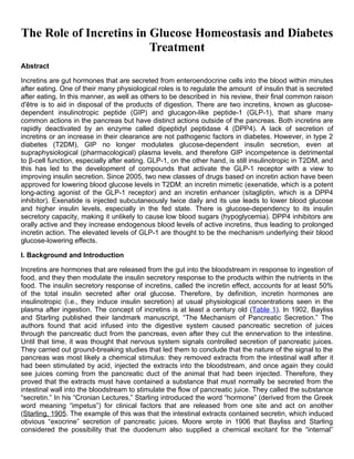
  • 14. of GLP-1 in T2DM is better preserved, in contrast to that of GIP (Nauck et al., 1993a), because an infusion of GLP-1 in T2DM so as to reach pharmacologic concentrations in plasma can normalize fasting (Nauck et al., 1993b; Gutniak et al., 1997; Rachman et al., 1997) and postprandial (Gutniak et al., 1997; Rachman et al., 1997; Meier et al., 2003a) glucose concentrations, resulting from increase of glucose-stimulated insulin secretion, decrease of glucagon secretion and slowing of gastric emptying (Gutniak et al., 1992; Nauck et al., 1993b; Holst and Gromada, 2004). Continuous i.v. infusion of GLP-1 also lowers postprandial plasma glucose (PPG) levels in subjects with type 1 diabetes by delaying gastric emptying (Gutniak et al., 1992). These effects of GLP-1 have been consistently shown in a number of human studies (Nathan et al., 1992; Nauck et al., 1993a,b; Elahi et al., 1994). In particular, continuous subcutaneous infusion (4.8 pmol/kg/min) of GLP-1 for 6 weeks in T2DM subjects was associated with significant reductions in both fasting plasma glucose (FPG) and PPG as well as HbA1c with a slight decrease of body weight (Zander et al., 2002). Prolonging the GLP-1 infusion (3.2 pmol/kg/min) for 3 months in patients with T2DM resulted in a restoration of first- phase insulin secretion as well as an improvement of late-phase secretion during a glucose clamp, but no significant changes in body weight and plasma glucagon levels were noted (Meneilly et al., 2003). In another study, repeated i.v. infusion (1∼1.2 pmol/kg/min) of GLP-1 also normalized FPG in patients with T2DM (Nauck et al., 1997). Thus, there has been considerable interest in an incretin- based therapeutic approach for treating T2DM. However, continuous GLP-1 infusion or repeated GLP-1 injections are impractical and expensive ways to lower blood glucose and so the strategies mentioned above have been developed. FIG. 4. Hormone responses to oral glucose (75 g) in nondiabetic (green), glucose intolerant (blue), and newly diagnosed type 2 diabetic (red, T2DM) subjects within Baltimore Longitudinal Study of Aging: the impaired and diabetes subjects were matched for body mass index. These subjects were not taking any glucose-lowering medications. It is evident that glucose-mediated incretin secretion (GIP and GLP-1) is not deficient in newly diagnosed T2DM. IV. Incretin Receptors A. Glucose-Dependent Insulinotropic Peptide Receptor The presence of GIPRs was first demonstrated in a transplantable hamster insulinoma (Maletti et al., 1984) and an insulin-secreting hamster β-cell line In111 (Amiranoff et al., 1984, 1985), followed by
  • 15. cloning from rat cerebral cortex cDNA library (Usdin et al., 1993), and subsequently hamster (Yasuda et al., 1994) and human GIPRs (Yamada et al., 1995) were cloned. The gene encoding rat and mouse GIPRs contains 15 exons (Boylan et al., 1999). The human gipr gene comprises 14 exons that span approximately 14 kb (Yamada et al., 1995) and is localized to chromosome 19, band q13.3. Its protein exists as two isoforms of 466 and 493 amino acids. Northern blot, reverse transcription/polymerase chain reaction, and in situ hybridization studies have shown that the gipr gene is expressed in both α and β cells in pancreatic islets (Moens et al., 1996), GI tract, adipose tissue, adrenal cortex, pituitary, heart, testis, endothelium of major blood vessels, bone, trachea, spleen, thymus, lung, kidney, thyroid, and several brain areas (Usdin et al., 1993; Yasuda et al., 1994; McIntosh et al., 1996; Yip et al., 1998). Both GIP (1-42) and several GIP fragments, truncated at the N and C termini, bind to the GIPR with high affinity, but none of the related peptides from the glucagon family do so (Wheeler et al., 1995). The GIPR is a glycoprotein belonging to the class II G protein-coupled receptor superfamily that includes receptors for glucagon, GLP-1, secretin, vasoactive intestinal polypeptide, and pituitary adenylyl cyclase-activating protein. As with other GPCRs of this class, GIPR comprises an N-terminal extracellular domain that is essential for high- affinity GIP binding and receptor activation; a central transmembrane domain (the first transmembrane domain of which is important for receptor activation and cAMP coupling); and a C- terminal cytoplasmic domain that mediates intracellular signaling by physical association with Gs protein (Usdin et al., 1993; Gelling et al., 1997; Wheeler et al., 1999). Although the majority of the cytoplasmic domain of the GIPR mediates intracellular signal transduction, a minimum length of approximately 405 amino acids is required for efficient transport and plasma membrane insertion (Baggio and Drucker, 2007). Ligand binding to the GIPR activates a heterotrimeric Gs protein that in turn activates adenylate cyclase, elevates intracellular cAMP and Ca2+ levels, and activates PKA, as well as a host of other signaling pathways, including PI-3K, PKB, MAPK, and phospholipase A2, which result in a cascade of intracellular events that mediate the potentiation of glucose- and depolarization-stimulated exocytosis of insulin-containing granules. The regulators of GIPR expression are not all known. Rat gipr gene contains a TATA-less promoter, and the first 100 bp of this promoter directed high levels of gene expression (Boylan et al., 1999). The 5′-flanking region of the gipr gene is sufficient to direct transcription in a rat insulinoma cell line 2 (RIN38), and this region contains negative regulatory sequences distal to the transcription start site controlling the cell-specific expression of the gipr gene (Boylan et al., 1999). In addition, the 5′- flanking region of gipr gene contains the binding sites for Oct-1, Sp1, and Sp3 transcription factors; binding of these transcription factors is critical not only for high levels of gipr transcription but also for cell-specific expression of the GIPR (Boylan et al., 2006). B. Glucagon-Like Peptide-1 Receptor GLP-1R is in the same class of receptors as GIPR. Although, as stated above, GLP-1, GLP-2, and glucagon result from post-translational modifications of the proglucagon molecule encoded by one gene, ligand binding of the three hormones to their unique receptors is highly specific with no relevant cross-reactivity to receptors for the other two peptides. Glucagon, for example, binds GLP-1R with 100- to 1000-fold less affinity than does GLP-1 (Thorens, 1992; Fehmann et al., 1994; Doyle and Egan, 2007). Ex-4 and its N-terminally truncated peptide exendin (9-39) (Ex-9-39) bind GLP-1R and act as potent and specific GLP-1R agonists and antagonists, respectively (Göke et al., 1993). Thus Ex-9-39 is also used as an investigational tool to uncover the physiological effects of GLP-1 signaling. The glp-1r gene was first cloned from rat pancreatic islets cDNA library in 1992 (Thorens, 1992), and the highly homologous human receptor was next cloned from a human pancreatic insulinoma (Dillon et al., 1993; Thorens et al., 1993) and a gut tumor cell line (Graziano et al., 1993). The human glp-1r gene is located on chromosome 6p21.1 (Stoffel et al., 1993). The rat and human GLP-1Rs are 463 amino acids in length and are 90% identical (Thorens, 1992; Thorens et al., 1993), differing at 42 amino acid positions (Tibaduiza et al., 2001). Although alternate splicing results in two different
  • 16. transcripts for GLP-1R in rat and human (Thorens, 1992; Dillon et al., 1993), and numerous attempts have been made to identify alternative GLP-1R, only one structurally and functionally identical GLP-1R has been described. Ex-4 is also a ligand for the known GLP-1R and yet the lizard also synthesizes GLP-1 in its gut, but it does not have a unique Ex-4 receptor. GLP-1R has, over the years, been reported to be expressed in a variety of tissues: pancreatic ducts, many cell types within pancreatic islets, thyroid C cells, kidney, lung, heart, gastrointestinal track, skin, pituitary, and multiple regions of the peripheral and central nervous system, including hypothalamus, hippocampus and cortex. In islets, it is clear that GLP-1Rs are expressed in all β cells, whereas much contradictory information has been published with regard to their presence in the other islet cell types (Fehmann and Habener, 1991; Heller and Aponte, 1995; Heller et al., 1997). A recent study using in situ hybridization and double and triple fluorescence microscopy in mouse, rat, and human pancreas seems to have cleared the waters regarding which islet cell types express GLP-1Rs (Tornehave et al., 2008). Judging from those data, GLP-1R may be almost exclusively restricted to the β cells, because it was not observed in α cells and was rarely observed in δ (somatostatin-secreting) cells, and is present in cells lining the pancreatic ducts. Therefore, additional studies are required to determine whether GLP-1R actually exists in δ cells [The presence of GLP-1Rs (or GIPRs for that matter) on pancreatic polypeptide- or ghrelin-expressing cells in islets was not investigated.] It is noteworthy that functional GLP-1Rs are also detected on intragemmal nerve fibers of taste buds of rodent and monkey (Shin et al., 2008). There is considerable controversy with respect to the presence of functional GLP-1Rs in human and rodent muscle, adipose tissue, and liver, although GLP-1R expression was detected in muscle and adipose tissue of dog (Sandhu et al., 1999) and in muscle, liver, and fat of rodents (Campos et al., 1994; Egan et al., 1994). GLP-1R mRNA transcripts have also been detected in spleen, thymus, and lymph nodes from nondiabetic and diabetic mice (Hadjiyanni et al., 2008). In brain, activated GLP-1Rs are involved in regulation of satiety and food intake, memory and learning, and, similar to GIP, hippocampal cell turnover. Like other GIPRs in its superfamily, GLP-1R comprises an N-terminal extracellular region that is important for GLP-1 recognition and binding, seven α-helical transmembrane domains (the second and fourth transmembrane domains is also important for GLP-1 binding), and a C-terminal, cytoplasmic region that contains the major determinants required for specific G protein coupling. GLP-1R is capable of signaling through Gαs subunit as well as additional G protein subunits such as Gαq, Gαo, and Gαi (Montrose-Rafizadeh et al., 1999; Hällbrink et al., 2001). Under physiological conditions studied to date, however, it seems that GLP-1R activation leads to increased intracellular cAMP and Ca2+ concentrations and activation of downstream pathways, including PKA, PKC, PI-3K, Epac2, and MAPK signaling pathway (Drucker et al., 1987; Thorens, 1992; Wheeler et al., 1993; Holz et al., 1995; Montrose-Rafizadeh et al., 1999). Distinct domains within the third intracellular loop of GLP-1R are responsible for the activation of the different G proteins (Hällbrink et al., 2001). However, the coupling of additional G proteins to GLP-1R in vivo remains to be clarified, although the coupling of the receptor to specific G proteins could be explained by the existence of subtypes of GLP-1R (to be discussed in section VII.B.3) (which have not been shown to be present in humans), or could result from a consequence of tissue-specific distribution of G proteins (Hällbrink et al., 2001). It is likely that glycosylation of GLP-1R also regulates and optimizes its function by facilitating its correct insertion into the cell membrane. Treatment with tunicamycin, which prevents glycosylation, resulted in a decrease in the number of GLP-1 binding sites in the membrane of rat insulinoma RINm5F cells without an inhibition of transcription of mRNA but a reduction in cAMP production occurred in response to GLP-1 stimulation (Göke et al., 1994). The signal transduction activity of GLP-1R is also regulated by palmitoylation at the cysteine 438 residue (Vázquez et al., 2005b). However, the significance of these effects in vivo is unknown. V. The Incretin Effect A. The Incretin Effect of Glucose-Dependent Insulinotropic Peptide and Its Impact in Type 2 Diabetes
  • 17. The incretin function of GIP has been identified in studies using GIP antagonists (Tseng et al., 1996b; Lewis et al., 2000) and GIPR antisera (“immunoneutralization studies”) (Ebert and Creutzfeldt, 1982). Treatment of animals with these compounds caused a reduction in the insulin response to oral glucose that resulted in impaired glucose tolerance. In vitro studies of treatment of isolated islets or perfused pancreas with GIP indicate that it increases insulin secretion (Dupre et al., 1973; Schauder et al., 1975). Other studies demonstrate that the elevated GIP concentrations elicited by oral glucose can almost completely account for the added insulin secretory response seen in oral versus intravenous glucose administration (Nauck et al., 1989). Studies using the specific GIPR antagonist (Pro3)GIP in Lepob/Lepob mice (mice with defective leptin that become obese) show that daily administration of (Pro3)GIP reduced pancreatic insulin content (Gault et al., 2005) and that GIP might be responsible for as much as 80% of the incretin effect seen after oral ingestion (Gault et al., 2002, 2003). This seems to confirm the pre-eminence of GIP as an incretin. GIPR(-/-) mice have fasting blood glucose levels comparable with those of wild-type mice, and after intraperitoneal glucose challenge, glucose-stimulated insulin secretion also seems comparable with that of wild-type mice. In addition, glucose-stimulated insulin secretion from isolated islets is preserved in GIPR(-/-) mice (Miyawaki et al., 1999). However, in response to oral glucose administration, GIPR(-/-) mice have impaired glucose tolerance as a result of a 50% reduction in insulin secretion compared with wild-type animals. These results demonstrate that GIP's primary role is that of an incretin and that insulin secretion from β cells is regulated not only by glucose but also by GIP, especially in the postprandial state (Miyawaki et al., 1999). It is noteworthy that isolated islets from GIPR(-/-) mice show increased responsivity to GLP-1, although serum GLP-1 levels in GIPR(-/-) mice are unaltered (Pamir et al., 2003). Furthermore, these mice exhibit a decrease in intrapancreatic insulin content and insulin gene expression (Pamir et al., 2003). Although it has been reported that intracerebroventricular administration of GIP does not affect food intake (Woods et al., 1981) and there was no significant difference in food intake between WT and GIPR(-/-) mice, GIPR(-/-) mice fed a high-fat diet do not become obese or insulin-resistant. Moreover, GIPR(-/-) mice with defective leptin [GIPR(-/-), Lepob/Lepob] are protected from diet-induced obesity and its associated complications that occur with defective leptin signaling, including T2DM and fatty liver (Miyawaki et al., 2002). The incretin effect of GIP in stimulating insulin secretion is almost lost in T2DM (Nauck et al., 1993a; Vilsbøll et al., 2002), and many studies indicate the existence of a specific defect in GIP action in these patients. It has been speculated that this loss of insulinotropic action of GIP may occur as a result of either chronic desensitization of GIPRs (Tseng et al., 1996a) or a reduction in the expression of GIPRs on pancreatic β cells (Holst et al., 1997; Lynn et al., 2001; Xu et al., 2007; Zhou et al., 2007). Several studies have examined the expression of GIPR in experimental models of diabetes. Indeed, it has been shown that the expressions of both GIPR mRNA and protein are decreased in β cells of diabetic-fatty Zucker (ZDF) rats containing a leptin receptor missense mutation (Lynn et al., 2001; Piteau et al., 2007). Another study suggested a role for circulating free fatty acids in down- regulation of GIPR expression (Lynn et al., 2003). Zhou et al. (2007) suggested that ubiquitination may also be critical for the hyperglycemia-associated down-regulation of GIPR expression. Hyperglycemia triggered the association of GIPR and ubiquitin ligase complexes, resulting in reduction of GIPR protein levels on β-cell membranes (but not GLP-1R) and down-regulation of GIP action. Receptor desensitization has been described for several receptors, including glucagon, somatostatin, β-adrenergic receptor, and GLP-1. The GIPR is susceptible to very rapid and reversible homologous desensitization in vitro (Jones et al., 1989; Tseng et al., 1996a; Wheeler et al., 1999). This desensitization process has been shown to be mediated by a number of separate mechanisms, including receptor internalization, down-regulation, and uncoupling from G proteins. Upon agonist stimulation, GIPRs are phosphorylated by protein kinase, resulting in their uncoupling from interaction with G protein (Premont et al., 1995), and regulators of G protein signaling and G protein receptor kinase 2 proteins have been demonstrated to mediate this desensitization mechanism (Druey et al.,
  • 18. 1996; Koelle and Horvitz, 1996; Neill et al., 1997; Tseng and Zhang, 1998). Functional studies on the role of the COOH-terminal tail (CT) of GPCRs have been implicated in receptor desensitization and endocytosis (Reneke et al., 1988; Hausdorff et al., 1990), and the CTs of glucagon receptor (Buggy et al., 1997) and GLP-1R (Widmann et al., 1997) have been shown to be required for receptor desensitization. In the case of GIPRs, CT deletion analyses demonstrate that the majority of the CT of GIPR is important for regulating the rate of receptor internalization but not for GIPR expression and signaling (Wheeler et al., 1999). Specific serine residues within the CT, particularly serines 426 and 427, play an important role in regulating the rate of receptor internalization, whereas serines 406 and 411 are important for receptor desensitization (Wheeler et al., 1999; Baggio and Drucker, 2007). In summary, the incretin effect of GIP is deficient in T2DM; a unifying consensus has not yet emerged, but increasing evidence suggests that persistent hyperglycemia leads to reduced GIPR protein levels. A recent study in ZDF animals supports the role of hyperglycemia in GIPR down-regulation. Piteau et al. (2007) treated ZDF rats with phlorizin, bringing blood glucose levels down from 28 to 10 mM. Nontreated ZDF rats had GIPR mRNA levels that were 94% less than those of lean rats, and insulin secretion did not increase in response to exogenous GIP in the ZDF animals. But after phlorizin treatment, mRNA levels returned to those of lean rats. In addition, insulinotropic responsiveness to GIP was restored in the treated rats. GIP also promotes insulin biosynthesis as well as β-cell proliferation and survival, and whether these pleiotropic effects (to be discussed in section VI.A and Table 2) as well as the insulinotropic effects of GIP are also deficient in humans with T2DM is not known. Actions of GIP and GLP-1 that affect blood glucose levels GIP GLP-1 Islets    Insulin secretion ↑ ↑    Insulin synthesis ↑ ↑    Insulin, glucokinase and GLUT2 ↑ ↑ expression    Glucagon secretion ↑ ↓ (indirect)    Somatostatin secretion ↑ ↑ (indirect)    β cell proliferation ↑ ↑    β cell apoptosis ↓ ↓ Sweet taste modulator — Yes Gastrointestinal tract    Gastric emptying — ↓    Gastric acid secretion ↓ ↓    Motility — ↓ Central nervous system    Food intake — ↓    Satiety — ↑ Muscle
  • 19. GIP GLP-1    Glucose uptake — ↑ Liver    Glucose production ↓ ↓ (indirect (indirect ) ) • ↑, increase; ↓, decrease; —, no effect or not reported B. The Incretin Effect of Glucagon-Like Peptide-1 and Its Impact in Type 2 Diabetes Blockade of endogenous GIP action by injection of its antiserum resulted in partial preservation of incretin activity. This observation led to suspicion of the existence of additional incretin hormones, and insulinotropic effects of GLP-1 were first described in rodent in 1985 (Schmidt et al., 1985). In humans, insulinotropic effects of GLP-1 were first described in 1987 (Kreymann et al., 1987). In that study, the ability of GLP-1 to enhance glucose-dependent insulin secretion was of such magnitude that the authors believed that its effects, in conjunction with those of GIP, were sufficient to account for the total incretin effect. GLP-1 (7-37) and GLP-1 (7-36) amide seem to be equipotent insulinotropic fectors (Orskov et al., 1986). Although GLP-1 is circulating in much lower concentrations than GIP, GLP-1 is one of the most potent insulinotropic factors that stimulate insulin secretion in a glucose- dependent manner, far exceeding GIP's effects on a molar basis. In animals and humans, it is essential for normal postprandial glucose homeostasis and the complete early/first-phase insulin response (Otonkoski and Hayek, 1995). Secretion of GLP-1 highly correlates with glucose level and insulin secretion, and its insulinotropic effects are observed at increased circulating glucose concentrations, but disappear once plasma glucose returns to normal (Nauck et al., 2002). Evidence for the functional importance of GLP-1 as a physiologically relevant incretin derives from studies that have inactivated GLP-1 signaling using immunoneutralizing antisera, or GLP-1R antagonists or knockout mice. A specific antibody to immunoneutralize circulating GLP-1 and the GLP-1R antagonist Ex-9-39 have been used to demonstrate the essential physiological role of endogenous GLP-1 in the control of glucose-dependent insulin secretion and glucose homeostasis in rodent and human subjects. Removal of GLP-1 action, whether by Ex-9-39 or by its immunoneutralization, causes increases in both fasting and postprandial glycemia and reduces glucose-stimulated insulin release in both animals and humans (Kolligs et al., 1995; Wang et al., 1995; D'Alessio et al., 1996; Edwards et al., 1999; Baggio et al., 2000). Administration of Ex-9-39 to rodents or humans results in a suppression of the incretin effect by 50 to 70% (Kolligs et al., 1995; Flamez et al., 1999; Schirra et al., 2006). In agreement with these results, GLP-1R(-/-) mice with a targeted deletion of all GLP-1Rs have mild fasting hyperglycemia and abnormally high blood glucose levels after oral and intraperitoneal glucose challenges that are associated with reduced circulating levels of glucose-stimulated insulin secretion, whereas GLP-1R(-/-) mice have normal body weight, food intake, and fasting and postprandial plasma levels of glucagon (Scrocchi et al., 1996; Scrocchi et al., 1998). However, GLP-1R signaling is not required for actual glucose responsiveness of pancreatic β cells, as demonstrated by the fact that isolated islets from GLP-1R(-/-) mice still have a well preserved glucose response in terms of insulin secretion, and both islet and total pancreatic insulin content are not significantly lower than in wild-type mice (Flamez et al., 1998). Pancreatic insulin mRNA transcripts are also similar in wild-type and GLP-1R(-/-) mice (Scrocchi et al., 1998). Moreover, GLP-1R(-/-) islets exhibit reduced basal but enhanced GIP-stimulated cAMP production and abnormalities in basal and glucose-stimulated intracellular Ca2+ concentration (Flamez et al.,
  • 20. 1998). In these mice, the lack of GLP-1R signaling is compensated for, at least in part, via increased GIP secretion and up-regulation of the GIP-insulin axis on pancreatic islets (Pederson et al., 1998). In addition, GIPR(-/-) mice exhibit an enhanced GLP-1-mediated insulin secretory response in pancreatic islets (Pamir et al., 2003). Thus, disruption of single incretin receptor genes in mice causes only modest glucose intolerance, with a well preserved insulin-secretory response to glucose that is partially modified as a result of complementary up-regulation of the remaining intact incretin hormone. However, double incretin receptor knockout mice, which have disruption of both glp-1r and gipr, surprisingly show only a modest and obviously incomplete perturbation in glucose homeostasis, suggesting that other mechanisms are capable of compensating for the absence of the actions of GIP and GLP-1 (Hansotia et al., 2004; Preitner et al., 2004). The GLP-1R is no exception to the susceptibility of members of its subfamily to undergo desensitization, at least in vitro. It undergoes rapid internalization, primarily via mechanisms dependent on clathrin-coated pits (Widmann et al., 1995, 1997; Vázquez et al., 2005a) and caveolin (Syme et al., 2006). This internalization is mediated by phosphorylation of specific residues in the GLP-1R C-terminal tail (Widmann et al., 1997) that regulates receptor subcellular localization, trafficking, and signaling. In addition, GLP-1R also undergoes homologous and heterologous desensitization in vitro, which correlates with phosphorylation of GLP-1R C-terminal tail (Widmann et al., 1996a,b, 1997; Baggio et al., 2004c). Xu et al. (2007) suggested that protein levels of GLP-1R are significantly decreased in β cells exposed to high blood glucose concentrations in vivo and in vitro, and insulin response to GLP-1 is reduced in this system. But research in our laboratory (Zhou et al., 2007) did not find this to be the case in human islets. Even after long-term GLP-1R agonist administration, such as with exenatide treatment, there does not seem to be any attenuation of GLP-1R-dependent effects on glucose homeostasis in vivo (Klonoff et al., 2008), and an elevated steady state of plasma GLP-1 for 6 to 12 weeks in clinical trials has resulted in effective reduction in blood glucose levels without any loss of potency (Zander et al., 2002; Meneilly et al., 2003). Therefore, no physiological significance for either the homologous or heterologous in vitro desensitization has been established in vivo. In T2DM, the insulinotropic properties of GLP-1 seem to be largely intact (Nauck et al., 1993a). In addition, in support of fully functional GLP-1Rs in T2DM, an acute infusion of exenatide perfectly restores first-phase insulin secretion in response to i.v. glucose in subjects with T2DM (Fehse et al., 2005). In addition to stimulating insulin secretion, GLP-1 also promotes insulin biosynthesis as well as β-cell proliferation and survival, thereby augmenting supplies of insulin for secretion. It should be noted that GLP-1, and not GIP, also suppresses glucagon secretion in a glucose-dependent manner, contributing to the glucose-lowering effect of GLP-1. Deceleration of gastric emptying by GLP-1, but not GIP (Meier et al., 2004), also adds to the glucose-lowering effect of incretins after meal ingestion in humans. In addition, GLP-1 may enhance peripheral glucose disposal and insulin sensitivity (Todd and Bloom, 2007). GLP-1 and GIP in β-cell lines have additive effects on insulin secretion but only at concentration that have submaximal insulinotropic effects (Montrose-Rafizadeh et al., 1994). Therefore, the final pathway to insulin secretion is most likely a common one. But under usual postprandial physiological conditions, they are most likely each acting at submaximal concentrations. GLP-1 is at least 2 orders of magnitude more potent than GIP as an insulinotropic agent. There is some evidence that GLP-1 can recruit cells into a secretory mode in response to glucose (i.e., make the cells glucose- competent), whereas there is no such evidence for GIP (Holz et al., 1993; Montrose-Rafizadeh et al., 1994). It is conceivable, however, GLP-1 might prime the cells for GIP actions, although this was
  • 21. found not to be the case in elderly patients with T2DM (Meneilly and Elahi, 2006).  Incretin effect on insulin secretion-GIP + GLP-1 People with Type 2 diabetes 80 Control subjects 60 40 Incretin effect 20 m m U U u n u n s s ) ( ) ( / I / I i l i l 0 60 120 180 0 60 120 180 Time (min) Time (min) Oral glucose load Intravenous glucose infusion Nauck et al. Diabetologia. 1986 VI. The Pleiotropic Effect of Incretin in Pancreas A. The Pleiotropic Effects of Glucose-Dependent Insulinotropic Peptide in Pancreas 1. Effects on β Cells. GIP binding to its specific receptor on pancreatic β cells facilitates the secretion of insulin in the presence of PPG concentration via activation of proximal signal transduction pathways, and the ability of GIP to promote glucose-stimulated insulin secretion in pancreatic β cells is attributed to GIPR activation leading to membrane depolarization and increases in the intracellular Ca 2+ concentration as well as direct effects on insulin exocytosis (Fig. 5). Ligand binding to GIPR activates several mechanisms, including stimulation of the adenylyl cyclase/cAMP/PKA), inhibition of KATP channels, increases in intracellular Ca2+, activation of phospholipase A2 (PLA2) (Ehses et al., 2001), specific protein kinase signaling pathways, including PKB (Trümper et al., 2001), mitogen-activated protein kinases (Ehses et al., 2002), and stimulation of exocytosis (Ding and Gromada, 1997). The rise in cAMP significantly up-regulates activity of PKA, which leads to a cascade in which phosphorylation of regulatory proteins, including GLUT2 (a low Km glucose transporter), SUR1 (subunits of the pancreatic β-cell KATP channel), α-SNAP (a vesicle-associated protein), and other ion channels (Ca2+ channels, for example), induces membrane depolarization and subsequent exocytosis of insulin, as well as phosphorylation of critical transcription factors, which results in significant up-regulation of insulin gene promoter activity (Doyle and Egan, 2003). A PKA-independent mechanism is also involved in GIP-mediated insulin secretion. The cAMP-binding protein cAMP-GEFII (Epac2) mediates cAMP-dependent, PKA-independent insulin secretion, and this effect of Epac2 on insulin secretion is mediated by Rim2 and depends on intracellular Ca2+ as well as on cAMP (Kashima et al., 2001). GIP stimulation of insulin secretion from pancreatic β cells is also mediated by PLA2 (Ehses et al., 2001). Metabolism of glucose has been shown to induce hydrolysis of membrane phospholipids, leading to the accumulation of arachidonic acid (AA), which amplifies insulin secretion (Turk et al., 1987; Turk et al., 1993). GIP stimulates AA release from clonal β cells (βTC-3), and coupling of GIPR to AA production in β cells is mediated by cAMP and a Ca 2+-independent PLA2 (Ehses et al., 2001). Incretin- and glucose-stimulated insulin secretion is also mediated via opening of voltage-dependent Ca2+ channels as well as activation of several species of K+ channels, including KATP channels, Ca2+- activated K+ (KCA) channels, and voltage-gated K+ (KV) channels (for review, see Doyle and Egan,
  • 22. 2003). Although all K+ channels are present in pancreatic β cells, most of the studies have concentrated on studying KATP and KCA channels, and less is known about the mechanisms involved in insulin modulation by KV channels. However, one study identified a potential role for GIP in the regulation of KV1.4 endocytosis and GIP-induced down-regulation of KV1.4 via PKA-dependent phosphorylation and endocytosis of the channel protein, resulting in the modulation of insulin secretion (Kim et al., 2005b). A schematic representation of the main molecular events during incretin-induced insulin secretion from a β-cell. The binding of incretin ligands or agonists to the incretin receptors results in production of cAMP via adenylyl cyclase (AC) activation and subsequent activation of PKA and the Epac family of cAMP-regulated guanine nucleotide exchange factors (cAMP-GEFs), which leads to elevation of intracellular Ca2+ levels via a depolarization of plasma membrane by inhibition of KATP and KV channels after ATP generation from glucose and consequent opening of voltage-gated L-type Ca2+ channels. Intracellular Ca2+ levels are further increased via stimulation of IP3R and RyR on the ER. Long-term GLP-1 treatment also stimulates the expression of GLUT2 transporter and glucokinase (the β-cell glucose sensor), which lead to increased mitochondrial ATP synthesis. In addition, L-type Ca2+ channels are phosphorylated by PKA, resulting in an increase of their open probability and thus facilitation of enhanced Ca2+ influx. The changes in intracellular Ca2+ concentrations lead to fusion of insulin-containing vesicles to the plasma membrane and subsequent rapid exocytosis of insulin from β cells. In addition, the exocytosis of insulin-containing vesicles is directly regulated by PKA and Epac2 via interaction with the regulators of exocytosis and ATP. In addition to potentiating the release of insulin from β cells, GIP replenishes insulin in β cells by increasing insulin gene transcription and biosynthesis, and enhances the glucose-sensing system by increasing the expression of components of β-cell glucose sensors. Acute treatment of GIP synergizes with glucose, causing more insulin to be released from β cells both in vivo and in vitro, and the amount of insulin released from β cells is proportional to the rate of glucose metabolism (Newgard, 1994), which is determined by the plasma membrane glucose transporters, the enzymes responsible for glucose metabolism and their activity (Matschinsky et al., 1993). In the RIN 1046-38 rat insulinoma cell line, extended exposure to GIP up-regulates the expression of both GLUT1- mediated glucose uptake and hexokinases catalyzing the initial phosphorylation of glucose, allowing glycolysis to proceed (Wang et al., 1996). In addition, GIPR(-/-) mice exhibit decreased insulin gene transcription and protein biosynthesis (Pamir et al., 2003), confirming the role of GIP in insulin biosynthesis. Together, these results suggest that GIP confers long-term beneficial effects on insulin secretion and cellular health by replenishing insulin stores.
  • 23. Some evidence indicates that GIP acts synergistically with glucose as a growth- and antiapoptotic factor for β cells, providing another mechanism by which GIP promotes long-term accommodation of β-cell function to changing nutritional status. GIP potentiates glucose-induced β-cell proliferation and protects β cells from cell death induced by various stimuli, including exposure to wortmannin, STZ, glucolipotoxicity, or serum or glucose deprivation (Ehses et al., 2003; Kim et al., 2005c). Moreover, overexpression of a dominant-negative human GIPR in murine β cells results in diminished islet size (Herbach et al., 2005), and GIP reduces glucolipotoxicity-induced cell death in islets from wild-type but not GIPR(-/-) mice (Kim et al., 2005c). Recent studies have begun to unravel the molecular signaling pathways that mediate the proliferative and antiapoptotic actions of GIP in the β-cell, and it is clear that growth and antiapoptotic pathways rely on similar pathways, including activation of cAMP/PKA, PKA/CREB, MAPK, and PI-3K-dependent activation of PKB (Trümper et al., 2001, 2002; Ehses et al., 2002, 2003; Kim et al., 2005c) (Fig. 6). These signaling modules are dependent on glucose metabolism and Ca2+ influx, and cAMP-PKA-Rab1-extracellular signal-regulated kinase 1/2 pathway may also be one way in which GIP regulates β-cell proliferation (Ehses et al., 2002). In particular, GIP phosphorylates several transcription factors such as FoxO1 and p70 S6K, crucial regulators of translation known to be involved in glucose-induced β-cell mitogenesis, suggesting possible mediators of GIP-mediated β-cell growth (Ehses et al., 2002). FoxO1 has been shown to play a critical role in GIP-mediated β-cell survival and apoptosis. GIP improved cell survival in INS-1 (clone 832/13) cells and murine islets via GIP-induced phosphorylation of FoxO1, resulting in inactivation of the pro-apoptotic bax gene (Kim et al., 2005c). In addition, a 2-week infusion of GIP into diabetic (ZDF) rats was shown to significantly reduce β-cell apoptosis because of down- regulation of the bax gene and up-regulation of the antiapoptotic bcl-2 gene: these effects were mediated by activation of PI-3K/PKB, and subsequent phosphorylation and nuclear exclusion of FoxO1 (Kim et al., 2005c). The protective effects of GIP on β-cell apoptosis in the diabetic state are very intriguing to us. It is clear from many studies that GIP is not insulinotropic in T2DM and that in animal models, such as the ZDF rat discussed above (section V.A), GIP does not stimulate insulin secretion, and its receptor is severely down-regulated (94% reduction compared with lean nondiabetic rats) (Piteau et al., 2007). So, if the GIP receptor is so severely down-regulated in the ZDF rats, how did 2 weeks of GIP infusion have its antiapoptotic effects? Unlike the more recent work, in which the authors found that reduction in hyperglyemia by phlorizin treatment reversed GIPR down-regulation (Piteau et al., 2007), Kim et al. (2005c) did not first lower hyperglycemia before giving exogenous GIP. Is there a case to be made that the antiapoptotic effect, in vivo, is not through the known GIPR on β cells? Could this mean that by not developing GIPR agonists to treat T2DM, we are missing the other potential effect of GIPR stimulation? Is there a case to be made that GLP-1R and GIPR stimulation, together, might be beneficial in T2DM? In β-cell lines at least, a direct antiapoptotic effect of GIP seems to hold up, because another study from the same research group corroborated that GIP has antiapoptotic properties and that the antiapoptotic effect of GIP is, at the very least, partially due to GIP-stimulated increases in bcl-2 gene expression (Kim et al., 2008b). In that study, stimulation of INS-1 (clone 832/13) cells with GIP resulted in enhanced expression of bcl-2, and this was mediated by a pathway involving AMP-activated protein kinase, cAMP-responsive CREB activator 2, and CREB. GIP treatment also reduces caspase 3 activation via cAMP and MAPK regulation (Ehses et al., 2003; Kim et al., 2008b), and there is a possibility that GIP improves β-cell survival by reducing ER stress (Yusta et al., 2006).
  • 24. Schema outlining the incretin downstream signal transduction pathways in a β cell. GLP-1R activation (and GIPR activation, to some extent, but this has not been as well studied as GLP-1R activation) recruits signaling mechanisms that considerably overlap, leading to promotion of β-cell proliferation and prevention of β-cell apoptosis. Dashed line indicates mechanism that is not fully delineated. 2. Effects on Glucagon Secretion. Several studies have suggested that GIPR seems to be also present in pancreatic α cells (Moens et al., 1996) and compatible with this observation, exposure of α cells to GIP increases cAMP content to levels that are required for nutrient-induced glucagon secretion (Pipeleers et al., 1985; Moens et al., 1996), and GIP stimulates Ca2+-induced exocytosis in isolated rat α cells by a PKA-mediated mechanism (Ding and Gromada, 1997). However, several lines of evidence indicate that GIP stimulates glucagon secretion in human and animal models but only under certain circumstances. For example, a glucagonostatic GIP effect has been observed in patients with liver cirrhosis and basal hyperglucagonemia (Dupre et al., 1991) and in human and animal models during euglycemic conditions (Pederson and Brown, 1978; Meier et al., 2003b), but in healthy persons under hyperglycemic conditions or in T2DM, no GIP effects on glucagon secretion were reported (Nauck et al., 1993a; Meier et al., 2001; Vilsbøll et al., 2003b). Most experiments in the literature were carried out under hyperglycemic conditions to increase GIP-mediated insulin secretion, suggesting that an absence of GIP effect on glucagon secretion in those studies may be due to increased glucose concentrations and insulin-mediated counteraction of effects to stimulate glucagon secretion. B. Pleiotropic Effects of Glucagon-Like Peptide-1 in Pancreas 1. Effects on β Cells.
  • 25. GLP-1 is one of the most potent substances known to stimulate glucose-dependent insulin secretion from islet β cells and, like GIP, its stimulatory activity is exerted via binding to its receptor on β cells. This binding results in activation of adenylyl cyclase with consequent production of cAMP and subsequent activation of PKA and the Epac family, which led to inhibition of K ATP channels, elevation of intracellular Ca2+ levels, increases in mitochondrial ATP synthesis, and enhanced exocytosis of insulin from insulin-secretory vesicles (Fig. 5). PKA, a key component in the regulation of insulin secretion by cAMP, mediates many of the phosphorylation reactions required for insulin secretion including KATP channels, the L-type Ca2+ channels on the ER and GLUT2 glucose transporters. GLP-1-mediated activation of PKA results in phosphorylation of S1448 on the SUR1 KATP channel subunit via an ADP-dependent mechanism, facilitating its closure (Light et al., 2002). This is followed by membrane depolarization and triggering of the insulin secretory pathway. Treatment with the PKA inhibitor 8-bromoadenosine-3′,5′-cyclic monophosphorothioate, Rp-isomer (Gromada et al., 1998) or H89 (Light et al., 2002) abolishes GLP-1-induced inhibition of the KATP channels and SUR1(-/-) islets lack an insulin secretory response but exhibit a normal rise in cAMP to GLP-1 and GIP, implicating cAMP-dependent PKA-independent signal transduction pathway by which incretins exert their effects (Nakazaki et al., 2002; Shiota et al., 2002). It is now evident that the action of cAMP produced by GLP-1 signaling is mediated not just by PKA, but also by Epac2 (de Rooij et al., 1998; Kawasaki et al., 1998), and Epac2 also inhibits the function of KATP channels in rodent and human β cells via interaction with SUR1 (Shibasaki et al., 2004; Kang et al., 2006a). A recent study has also demonstrated that the scaffold protein β-arrestin-1 facilitates GLP-1-stimulated cAMP production via interaction with GLP-1Rs (Sonoda et al., 2008). GLP-1 signaling also antagonizes voltage-dependent K+ (Kv) channels via cAMP/PKA-dependent pathway in β cells, which prevents β-cell repolarization by reducing KV currents (MacDonald et al., 2002, 2003). However, Kv currents are not antagonized by treatment with cAMP analog or the constitutively active PKA catalytic subunit, and the Epac-selective activator 8-pT-2′-O-Me-cAMP, implying a role for additional signaling pathways required for antagonism of the K v current (MacDonald et al., 2003). This study has identified a role of PI-3K with subsequent activation of PKCζ in the antagonism of the Kv current by GLP-1 and that this occurs via epidermal growth factor receptor (EGFR) transactivation, not via the G protein-regulated isoform p110γ (MacDonald et al., 2003). L-type Ca2+ channels are also phosphorylated by PKA, leading to increase of their open probability and enhancement of Ca2+ influx (Lester et al., 1997; Fraser et al., 1998; Hulme et al., 2003). As well as effects on Ca2+ influx, activation of GLP-1R increases intracellular Ca2+ through two intracellular Ca2+-releasing receptors in the endoplasmic reticulum (ER): the inositol 1,4,5 triphosphate receptors (IP3R) activated by PKA and the ryanodine receptors (RyR) activated by Epac2 (Kang et al., 2003; Tsuboi et al., 2003; Doyle and Egan, 2007). Phosphorylation of IP3R by PKA alters Ca2+ release properties of IP3Rs (Bugrim, 1999), contributing to increasing intracellular Ca2+ concentrations. Pretreatment of β cells with ryanodine, and not with Xestospongin C, an IP3R inhibitor (Kang et al., 2001), blocked Ca2+ release from ER in response to 8-pT-2′-O-Me-cAMP (Kang et al., 2003), suggesting that Epac2 increases the release of Ca2+ from ER into cytosol through RyR. A recent study has indicated that GLP-1 elevates intracellular Ca2+ concentration via stimulation of the nicotinic acid adenine dinucleotide phosphate and cyclic ADP-ribose production (Kim et al., 2008a), catalyzed by cyclic ADP-ribose cyclases, that stimulates glucose-induced Ca2+ mobilization followed by insulin secretion (Kato et al., 1995, 1999). GLP-1-induced increase in intracellular Ca2+ concentrations is a key trigger for fusion of insulin-containing vesicles to the plasma membrane, and exocytosis of insulin from β cells rapidly follows. Direct effects of GLP-1 signaling on insulin-laden vesicles have also been described. PKA and Epac2 directly regulate exocytosis of insulin-containing vesicles via interaction with the regulators of exocytosis. Lester et al. (2001) proposed a mechanism by which changes in insulin secretion are