2. IMPLANT DENTISTRY / VOLUME 20, NUMBER 6 2011
Fig. 1. Preoperative pictures during initial examination.
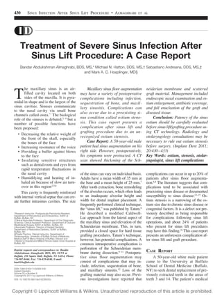
Fig. 2. Presurgical CT scan showing some remnants of the grafting material with thickened
Schneiderian membrane and ostium stenosis.
Fig. 3. Instrumentation: fiberoptic probe and inflatable device.
history included an allergy to penicillin. At this time, the patient was taking
multivitamins, Diazepam (SigmaAldrich, St. Louis, MO), Asmanex
(Merck & Co., Whitehouse Station,
NJ), Symbicort (AstraZeneca Pharmaceuticals, Wilmington, DE), and Albuterol (Mylan Pharmaceuticals Inc.,
Morgantown, WV). The latter three
medications were used to control reactive airway disease, whereas Diazepam was prescribed to control anxiety
and insomnia. A panoramic x-ray of
the patient showed alveolar atrophy
and sinus pneumatization in the areas
of teeth 3 and 14 (Fig. 1).
Treatment options were discussed
with the patient, and a mutual decision
was made to undergo sinus lift/
mineralized tissue graft procedures,
before implant placement. We decided
to provide care starting on the patient’s right side. The left side was
planned to be done at a future appointment. Risks, benefits, and alternatives
of the proposed procedure were discussed with the patient. All questions
were answered. The patient signed a
standard surgical consent form. Local
anesthesia was administered using one
carpule of 2% lidocaine, with epinephrine (1:50,000). A full-thickness flap
was reflected over the lateral aspect of
the right maxillary wall. An osseous
window was created in a standard
fashion using both rotary and piezoelectric surgery techniques. During the
procedure, the Schneiderian membrane was torn (Ͻ5 mm), and it was
closed using a collagen membrane
(Colla Tape; Zimmer, Carlsbad, CA)
with dimension of 1 ϫ 2.5 ϫ 7.5
mm.17 The intact side of the membrane
was fully reflected to achieve an ideal
space for grafting. Before graft placement (irradiated cancellous particulate
allograft bone; Rocky Mountain Tissue Bank, Aurora, CO), the membrane
perforation was checked for complete
coverage. Four grams of bone graft
material (irradiated cancellous particulate allograft bone; Rocky Mountain
Tissue Bank) was placed into the sinus lift
cavity. A collagen membrane (Conform,
Ace Surgical Supply, Brockton, MA)
was placed over the lateral aspect of the
bone window. The flap was replaced,
and 4.0 nonresorbable suture material
(Cytoplast PTFE; Osteogenics Biomedical, Lubbock, TX) was used to stabilize
the flap. This patient was prescribed 150
mg clindamycin four times per day for
10 days and 0.75 mg dexamethasone
four times per day for 6 days. The
patient started these medications one
day before surgery.
The first postsurgical week was uneventful. However, 2 weeks after the
431
surgery, the patient reported pain and
discomfort, with drainage from his nasal
cavity on the operative side. A periapical radiograph was taken which showed
that grafting material was intact.
Yellow mucus discharge from the
right nostril was cultured in standard
transport media. A mixture of aerobic
and anaerobic bacteria was noted. The
patient was prescribed clindamycin
300 mg along with metronidazole 250
mg to reduce the possibility of having
anaerobic bacterial infection. The patient showed no improvement, and he
was then prescribed tetracycline 500
mg, for 10 days. On the second day of
taking tetracycline (21 days after the
surgery), the patient reported swelling
in the right maxillary sinus area. There
was also pain on palpation, malaise,
and fever. Surgical intervention was
done via incision and drainage under
local anesthesia. Four days later, the patient reported that the swelling subsided,
with a decrease in nasal discharge. The
patient was monitored on a frequent basis. A CT scan was obtained which revealed that the bone graft was scattered
in the anterior and floor regions of the
right sinus area with most of the pieces
adhering to the soft tissue lining. Of
special note was that thickened soft tissue which completely blocked the right
maxillary sinus ostium. The treatment
plan was discussed. He agreed to have
the graft material removed from the
right maxillary sinus.
After several weeks, the intraoral
soft tissue stoma had closed. Under
local anesthesia, a full-thickness flap
was reflected over the right maxillary
sinus wall, and access was made
through the previous lateral window.
Findings included showed frank pus
accumulation and unattached bone
grafting material. The area was curetted and irrigated with saline. Suturing
was done using a 3.0 PTFE with interrupted technique.
After soft tissue healing of the
site, the patient noted discomfort and
tenderness over the right maxillary
sinus. A yellow nasal discharge persisted from the right nostril. We referred the patient for consultation with
an otolaryngologist. CT scans were
obtained, which revealed remnants of
3. 432
SINUS INFECTION AFTER SINUS LIFT PROCEDURE • ALMAGHRABI
ET AL
DISCUSSION
Fig. 4. Nasal endoscopy: ethmoid thickening with ostium stenosis.
Fig. 5. Sinus ostium after enlargement using inflatable device and shaving the ethmoid thickening.
Fig. 6. Polyps removed along with bone grafting material after maxillary sinus membrane
enucleation.
Fig. 7. Postsurgical radiographic image.
the grafting material and stenosis of
the right maxillary ostium (Fig. 2).
The otolaryngologist performed an
endoscopic examination under general
anesthesia (Fig. 3). Findings were consistent with stenosis of the right maxillary
sinus ostium (Fig. 4). Balloon catheterization and widening of the ostium were
completed (Fig. 5). Cultures were taken
during the surgery, and the sinus was
examined using a fiberoptic probe.
These cultures had shown presence of
Prevotella species and were identified
as Prevotella melaninogenica. The base
of the Schneiderian membrane on the
other hand appeared intact. No other abnormalities were noted.
The patient did improve after the
procedure and was less symptomatic.
Two months later, the patient developed copious clear mucus discharge
from the right nasal cavity and also
noted tenderness of the right maxillary
sinus. In addition, he reported intermittent blockage of the right nasal airway and difficulty with air flow
through the right nasal passage. Based
on the persistent symptoms and consultation with his otolaryngologist,
surgical exploration by an oral and
maxillofacial surgeon was offered to
the patient. The consulting otolaryngologist was not experienced in removing dental material from a grafted
sinus and asked that a dental surgeon
perform the procedure. Standard surgical consent was obtained. Under
local anesthesia, the oral and maxillofacial surgeon elevated a fullthickness mucoperiosteal flap over the
right lateral aspect of the maxilla. The
previous lateral window was used to
gain access into the base of the sinus.
The window was enlarged, and a thorough curettage of the graft material
was done. Multiple sinus polyps and
grafts material attached to the thickened Schneiderian membrane were
removed (Fig. 6). The sinus was thoroughly irrigated, and the osseous
window was covered with a collagen
membrane. The flap was brought
again to its original position and
closed with interrupted sutures. The
patient showed remarkable improvement (Fig. 7) and was symptom free on a
1-year follow-up.
Alveolar atrophy and maxillary sinus pneumatization after tooth extraction can create a significant challenge
for dental implant placement in the
posterior maxilla. To increase the
bone height of the posterior alveolar
areas of the maxilla, “sinus lift” procedures have become commonly used
in dental implant surgery.6 This communication describes a persistent postoperative maxillary sinusitis, which
can be traced to stenosis ostium of the
maxillary sinus.
Anatomical variation of the osteomeatal complex (which can include
narrowing of the ostium) can have
deleterious effects on sinus drainage.13,18 –20 Narrowing of the sinus
ostium can arise from congenital, inflammatory, or neoplastic sources.
Stenosis ostium has a prevalence of
24%16,21 and can be associated with
acute and chronic sinusitis where the
permeability of the ostium is altered.21–24 Mucosal edema can obliterate the ostium, along with nasal polyp
formation.25,26 A patient’s medical history is an important factor before
undertaking a sinus lift procedure, especially in regard to nasal symptoms.
This patient did have a history of reactive airway disease. He also had a
history of nasal problems, which were
not active at the time of the original
procedure. In fact, he had seen the
same treating otolaryngologist in the
past to stabilize the sinus symptoms
before proceeding with the sinus lift
procedure. Chronic sinusitis is more
prevalent among patients with reactive
airway disease.16 Although this patient
had an intraoperative tear of the Schneiderian membrane during the graft
procedure, we believe that the persistent sinusitis was indeed related to stenosis ostium. The patient had adequate
pre- and postoperative antibiotic coverage for the procedure. The incidence
of membrane perforation varies
widely in the literature.27,28 It is not
uncommon and is usually dealt with as
described above.3,27,29
Schneiderian membrane perforation can be associated with postoperative complications, such as acute or
chronic sinus infection, wound dehiscence, loss of the graft material, and a
4. IMPLANT DENTISTRY / VOLUME 20, NUMBER 6 2011
disruption of normal sinus physiologic
function.3,30 –33 Becker et al,27 however,
found that sinus perforation per se did
not necessarily result in implant loss,
displacement of graft material, or infectious complications. The authors believe
that this patient’s protracted course was
indeed due to ostium stenosis.
CONCLUSION
Ostium stenosis cannot be visualized on intraoral dental radiographs or
panoramic films. On the basis of this
case, we now strongly recommend the
use of a CT scan before proceeding
with sinus lift procedures. Patency of
the ostium should be carefully evaluated, along with any preexisting sinus
disease or other aberrant anatomical
factors of the sinus. All of these issues
must be taken into account before
commencing a sinus lift/grafting
procedure. If the dental clinician is
unfamiliar with reading a CT of the
paranasal sinuses, we recommend a
radiologist review the scan. An otolaryngologist should be consulted preoperatively if there are issues with the
CT or the patient has a history of sinus
ailments. On the basis of this case, we
now strongly believe that a patent ostium should be verbally included in
any consultant’s report.
DISCLOSURE
The authors claim to have no financial interest in any company or any of
the products mentioned in this article.
REFERENCES
1. Phillips JE, Ji L, Rivelli MA. Threedimensional analysis of rodent paranasal
sinus cavities from X-ray computed tomography (CT) scans. Can J Vet Res.
2009;73:205-211.
2. Norris AM, Laing EJ. Diseases of
the nose and sinuses. Vet Clin North Am
Small Anim Pract. 1985;15:865-890.
3. van den Bergh JP, ten Bruggenkate CM, Disch FJ. Anatomical aspects of
sinus floor elevations. Clin Oral Implants
Res. 2000;11:256-265.
4. Ritter FN. The Paranasal Sinuses:
Anatomy and Surgical Technique. Saint
Louis, MO: Mosby; 1973:ix, 153.
5. Blanton PL, Biggs NL. Eighteen hundred years of controversy: The paranasal sinuses. Am J Anat. 1969;124:135-147.
6. Wagenmann M, Naclerio RM. Ana-
tomic and physiologic considerations in sinusitis. J Allergy Clin Immunol. 1992;90:
419-423.
7. Small SA, Zinner ID, Panno FV, et
al. Augmenting the maxillary sinus for
implants: Report of 27 patients. Int J Oral
Maxillofac Implants. 1993;8:523-528.
8. Tatum H Jr. Maxillary and sinus implant reconstructions. Dent Clin North Am.
1986:30:207-229.
9. Smiler DG, Johnson PW, Lozada
JL, et al. Sinus lift grafts and endosseous
implants. Treatment of the atrophic posterior maxilla. Dent Clin North Am. 1992;36:
151-186; discussion 187-188.
10. Schwartz-Arad D, Herzberg R,
Dolev E. The prevalence of surgical complications of the sinus graft procedure and
their impact on implant survival. J Periodontol. 2004;75:511-516.
11. Proussaefs P, Lozada J, Kim J, et
al. Repair of the perforated sinus membrane with a resorbable collagen
membrane: A human study. Int J Oral Maxillofac Implants. 2004;19:413-420.
12. Timmenga NM, Raghoebar GM,
van Weissenbruch R, et al. Maxillary sinusitis after augmentation of the maxillary
sinus floor: A report of 2 cases. J Oral Maxillofac Surg. 2001;59:200-204.
13. Timmenga NM, Raghoebar GM,
Boering G, et al. Maxillary sinus function
after sinus lifts for the insertion of dental
implants. J Oral Maxillofac Surg. 1997;55:
936-939; discussion 940.
14. Raghoebar GM, Batenburg RH,
Timmenga NM, et al. Morbidity and complications of bone grafting of the floor of the
maxillary sinus for the placement of endosseous implants. Mund Kiefer Gesichtschir.
1999;3:S65–S69.
15. Timmenga NM, Raghoebar GM,
Liem RS, et al. Effects of maxillary sinus floor
elevation surgery on maxillary sinus physiology. Eur J Oral Sci. 2003;111:189-197.
16. Beaumont C, Zafiropoulos GG,
Rohmann K, et al. Prevalence of maxillary
sinus disease and abnormalities in patients
scheduled for sinus lift procedures. J Periodontol. 2005;76:461-467.
17. Testori T, Wallace SS, Del Fabbro
M, et al. Repair of large sinus membrane
perforations using stabilized collagen barrier membranes: Surgical techniques with
histologic and radiographic evidence of
success. Int J Periodontics Restorative
Dent. 2008;28:9-17.
18. Bertrand BM, Robillard TA. Comparative study of standard radiology, sinuscopy and sinusomanometry in the
maxillary sinus of the adult (about 465
maxillary sinuses). Rhinology. 1985;23:
237-246.
19. Aust R, Drettner B. The functional
size of the human maxillary ostium in vivo.
Acta Otolaryngol. 1974;78:432-435.
20. Aust R, Stierna P, Drettner B. Basic
433
experimental studies of ostial patency and
local metabolic environment of the maxillary sinus. Acta Otolaryngol Suppl. 1994;
515:7-10; discussion 11.
21. Gilbert JG. Antroscopy in maxillary
sinus disease associated with nasal polyposis. J Laryngol Otol. 1989;103:861-863.
22. Stierna P, Soderlund K, Hultman E.
Chronic maxillary sinusitis. Energy metabolism in sinus mucosa and secretion. Acta
Otolaryngol. 1991;111:135-143.
23. Aust R, Drettner B. Oxygen tension
in the human maxillary sinus under normal
and pathological conditions. Acta Otolaryngol. 1974;78:264-269.
24. Carenfelt C, Lundberg C. Purulent
and non-purulent maxillary sinus secretions with respect to pO2, pCO2 and pH.
Acta Otolaryngol. 1977;84:138-144.
25. Drettner B. The permeability of the
maxillary ostium. Acta Otolaryngol. 1965;
60:304-314.
26. Melen I, Friberg B, Andreasson L,
´
´
et al. Ostial and nasal patency in chronic
maxillary sinusitis. A long-term posttreatment study. Acta Otolaryngol. 1986;
102:500-508.
27. Becker ST, Terheyden H, Steinriede A, et al. Prospective observation of
41 perforations of the Schneiderian membrane during sinus floor elevation. Clin Oral
Implants Res. 2008;19:1285-1289.
28. Jensen J, Sindet-Pedersen S, Oliver AJ. Varying treatment strategies for reconstruction of maxillary atrophy with
implants: Results in 98 patients. J Oral
Maxillofac Surg. 1994;52:210-216; discussion 216-218.
29. van den Bergh JP, ten Bruggenkate CM, Krekeler G, et al. Maxillary sinusfloor elevation and grafting with human
demineralized freeze dried bone. Clin Oral
Implants Res. 2000;11:487-493.
30. Chanavaz M. Maxillary sinus: Anatomy, physiology, surgery, and bone grafting related to implantology—Eleven years
of surgical experience (1979–1990). J Oral
Implantol. 1990;16:199-209.
31. van den Bergh JP, ten Bruggenkate CM, Groeneveld HH, et al. Recombinant human bone morphogenetic
protein-7 in maxillary sinus floor elevation
surgery in 3 patients compared to autogenous bone grafts. A clinical pilot study.
J Clin Periodontol. 2000;27:627-636.
32. Aimetti M, Romagnoli R, Ricci G, et
al. Maxillary sinus elevation: The effect of
macrolacerations and microlacerations of
the sinus membrane as determined by endoscopy. Int J Periodontics Restorative
Dent. 2001;21:581-589.
33. Cho SC, Wallace SS, Froum SJ, et
al. Influence of anatomy on Schneiderian
membrane perforations during sinus elevation surgery: Three-dimensional analysis.
Pract Proced Aesthet Dent. 2001;13:160163.