Anatomy of the Lens
•Télécharger en tant que PPT, PDF•
7 j'aime•4,330 vues

The document provides an overview of the anatomy, histology, development, functions, and pathology of the lens. It discusses the lens's gross anatomy, microscopic anatomy consisting of the capsule, epithelium, fibres and cement substance. Development of cataracts due to various etiologies is described. Classification of cataracts by location, morphology, etiology and associated conditions is also summarized.

Recommandé
Contenu connexe
Tendances
Tendances (20)
Similaire à Anatomy of the Lens
Similaire à Anatomy of the Lens (20)
Plus de College of Medicine, Sulaymaniyah
Plus de College of Medicine, Sulaymaniyah (20)
Dernier
Dernier (20)
Anatomy of the Lens
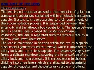
- 1. ANATOMY OF THE LENS Gross Anatomy The lens is an intraocular avascular biconvex disc of gelatinous transparent substance: contained within an elastic transparent capsule. It alters its shape according to theI requirements of the accommodative process. It is suspended between the iris anteriorly and the vitreous body posteriorly. The space between the iris and the lens is called the posterioer chamber. Posteriorly, the lens is separated from the vitreous face by a narrow retro-lental fluid space. The lens is maintained in position within the eye by a special suspensory ligament called the zonule, which is attached to the ciliary body and to the lens capsule. The suspensory ligament forms a delicate membrane covering the inner surface of the ciliary body and its processes. It then passes on to the lens dividing into three layers which are attached to the anterior capsule, the equator and the posterior capsule of the lens.
- 2. ANATOMY
- 3. The lens measures about 9 mm. diameter and 3 - 4 mm. in thickness. At birth, the lens weighs about 60 mg which increases slowly in a linear progression to a weight of approximate 250 mg at the age of 70 years. Its anterior surface is less convex than the posterior. The radius of curvature of the anterior surface is10 mm. while that of the posterior surface is 6 mm. The refractive index of the lens cortex is 1.38 and that the nucleus is 1.40. The refractive (coverging) power of the lens is approximaely 16 - 22 dioptres when inside the eye but is approximately 60 - 70 dioptres when in air. Microscopic Anatomy Histologically, the lens consists essentially of a mass of transparent cells, called the lens fibres, enclosed in an elastic membrane, called the lens capsule. The lens is composed of three distint portions :
- 5. The Lens Capsule . —This is a very elastic transparent non-cellular membrane which completely surrounds the lens. Its thickness is not uniform, being thickest near the equator and thinnest at the anterior and posterior poles of the lens. The Lens Epithelium . —The anterior epithelium consists of a single layer of cubical cells covering the anterior surface of the lens substance and lying between the latter and the capsule. There is no corresponding posterior epithelium.
- 6. The Lens Fibres and Cement Substance . —The bulk of the lens is composed of successive laminae of fibres, but between these there is some kind of cement substance having the same refractive index as the fibres and gluing them together. The lens substance comprises a cortex and a nucleus. The lens cortex consists of concentric lamellae of long hexagonal transparent fibres which are arranged in structure of an onion. The nucleus consists of the compressed central portions of lens cortex which gradually undergoes a process of sclerosis and becomes optically denser and harder than the cortical fibres.
- 7. The Suspensory Ligament of the Lens (the Zonule). —The zonule is a band-shaped gel-structure stretching from the ciliary body to the periphery of the lens. The zonule is inserted into the zonular lamellae in a belt running concentrically round the equator of the lens. Its anterior surface runs straight from the lens to meet the ciliary processes just behind their apices. Its posterior surface is bow-shaped curving along the inner surface of the body
- 8. Functions of the Lens Static Dioptric Function . —Together with the cornea, the lens forms the eye's dioptric system, which to converge parallel light rays from a distant object to a focus on the photoreceptor layer of the retina. Dynamic Dioptric Function .— The refractive power of the lens varies with the distance of the object of regard so that a perfect image is formed on the retina at all distances. This dynamic alteration in the refractive power of the lens to see clearly at all distances, known as accommodation, is achieved by a change in the curvature of the lens, mainly its anterior surface.
- 9. Protective Function .— The lens also protects the retina by absorbing the ultra violet rays. CHEMISTRY OF THE LENS The adult lens contains approximately 65% water and 34% proteins. The remaining 1 % is made up of inorganic compounds. Lens dehydration is maintained by an active sodium pump in the epithelium.
- 10. METABOLISM OF THE LENS The lens, although avascular, is a living structure with definite metabolic needs. It requires very little energy. This is because its energy requirements are limited to the following metabolic needs : 1. The maintenance of its transparency. 2. The maintenance of the elasticity of its capsule. 3. The development and growth of new lens fibres. Glucose seems to be the only substrate for the lens energy requirements. With restricted oxygen supply, most of the metabolism of glucose is through anaerobic glycolysis. Pyruvic acid is the end point of glycolysis and is further converted anaerobically to lactic acid, which diffuses out to the aqueous.
- 11. As the lens is avascular, the transport of nutrient materials and waste products to and away from the lens takes place by exchange between the lens susbtance and the aqueous humour. Therefore, it is important for the normal metabolism of the lens to have a normal aqueous humour and a normal permeability of the lens capsule, the capsule acts as an inert, non-selective, semi-permeable membrane, freely permeable to water and electrolytes but impermeable to large complex molecules.
- 13. ACCOMMODATION Accommodation is the act of altering the dioptric power of the lens in order to keep the image in sharp focus on the retina when the gaze is directed from far to near objects. This is accomplished by means of increasing the curvature of the lens surfaces particularly the anterior surface, and thus changing its refractive power. The degree of accommodation varies with the distance of the object of regard. Accommodation is most active in children and decreases gradually throughout life. This is probably due to the fact that as age advances the ciliary muscle atrophies aod lens becomes less elastic, and thus changes its shape with difficulty. Mechanism of Accommodation Accommodation comprises two mechanisms, namely, an active contraction of the ciliary muscle, followed by passive change of the shape of the lens.
- 15. CATARACT General Considerations A cataract means cloudiness or opacity of the lens substance or its capsule. This definition includes vacuoles, water clefts, dense areas reflecting or refracting light, and punctuate microscopic spots in the lens substance. Cataracts most commonly develop as part of the normal aging process and are called senile cataracts, but sometimes they are developmental. Cataracts may also be acquired as a result of ocular pathology, metabolic defects, systemic disease, toxins or trauma. Depending on the location and the extent of lenticular opacity, light rays passing through the lens may be blocked or scattered, resulting in a blurred retinal image or a bothersome glare.
- 16. Pathology of Cataract Formation The exact cause of cataractogenesis is obscure, the loss of transparency is due to a disturbance of the structure of the lens. The disturbance may be of two types : Hydration Opacity. — Biochemical, electrolytic and permeability factors produce abnormal osmotic gradiant leading to an influx of water and sodium ions and an egress of potassium ions. Coagulative Opacity. —An irreversible chemical change whereby the proteins become coagulated and insoluble. Denaturization of the lens proteins may initiate or potentiate the process of water influx and cellular fragmentation. Several factors may be responsible for loss of transparency of the lens such as :
- 17. Diagnostic Methods for Catarac t Examination of the Visual Acuity. —Distant and near vision with the appropriate glasses would be tested. Nuclear cataracts affect vision more than peripheral cortical cataracts. Examination of the Pupillary Responses . —The direct and consensual pupillary responses are usually affected to a slight extent by the lens opacities. Examination of the Lens by Direct Focal Illumination. —Lens opacities appear in the pupillary area as grey or white areas against a black background. Slit-lamp Biomicroscopy .—The extent, density, type and location of the cataract can accurately determined by slit-lamp biomicroscopy. Examination of the Red Reflex. Examination of the Iris Shadow by Oblique Focal Illumination. —
- 18. Refraction and Retinoscopy. —Retinoscopy often confirms that lens opacities are the cause of a patient's poor vision. A-Scan and B-Scan Ultrasonography.
- 19. CLASSIFICATION OF CATARACT DEVELOPMENTAL SENILE ACQUIRED : Traumatic Complicating Ocular Pathology Associating Systemic Disease
- 27. Classification according to maturity Immature Mature Hypermature Morgagnian
- 29. Mature cataract
- 30. DEVELOPMENTAL CATARACT Congenital cataracts are present at birth or within 3 months after birth. Developmental cataracts are not evident at birth but may form during infancy or adolescence. They normally remain stationary throughout life. Etiology. —The cause may be either one of the following : 1. A hereditary defect, usually of the dominant type, which is transmitted by the father or mother. It is due to an anomaly in the chromosomal pattern. 2. A maternal nutritional deficiency during the process of development leading to a lowered blood calcium. It may be accompanied by signs of rickets. 3. A maternal infection during the early months of pregnancy, e.g. rubella. 4. A deficient oxygenation, e.g. due to repeated placental haemorrhages. 5. A familial incidence, which is dependent upon a genetic influence or some maternal abnormality. Symptoms .—The child is usually brought for examination because the parents may notice that the pupil is white or that the child holds things too close to his eyes.
- 31. S ENILE CATARACT Senile cataract denotes an age-related bilateral progressive opacification of the lens affecting elderly people not suffering from local or systemic disease. It commonly affects persons over 50 years of age and is due to a process of aging and degeneration. Sometimes, there appears to be a familial tendency to cataract formation in which case the lens opacity may occur at an earlier age in successive generations. It is controversial whether the cataract is genetically determined or environmentally influenced.Ccataract is usually bilateral, but often one side is more advanced than the other.
- 32. Clinical Picture Symptoms. — The patients may complain of one or more of the following symptc Gradual Diminution of Vision without Pain or Discharge. Uniocular Diplopia or Polyopia. Myopia. Positive Scotomata. Glare. Altered Colour Perception Signs. —Senile cataract is essentially a process in which the transparency of is impaired by changes either in the cortex or in the nucleus. The principal sign is a whitish opacity within the pupillary area, the eye appearing otherwise quiti A white pupil (leucocoria) is usually seen as a late manifestation of cataract.
- 34. Intumescent Cataract .— Sometimes, during the immature stage of cataract formation, the lens absorbs an increasing amount of fluid from the aqueous and becomes swollen, it intumescent. The swollen lens pushes the iris forwards, reduces the depth of the anterior chamber and may block the angle, hence there is a tendency to secondary glaucoma. The Mature Stage. —A cataract is called mature, when complete opacification of the fibres extends to the capsule.
- 36. TREATMENT OF SENILE CATARACT Th e only treatment of senile cataract is by surgical removal of the opaque lens. The action should be undertaken on the worse eye as soon as the vision in the better eye less than 6/18, or when the patient's ability to work is threatened. It is no longer necessary to wait until the cataract is mature. With modern surgical techniques, an immature presents no difficulty in its removal. However, if the cataract shows signs of maturity, it should be extracted even if the vision in the better eye is still perfect.
- 37. COMPLICATED CATARACTS ASSOCIATED WITH SYSTEMIC DISEASE: Cataracts Associated with Metabolic Disturbances (Metabolic Cataracts): (a) Diabetic cataract. (b) Galactosemic cataract. (c) Hypocalccemic cataract. (d) Hypothyroidic cataract. (e) Myotonic cataract. (f) Deficiency cataract. Cataracts Associated with Skin Diseases: Atopic dermatitis (Eczema). Poikiloderma atrophicans (Rothmund Syndrome). Sclero-Poikiloderma (Werner Syndrome). Anhidrotic ectodermal dysplasia.
- 38. Extracapsular cataract extraction 1. Anterior capsulotomy 2. Completion of incision 3. Expression of nucleus 4. Cortical cleanup 6. Polishing of posterior capsule, if appropriate 5. Care not to aspirate posterior capsule accidentally
- 39. 8. Grasping of IOL and coating with viscoelastic substance Extracapsular cataract extraction ( cont. ) 7. Injection of viscoelastic substance 9. Insertion of inferior haptic and optic 11. Placement of haptics into capsular bag 10. Insertion of superior haptic 12. Dialling of IOL into horizontal position and not into ciliary sulcus