
Recommended
More Related Content
What's hot
What's hot (20)
Viewers also liked
Similar to Oral pathology dr faeza
Similar to Oral pathology dr faeza (20)
More from eliasmawla
More from eliasmawla (20)
Recently uploaded
Recently uploaded (20)
Oral pathology dr faeza
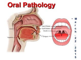
- 1. Oral PathologyOral Pathology • M a r c h . 2 2 . 2 0 1 5
- 2. ORAL PATHOLOGY • INFECTIONS: HSV, VIRAL, FUNGI • LEUKOPLAKIA/” • SQUAMOUS TUMORS: • ODONTOGENIC CYSTS/TUMORS
- 3. Herpetic vesicles Herpesvesicle with ulceration & secondary inflammation
- 7. •Syphilis “1, 2, 3”.
- 8. Aphthous ulcer Aphthous ulcer. Single ulceration with an erythematous halo surrounding a yellowish fibrinopurulent membrane.
- 11. Tonsillitis & peritonsillar abscess “Quinsy” Oropharynx
- 13. Oral Pathology Skin diseases can affect oral mucosa Lichen planus Erythema multiforme Pemphigus vulgaris Pemphigoid. Pigmentation of lip & oral mucosa. Peutz–Jeghers syndrome
- 14. Oral Pathology Polypoid nodules in the mouth • Pyogenic granuloma. • Fibroepithelial polyp Leukoplakia. Sq C Ca.
- 15. “Irritation” Fibroma Fibroma. Smooth pink exophytic nodule on the buccal mucosa.
- 16. PYOGENIC GRANULOMA Pyogenic granuloma. Erythematous, hemorrhagic, and exophytic mass arising from the gingival mucosa
- 17. • Leukoplakia is defined by the WHO as; • “a white patch or plaque that cannot be scraped off and cannot be characterized clinically or pathologically as any other disease.”
- 18. • Erythroplakia (red area), associated with a greater risk of malignant transformation than leukoplakia. • 40 and 70 years, 2 : 1 male preponderance. • Tobacco is the most common risk factor for leukoplakia and erythroplakia.
- 19. leukoplakia
- 20. Tobacco Alcohol HPV Family history chronic irritation 95% are Sq C Ca.
- 21. the sites of origin of Sq C Ca of the oral cavity, in numerical order of frequency the favored locations are the ventral surface of the tongue, floor of the mouth, lower lip, soft palate, and gingiva
- 23. 3 exophytic masses “SqcCa”, the hyperkeratotic area is lichen planus.
- 24. ODONTOGENIC CYSTS/TUMORS • INFLAMMATORY CYSTS (e.g., “Radicular”[periapical] most common) • DEVELOPMENTAL CYSTS • MALIGNANT TUMORS of ODONTOGENIC ORIGIN (AMELOBLASTOMAS) (rare)
- 25. Odontogenic Cysts • Epithelial-lined cysts are common in the jaws • derived from remnants of odontogenic epithelium present within the jaws. • these cysts are; 1. inflammatory or 2. developmental
- 26. Salivary glands
- 27. Salivary glands
- 29. Mucocele of an accessory salivary gland duct
- 30. Mucocele of an accessory salivary gland duct
- 31. Sialadenitis
- 33. DISEASES OF SALIVARY GLANDS • Xerostomia • autoimmune disorder Sjِgren syndrome • Rtx • Drugs; anticholinergic, antidepressant/ antipsychotic, diuretic, antihypertensive, sedative, muscle relaxant, analgesic, & antihistaminic agents • dental caries & candidiasis, difficulty in swallowing & speaking
- 34. Oral Pathology Salivary glands tumors; • are mostly Benign & affect parotid.parotid. - “Pleomorphic adenoma” or “ Mixed tumor” - Adenolymphoma or “Warthin’s tumor” of parotid. • Malignant; • Affect Minor salivary glands. • “Mucoepidermoid ca”. • “Adenoid cystic carcinoma” • Others.
- 35. P A R O T I D
- 37. PLEOMORPHIC ADENOMA i.e., MIXED TUMOR
- 38. Pleomorphic Adenoma • pleomorphic adenoma contains both epithelial (E) and stromal (S) components.
- 39. Pleomorphic Adenoma • This neoplasm is typically encapsulated, although tumor islands may be found within the fibrous capsule.
- 40. WARTHIN TUMOR
- 41. Warthin's Tumor • Warthin's tumor (benign papillary cystadenoma lymphomatosum) • the second most common benign tumor of the parotid gland
- 42. Mucoepidermoid Ca. • MECs contain two major elements: • mucin-producing cells and epithelial cells of the epidermoid variety • Divided into low-grade (well differentiated), & High-grade (poorly differentiated).
- 43. Adenoid Cystic Carcinoma • Adenoid cystic carcinoma with Swiss cheese pattern. • It is the second-most common malignant tumor of the salivary glands. • ACC is the most common malignant tumor found in the submandibular, sublingual, and minor salivary glands.
- 44. Adenoid Cystic Carcinoma • Nerve (N) invaded by adenoid cystic carcinoma (the blue area surrounding the nerve). • Spread may occur by emboli along the nerve lymphatics
- 45. • Behcet's (beh-CHETS) disease, also called Behcet's syndrome, is a rare disorder that causes inflammation in blood vessels. • The signs and symptoms of Behcet's disease — which may include mouth sores, eye inflammation, skin rashes and lesions, and genital sores — vary from person to person and may come and go on their own. • The exact cause of Behcet's is unknown, but it may be an autoimmune disorder, • Both genetic and environmental factors may be responsible for Behcet's disease. • Treatment aims to reduce the signs and symptoms of Behcet's disease and to prevent serious complications, such as blindness.
Editor's Notes
- Common pathologic conditions of the oral cavity
- Herpetology is the study of creepy critters, reptiles and amphibians. Herpetic vesicles “creep” over mucosal surfaces. Like CMV and V-Z viruses, the various herpes viruses (mainly type 1 and 2) are, amazingly, in the “herpes” family of viruses. Early lesions crop up as vesicles, after a few days, these vesicles can be irritated, ulcerated, inflamed, and secondarily pustulated. Classically type 1 was predominantly oral mucosa, and the slightly nastier type 2 was more genital, but nowadays, crossover is so common, who cares any more? The virus recurs often for many years, triggered off by god-knows-what, and the newer antiviral agents have shown amazing efficacy in preventing recurrences. Just about everybody had been exposed to herpes of some type.
- Herpes zoster (or simply zoster), commonly known as shingles and also known as zona, is a viral disease characterized by a painful skin rash with blisters in a limited area on one side of the body (left or right), often in a stripe.
- Monilia, thrush-mouth, candida: they all mean the same thing. Look for a whitish oral film without much underlying inflammation (i.e., redness). Common in babies, diabetics, immunocompromised people. Candida (almost always ablicans) always affects moist, usually non-keratinized, stratified squamous mucosa, i.e., mouth, vagina, moist genital skin areas. Everybody has it lying around waiting for an immunocompromised condition to occur.
- Obscure etiology, 40% of us have had them, painful, and you can bet there are inflammatory cells at the base. You know what they are. You’ve had them. Things related to them include: stress, fatigue, illness, injury from accidental biting, hormonal changes, menstruation, sudden weight loss, food allergies, and deficiencies in vitamin B12, iron, and folic acid (wikipedia). Whenever a condition is associated with LOTS of things, like this, we call this an “obscure” etiology ;)
- Inflammatory endpoint or true neoplasm? Who cares? Often the terms “inflammatory” or “reactive” refers to this type of “fibroma”.
- Granuloma or neoplasm or granulation tissue? Who cares? A pyogenic granuloma pops out like a “tumor” and is 100% indistinguishable from normal granulation tissue, and looks nice and pink and healthy like granulation tissue, i.e., organizing inflammation, too. The least thing it can be called is a granuloma, because it rarely has clusters of macrophages or giant cells. Would you expect a pyogenic granuloma to “blanch” and a fibroma NOT to “blanch”? Answer: YES
- “Hairy” leukoplakia however, is usually a sign of HIV.
- Classical radiologic and histologic image of a dentigerous cyst.
- Common pathologic conditions of the “non-accessory” salivary glands, i.e., submandibular (mixed), parotid (99% serous), sunlingual (99% mucinous).
- Batsakis- pg 6
- Viral parotitis, i.e., mumps
- Mucocele of an accessory salivary gland duct, just a big cyst filled with mucin and lined by mucinous columnar epithelium, often inflamed and/or squamous metaplastic
- The classic place for ANY visible parotid swelling or tumor to present, between the tip of the ear and the tip (angle) of the mandible.
- Pleomorphic adenoma. A, Slowly enlarging neoplasm in the parotid gland of many years duration. B, The bisected, sharply circumscribed, yellow-white tumor can be seen surrounded by normal salivary gland tissue.
- Mixed tumors are generally benign, have BOTH connective tissue (i.e., usually cartilagenous) components as well as glandular components, hence the name pleomorphic or mixed, they generally look and feel like little round soft cartilage balls.
- Batsakis-Pg 6
- Batsakis-Pg 8
- Why is muco-epidermoid carcinoma a perfect name for this salivary gland malignancy?