2. CONSORT Clinical Trial
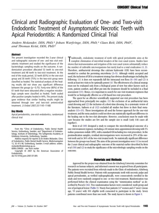
TABLE 1. Distribution of Teeth by Randomization Factors
One-visit One-visit Two-visit Two-visit
Tooth group pre treatment follow-up pre treatment follow-up
(n 53) (%) (n 49) (%) (n 48) (%) (n 40) (%)
Incisors and canines 26 (49) 25 (51) 27 (56) 22 (55)
Bicuspids 17 (32) 15 (30) 12 (25) 10 (25)
Molars 10 (19) 9 (19) 9 (19) 8 (20)
Size of lesion
2 mm 5 (9) 5 (10) 5 (10) 5 (13)
2-5 mm 29 (55) 26 (53) 24 (50) 22 (55)
5 mm 19 (36) 18 (37) 19 (40) 13 (32)
Intracanal Procedures For the one-visit group, to remove the smear layer, the canals were
Each tooth was isolated with a rubber dam and disinfected with filled with Tubulicid Plus (Dental Therapeutics AB, Nacka, Sweden) for
30% hydrogen peroxide and 10% iodine tincture according to the pro- 20 seconds, dried with paper points, and refilled for an additional 20
tocol proposed by Möller (17). After access preparation, the working seconds. Subsequently, the canals were filled with 5% IPI solution for 10
length was established radiographically. minutes. Before root filling, a postmedication microbiological sample
To expose the apical part of the root canals to microbiological was taken (10). Finally, root canals were filled with gutta percha cones
sampling, the canals were sequentially enlarged with nickel-titanium using cold lateral condensation technique including rosin chloroform
instruments for rotary (GT/ Profile; Dentsply-Maillefer, Ballaigues, Swit- as sealer. The root-filling quality was radiographically checked.
zerland) and/or hand use (Nitiflex, Dentsply-Maillefer), reaching size For the two-visit group, CH was placed meticulously by means of a
ISO #20 at the working length. Before further instrumentation of the Lentulo spiral, and the access cavity sealed with Coltosol (Coltène
canals, initial microbiological samples were taken (10). The canals Whaledent, Cuyahoga Falls, OH). After a week, CH was removed by hand
were then enlarged, and apical boxes were prepared between size ISO instruments and irrigation with VMGA I (17). A postmedication micro-
#40 and #60, depending on the size of the root. During instrumentation, biological sample was obtained (10), and canals obturated with gutta
the canals were irrigated with 0.5% NaOCl (Dakins solution). Immedi- percha as described earlier. The treatments were performed by four
ately after the completion of the chemomechanical preparation, postin- endodontic specialists.
strumentation microbiological samples were obtained (10). Up to this
point, treatment procedures were identical for both groups. Follow-up
The healing results were clinically and radiographically evaluated
2 years postoperatively.
Radiographic Assessments
All radiographic films obtained preoperatively and at follow-up
were coded blind and organised in random order. Two examiners in-
dependently evaluated all radiographs. Films were examined under
moderate illumination at a light table using a magnifying viewer
equipped with a masking frame with the size of a dental film. To mini-
mize the false-positive diagnoses, observers used a strict definition of
periapical disease and reported a positive finding only when absolutely
certain (18). In case of disagreement, joint re-evaluation was per-
formed until a consensus was reached. The size of periapical radiolu-
cency was assessed by measuring with a ruler (to the nearest millime-
ter) its largest horizontal and vertical width.
Outcome Classification
Outcome of treatment was classified by using a modification of the
Strindberg (19) criteria. Teeth with symptoms of persisting periapical
inflammation were scored as not healed as were cases with unchanged
or increased size of the periapical radiolucency. Teeth with a reduced
TABLE 2. Distribution of Teeth According to Outcome Classification in the
Two Experimental Groups*
One visit Two visit Total
Healed 32 30 62
Uncertain healing 13 5 18
Not healed 4 5 9
Total 49 40 89
p 0.7532.
2
Figure 1. Flow diagram of the progress of the phases of the trial. *Mantel-Haenszels test was used to test trends in the contingency table.
1146 Molander et al. JOE — Volume 33, Number 10, October 2007
3. CONSORT Clinical Trial
TABLE 3. Distribution of Teeth According to Outcome Classification and Post- so-called minimization method was used. Twelve teeth (12%) were lost
medication Sample Results (PMS)* to the 2-year follow-up. However, the loss did not alter the situation to an
Positive Negative extent that would render statistical comparisons and conclusions in-
Total valid (Table 1).
R-sample R-sample
Healed 12 49 61 Clinical symptoms were rare during the follow-up period (only 4
Uncertain healing 14 4 18 cases). Thus, the outcome was classified mainly on the radiographic
Not healed 1 8 9 evaluation. The radiographic image of periapical bone lesions develops
Total 27 61 88 from being impossible or difficult to see to being easily distinguished
p 0.1202. from the background (20). Radiographic diagnosis of apical periodon-
*Mantel-Haenszels 2
test was used to test trends in the contingency table. titis, therefore, may be regarded as a signal-detection task (18). The
actual prevalence of apical periodontitis in a cohort is difficult to reveal
size of the periapical rarefaction (sum of horizontal and vertical reduc- by radiographic means (21). But, if false-positive diagnoses can be
tion 2 mm) were judged as uncertain. Teeth with complete restitution minimized, chances will increase to disclose the true relation between
of the periodontal contours were judged as healed. In teeth with more investigated factors or populations. Therefore, in the present study, a
than one root, the least favorable outcome was registered. periapical radiolucency was reported by the observers only when ab-
solutely certain. This implies that stated healing frequencies should not
Microbiological Examination be given an absolute meaning but only a relative one.
Details of laboratory procedures and microbiologic analyses can No statistically significant difference in terms of healing was ob-
be found in Kvist et al (10). served between the one- and two-visit groups. This finding corroborates
the results of four previous studies (9, 13, 14, 22) (Table 4). It should
Statistical Methods be pointed out that beside the study by Friedman et al (22) all cited
A Mantel-Haenszels chi-square test was used to test trends in con- investigations report on small clinical materials. On the other hand,
tingency tables. All hypothesis tests were conducted at the 0.05 level of Friedman et al did not analyze a randomized sample. Thus, published
significance. studies including the present one have failed to show any statistically
significant difference in the outcome between one-visit and two-visit
Results root canal therapy.
The randomization procedure allocated 53 teeth to one-visit treat- Clinical outcome studies take a long time to monitor, demand
ment and 48 teeth to two-visit treatment. Twelve teeth, eight in the substantial economic resources, and run the risk of losing patients at
two-visit and four in the one-visit group, respectively, were lost to fol- follow-ups. Therefore, it is desirable to find simpler but accurate sur-
low-up. Reasons for drop out are given in Figure 1. At the end of the rogate endpoints for such investigations. When assessing the present
study period, 32 teeth (65%) in the one-visit group and 30 teeth (75%) material, the identical overall conclusion was drawn after both the mi-
in the two-visit group were classified as healed (Table 2). The number crobiological (10) and the clinical/radiographic evaluation; no statis-
of cases classified as uncertain was higher in the one-visit group, 13 tically significant difference was found between the one- and two-visit
(27%) as compared with 5 (13%). Four teeth in the two-visit group treatment regimens. Hence, at this level, support was given to the notion
showed clinical symptoms before the 2-year follow-up and were clas- that postmicrobiologic sampling could replace radiographically based
sified as not healed. The statistical analysis of the healing results did not long-time studies and be used as a surrogate endpoint. However, at a
show any significant difference between the groups (p 0.75). Forty- more case-specific level, the relation between the results of microbio-
nine (80%) of the 61 teeth that were obturated after a negative mico- logic analysis and outcome was not as clear.
biologic sample were classified as healed (Table 3). Teeth sealed after There was a tendency toward a more favorable outcome in teeth
positive samples healed in 44%. Notably, teeth with positive samples yielding a negative culture immediately before root filling (p 0.1202).
were classified as uncertain more often (52%) than teeth with no sign of This finding is in concordance with Bender et al (23), Heling and
cultivable microorganisms (7%). However, the presence or absence of Shapira (24), and Peters et al (9). Others have indeed reported that
detectable microbes just before obturation did not influence the healing presence of microbes at the time of root filling will adversely affect the
results at a statistically significant level (p 0.12). outcome (25–29). The idea that absence of cultivable microbes at the
time of obturation will favor healing is consistent with the idea that
Discussion microorganisms are the prime reason for persistent apical periodonti-
The present investigation was designed as a randomized study of tis. Recently, Fabricius et al (30) reported results from an extensive
the effect of one-visit and two-visit treatment procedures on periapical experiment conducted on 175 root canals in monkey teeth in support of
healing. Because the material was small and important prognostic fac- such an assumption. However, in this context, it must be understood
tors might be unevenly distributed among the groups, Pocock=s (16) that the methodology of microbiological root canal sampling is complex
TABLE 4. Data Summary of Included Studies
Healing rate (%)*
All types Observation
Citation Randomization No. of cases single versus
of teeth Time (y)
multiple visit
Friedman et al (1995) No Yes 378 1.5 86 vs 75
Trope et al (1999) Yes No 76 1 64 vs 74
Weiger et al (2000) Yes Yes 67 0.5-5 83 vs 71
Peters and Wesselink (2002) Yes No 38 4.5 81 vs 71
Molander et al (2007) Yes Yes 89 2 65 vs 75
*Teeth with complete healing.
JOE — Volume 33, Number 10, October 2007 One- and Two-visit Treatment of Asymptomatic Necrotic Teeth with Apical Periodontitis 1147
4. CONSORT Clinical Trial
and that the diagnostic accuracy is poorly known (7, 31–33). For ex- 14. Weiger R, Rosendahl R, Löst C. Influence of calcium hydroxide intracanal dressings
ample, microorganisms hiding in biofilms or in untreated parts of the on the prognosis of teeth with endodontically induced periapical lesions. Int Endod
J 2000;33:219 –26.
canal system may be hard to sample, and remnants of the medication 15. Peters LB, Wesselink PR. Periapical healing of endodontically treated teeth in one and
might depress laboratory growth. two visits obturated in the presence or absence of detectable microorganisms. Int
In conclusion, the present study gave evidence that, given a metic- Endod J 2002;35:660 –7.
ulously instrumented root canal, a one-visit antimicrobial treatment 16. Pocock SJ. Clinical Trials. A Practical Approach. Chichester, UK: John Wiley & Sons;
1983.
including 10 minutes of dressing with 5% IPI is as effective as a two-visit 17. Möller ÅJR. Microbial examination of root canals and periapical tissues of human
procedure using CH. Hence, at this level, support was given to the notion teeth. Odontol Tidskr 1966;74:1–380.
that postmicrobiologic sampling could replace radiographically based 18. Reit C, Gröndahl HG. Application of statistical decision theory to radiographic diag-
long-time studies and be used as a surrogate endpoint. nosis of endodontically treated teeth. Scand J Dent Res 1983;91:213– 8.
19. Strindberg LZ. The dependence of the results of pulp therapy on certain factors. Acta
Odontol Scand 1956;14(suppl 21).
References 20. Bender IB, Seltzer S. Roentgenographic and direct observation of experimental le-
1. Byström A, Sundqvist G. Bacteriologic evaluation of the efficacy of mechanical root sions in bone: I and II. J Am Dent Assoc 1961;62:152– 60.
canal instrumentation in endodontic therapy. Scand J Dent Res 1981;89:321– 8. 21. Brynolf I. Histological and roentgenological study of periapical region of human
2. Dalton BC, Ørstavik D, Phillips C, Pettiette M, Trope M. Bacterial reduction with upper incisors. Odontol Revy 1961;18(suppl 11).
nickel-titanium rotary instrumentation. J Endod 1998;24:763–7. 22. Friedman S, Löst C, Zarrabian M, Trope M. Evaluation of success and failure after
3. Byström A, Claesson R, Sundqvist G. The antibacterial effect of camphorated par- endodontic therapy using a glass ionomer cement sealer. J Endod 1995;21:384 –90.
amonocholorophenol, camphorated phenol and calciumhydroxide in the treatment 23. Bender IB, Seltzer S, Turkenkopf S. To culture or not to culture. Oral Surg
of infected root canals. Endod Dent Traumatol 1985;1:170 –5. 1964;18:527– 40.
4. Sjögren U, Figdor D, Spångberg L, Sundqvist G. The antibacterial effect of calcium 24. Heling B, Shapira J. Roentgenologic and clinical evaluation of endodontically treated
hydroxide as a short-term intracanal dressing. Int Endod J 1991;24:119 –25. teeth, with or without negative culture. Quintessence Int 1978;11:79 – 85.
5. Shuping GB, Ørsatvik D, Sigurddsson A, Trope M. Reduction of intracanal bacteria 25. Engström B, Hård af Segerstad L, Ramström G, Frostell G. Correlation of positive
using nickel-titanium rotary instrumentation and various medications. J Endodon cultures with the prognosis for root canal treatment. Odontol Revy 1964;15:257–70.
2000;26:751–5. 26. Sjögren U, Figdor D, Persson S, Sundqvist G. Influence of infection at the time of root
6. Akpata ES. Effect of endodontic procedures on the population of viable microorgan- filling on the outcome of endodontic treatment of teeth with apical periodontitis. Int
isms in the infected root canal. J Endod 1976;2:369 –73. Endod J 1997;30:297–306.
7. Reit C, Dahlén G. Decision making analysis of endodontic treatment strategies in teeth 27. Sundqvist G, Figdor D, Persson S. Microbiologic findings of teeth with failed end-
with apical periodontitis. Int Endod J 1988;21:291–9. odontic treatment and the outcome of conservative re-treatment. Oral Surg
8. Ørstavik D, Kerekes K, Molven O. Effects of extensive apical reaming and calcium 1998;85:86 –93.
hydroxide dressing on bacterial infection during treatment of apical periodontitis: a 28. Katebzadeh N, Sigurdsson A, Trope M. Radiographic evaluation of periapical healing
pilot study. Int Endod J 1991;24:1–7. after obturation of infected root canals: an in vivo study. Int Endod J 2000;33:60 – 6.
9. Peters LB, van Winkelhoff AJ, Buijs JF, Wesselink PR. Effects of instrumentation, 29. Waltimo T, Trope M, Haapasalo M, Örstavik D. Clinical efficacy of treatment proce-
irrigation and dressing with calcium hydroxide on infection in pulpless teeth with dures in endodontic infection control and one year follow-up of periapical healing.
periapical bone lesions. Int Endod J 2002;35:13–21. J Endod 2005;31:863– 6.
10. Kvist T, Molander A, Dahlén G, Reit C. Microbiological evaluation of one- and two-visit 30. Fabricius L, Dahlén G, Sundqvist G, Happonen R-P, Möller ÅJR. Influence of residual
endodontic treatment of teeth with apical periodontitis: a randomized, clinical trial. bacteria on periapical tissue healing after chemomechanical treatment and root
J Endod 2004;30:572– 6. filling of experimentally infected monkey teeth. Eur J Oral Sci 2006;114:278 – 85.
11. Sackett D. Evidence Based Medicine: How to Practice and Teach EBM, ed 2. Edin- 31. Morse DR. The endodontic culture technique: a critical evaluation. Oral Surg
burgh: Churchill Livingstone; 2000. 1970;30:540 – 4.
12. Sathorn C, Parashos P, Messer HH. Effectiveness of single- versus multiple-visit end- 32. Molander A, Reit C, Dahlén G. Microbiological evaluation of clindamycin as a root
odontic treatment of teeth with apical periodontitis: a systematic review and meta- canal dressing in teeth with apical periodontitis. Int Endod J 1990;23:113– 8.
analysis. Int Endod J 2005;38:347–55. 33. Reit C, Molander A, Dahlén G. The diagnostic accuracy pf microbiologic root canal
13. Trope M, Delano EO, Örstavik D. Endodontic treatment of teeth with apical periodon- sampling and the influence of antimicrobial dressings. Endod Dent Traumatol
titis: single vs. multivisit treatment. J Endod 1999;25:345–50. 1999;15:278 – 83.
1148 Molander et al. JOE — Volume 33, Number 10, October 2007