Case Review #6: 53 year old woman with Adult Scoliosis
•
1 j'aime•2,000 vues

A 53 year old woman, with an 85° thoracic curve, and a 75° lumbar curve. Dr. Pashman treated her with an Anterior fusion followed by a Posterior Spinal Fusion from T1 to the Pelvis. Curve was a KIM/SRP Classification 3.

Recommandé
Recommandé
Contenu connexe
Tendances
Tendances (20)
En vedette
En vedette (18)
Similaire à Case Review #6: 53 year old woman with Adult Scoliosis
Similaire à Case Review #6: 53 year old woman with Adult Scoliosis (15)
Plus de Robert Pashman
Plus de Robert Pashman (12)
Dernier
Dernier (20)
Case Review #6: 53 year old woman with Adult Scoliosis
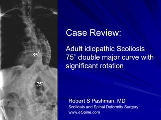
- 1. Case Review: Adult idiopathic Scoliosis 85˚ 75˚ double major curve with significant rotation 75˚ Robert S Pashman, MD Scoliosis and Spinal Deformity Surgery www.eSpine.com
- 2. Patient History 53 year old female Presented with 85˚ thoracic and 75˚ lumbar curvature Right thoracic/left lumbar curve with flank rotation, a small rib hump. She is well balanced on frontal and sagittal plane, has significant asymmetric skin folds but her shoulders and pelvis appear balanced. She is in good health and is very lean. Right leg pain Spinal Stenosis and Facet dislocation at L4/5 Failed conservative therapy
- 3. Pre-op X-rays The patient has a well documented history of 85˚ scoliosis progression, and has obtained several opinions about the treatment options. 75˚
- 4. Bending X-rays L R
- 5. Bending Films L R
- 6. Indications for Surgery Severe low back pain and radicular pain 75˚ adult idiopathic scoliosis, double major curve, with significant rotation. Degenerative disc disease, L4-5 and L5-S1. Lateral recess stenosis, and instability lumbar spine. Failed conservative therapy.
- 7. Surgical Strategy STAGE 1 Radical diskectomy with epidural decompression, L4-5 and L5-S1. Subtotal vertebrectomy for placement of screw fixation, L5. Anterior interbody fusion with FRA device and putty graft, L5-S1 and L4-5, Anterior screw fixation, L4-5 and L5-S. STAGE 2 Segmental spinal instrumentation using 5.5 stainless steel Legacy thoracic tented pelvis. This is an 8-level fusion. Sacral pelvic fixation with bilateral exposure of the iliac crest. Posterior spinal fusion T10 to the pelvis using locally harvested autogenous bone. 7-level osteotomy through ankylosed spine, Smith-Peterson osteotomy T11 to the sacrum. Subtotal laminectomy T12 to L5. STAGE 3 T2 to L2 12-level segmented spinal instrumentation Posterior spinal fusion, T2 to L2, using locally harvested autogenous bone and rhBMP. Spinal osteotomy, T4-T5, T5-T6, T6-T7, T7-T8, T9-T10, and T11-T12 for rigid adult idiopathic scoliosis. These are Smith-Peterson osteotomies through ankylosed and rigid spine.
- 8. Findings during surgery At the time of operation, severe rigidity ankylosing especially in the concave side of the spine. Each joint was fused, this in the concavity from L1 to L5 with the joints ankylosed, a few solid to the pars, through big degenerative changes. This required multiple level osteotomy as indicated in the procedure section. The patient was mobilized after that. The surgery took significantly longer than expected because of the need for multiple level surgery and therefore the third stage will be completed on an interval basis.
- 9. Post-Op Films X-rays show excellent balance in frontal and sagittal plane, good correction of the curve. All the instrumentation looks intact.
- 10. Pre-Op/Post-op Comparison The patient has no post-operative radiculopathy, and minimal pain. She is not taking pain medication. Overall, she is doing quite well.