Basic Hematology
•
23 j'aime•6,235 vues

This slide is an overview of basic hematology and blood physiology including RBCs, WBCs, and platelets

Recommandé
Contenu connexe
Tendances
Tendances (20)
Similaire à Basic Hematology
Similaire à Basic Hematology (20)
Plus de Fadel Muhammad Garishah
Plus de Fadel Muhammad Garishah (18)
Dernier
Dernier (20)
Basic Hematology
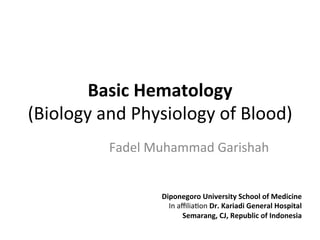
- 1. Basic Hematology (Biology and Physiology of Blood) Fadel Muhammad Garishah Diponegoro University School of Medicine In affilia:on Dr. Kariadi General Hospital Semarang, CJ, Republic of Indonesia
- 2. Component of blood Blood is the term for the liquid substance within vascular system. It consisted of plasma (55%), leukocytes and platelet (<1%) and red blood cells (45%).
- 5. Cellular basis of red blood cells
- 7. erythropoiesis Erythropoiesis is a sequence involving proliferaFon and differenFaFon of commiHed red marrow cells through the erythroblast and normoblast stages to the reFculocytes that are released into the bloodstream, and finally become erythrocytes.
- 8. RegulaFon of erythrocyte PO2 PO2 PO2 “Still Hemolysis Erythropoietin Kidney Bone marrow High altitude, etc. 2 Hemolysis 1 Hypoxia Spleen Blood Bone marrow RBC formation Life span: 120 days Erythrocytes Break- down B. Life cycle of red blood cellsA. Regulation of RBC production PO2 PO2 Erythropoietin Hemolysis Bone marrow 2 Hemolysis C. Erythrocyte parameters MCH, M
- 9. Life cycle of erythrocytes Plate4.1CompositionandFunctionofBlood “Still good” Bone marrow Lymph nodes Spleen Liver, etc. Spleen Blood Bone marrow Test RBC formation Phagocytosis by macrophages in: Sinus “Too old” Pulpal arteriole Life span: 120 days es Spenic pulp Break- down
- 10. Clinically related erythrocytes parameter 89 b a MCHC (mean Hb conc. in RBCs) Hb conc. = (g/LRBC) Hct MCV (mean volume of one RBC) Hct = (L/RBC) red cell count MCH (mean Hb mass/RBC) Hb conc. = (g/RBC) red cell count Normal: 320–360g/L Normal: 80–100fl Normal: 27–32pg Centrifugation Hemoglobin concentration (g/LBLOOD) Erythropoietin Bone marrow Lymph nodes Spleen Liver, etc. Sinus arteriole Red cell count (RCC) (quantity/LBLOOD) Hematocrit (Hct)=b/a (LRBC/LBlood) Blood sample C. Erythrocyte parameters MCH, MCV and MCHC
- 11. Iron intake and metabolism HCI Fe Fe Fe FeIII FeIII FeIII Fe2+ FeIII Fe2+ H + Stomach Liver Fe absorption: 3–15% of Fe intake Normal Fe intake: 10–20 mg/day 5–10 mg/day Lumen Ferritin Blood Lyso- some Cell turnover Mucosal transferrin Apo- transferrin Heme- FeII Mucosal cells (duodenum) Trans- ferrin Non-absorbed Fe in feces: Normally 85–97% of intake Heme FR FeIII 2 Fe absorption1 Iron intake A. Iron intake and metabolism
- 12. Iron storage and recycling Fe FeFe Fe stores Systemic blood Liver Bone marrow Hemo- siderin FerritinHeme Hb Erythrocytes Hemo- pexin Hapto- globin Ferritin Hemo- siderin Transferrin Macrophages in spleen, liver and bone marrow (extravascular) Already in bone marrow Vitamin B12 Folic acid 0.05mg/day Other organs NADP NADPH +H+ Dihydrofolate reductase Non-absorbed Fe in feces: Normally 85–97% of intake FeIII 3 Fe storage and Fe recycling B. Folic acid and vitamin B12 (cobalamins)
- 13. Folic acid and Cobalamin (B12) metabolism 7mg 1 mg Fe stores siderinHb Erythrocytes globin Macrophages in spleen, liver and bone marrow (extravascular) Already in bone marrow Liver Ileum Vitamin B12 0.001mg/day Folic acid 0.05mg/day Other organs Stores Bone marrow NADP NADPH +H+ Dihydrofolate reductase Folate regeneration Thymidylate synthase 7,8- dihydro- folate Tetrahydro- folate N5 -tetra- hydrofolate Deoxy- uridylate Deoxy- thymidylate Erythroblast DNA synthesis Erythropoiesis Stomach Erythrocytes Intrinsic factor Methyl- cobalamin B. Folic acid and vitamin B12 (cobalamins) Despopoulos, Color Atlas of Physiology © 2003 Thieme All rights reserved. Usage subject to terms and conditions of license.
- 14. The blood groups O A B AB O A B AB 5 10 15 20 2 (After Kownatzki) Days after first antigen exposure Compatible Incompatible (agglutination) Anti-A Anti-B Antigenonredbloodcells otrienes tors Antibody in serum ity
- 15. Biology of Rhesus 101 Pla + = A B AB Rh+ Rh+ Rh+ Rh+ Rh– Rh– Rh– Rh– Rh+ Incompatible (agglutination) Anti-A Anti-B Antigenonredbl Agglutination Hemolysis Severe hemolysis in fetus Father Mother Subsequent Rh+ childrenFirst Rh+ child Subsequent mismatched Rh+ transfusion Rh+ red cells High anti-Rh+ titer Damage Rh+ red cells Anti-Rh+ High anti-Rh+ titer Damage Anti-Rh+ antibodyBlood First mismatched Rh+ transfusion D. Rh sensitization of mother by child or by Rh-mismatched transfusion
- 16. Plasmaproteins wPropertiesofBlood 60% 4% 8% 12% 16% α1 α2 β γ 0 100 500 1000 Plasma Albumin Globulins Electrophoretic protein fractions 65–80g/L Proteins(100%) e diameter (µm) Blood Plasma Water ist effect B. Plasmaproteins of body fluids
- 17. Ion composiFon of body fluids Plate4.3FlowPropertiesofBlood 2 1 0 1 5 10 50 100 500 1000 Na+ K+ 142 4.3 2.6 (1.3*) 1.0 (0.5**) 153 4.6 2.8 (1.3) 1.0 (0.5) 145 4.4 2.5 (1.5) 0.9 (0.45) ca. 12 ca. 140 <0,001 1.6 150 162 153 ca. 152 Cl– HCO3 – *) Total plasma Ca: 2.5mmol/L; **) Total plasma Mg: 0.9mmol/L Na+ Ca 2+ ,Mg2+ Cl– HCO3 – HCO3 – K+ Ca2+ ,Mg2+ Na+K+ mval/L (mmol/L) 104 24 2 14 5.9 112 36 2.2 15 6.3 117 27 2.3 0.4 6.2 ca. 3 10 ca. 30 ca. 54 ca. 54 150 162 153 ca. 152 Electrophoretic protein fractions 65–80 Proteins(1 Cations Plasma Interstitium CytosolSerumIon Anions Proteins Misc. Sum Sum Proteins,phosphates, etc. Proteins– Interstitium Cytosol Cations Anions Cations Anions Viscosity Vessel inside diameter (µm) Plasma Water Free Ca2+ Inorganic phosphate Inorganic phosphate Misc. Free Mg 2+ C. Ion composition of body fluids
- 18. The immunoglobulin in serum IgA 2.25 IgM 1.15 IgD 0.03 IgE 0.0002 IgG 11.0 g/L 100 50 –3 181512963 IgM IgG IgA IgD IgE *) Total plasma Ca: 2.5mmol/L; **) Total plasma Mg: 0.9mmol/L 2 14 5.9 2.2 15 6.3 2.3 0.4 6.2 ca. 30 ca. 54 ca. 54 150 162 153 ca. 152 Birth Age (months) From Mother (After Hobbs) Anion Proteins Misc. Sum Inorganic phosphate %ofrespective serumconcentration inadult D. Concentrations of immunglobulins in serum Despopoulos, Color Atlas of Physiology © 2003 Thieme All rights reserved. Usage subject to terms and conditions of license.
- 19. White blood cells (Leukocytes)
- 20. The leukocytes Leukocytes. (a) Neutrophil. (b) Eosinophil. (c) Basophil. (d) Small lymphocyte. (e) Monocyte. In each case the leukocytes are surrounded by erythrocytes. Note also the three platelets above the lymphocyte in (d). (All 1600×)
- 21. The Leukopoiesis Leukocyte formaFon. Leukocytes arise from ancestral stem cells called hemocytoblasts. (a–c) Granular leukocytes develop via a sequence involving myeloblasts. (d–e) Monocytes, like granular leukocytes, are progeny of the myeloid stem cell and diverge from a myeloblast that can become either a neutrophil or a monocyte. Only lymphocytes arise via the lymphoid stem cell line.
- 22. The Thrombopoiesis The hemocytoblast gives rise to cells that undergo several mitoFc divisions unaccompanied by cytoplasmic division to produce megakaryocytes. The cytoplasm of the megakaryocyte becomes compartmentalized by membranes, and the plasma membrane then fragments, liberaFng the platelets. (Intermediate stages between the hemocytoblast and megakaryoblast are not illustrated.)
- 23. Clo]ng factors stops bleeding. Throm- ulation (or clotting) fac- l walls interact to seal The damaged vessel dothelin), and platelets puncture (and attract the leak by a platelet for sealing (ca. 2 to 4 ng time. Subsequently, m produces a fibrin ent cross-linking of fi- ot or thrombus that re- reinforcing the seal. f the vessel can be 103 per µL of blood; small non-nucleated d off from megakaryo- I Fibrinogen Half-life (h): 96 II K Prothrombin 72 III Tissue thromboplastin IV Ionized calcium (Ca2+ ) V Proaccelerin 20 VII K Proconvertin 5 VIII Antihemophilic factor A 12 IX K Antihemophilic factor B; plasma thromboplastin component (PTC); Christmas factor 24 X K Stuart–Prower factor 30 XI Plasma thromboplastin antecedent (PTA) 48 XII Hageman factor 50 XIII Fibrin-stabilizing factor (FSF) 250 – Prekallikrein (PKK); Fletcher factor – High-molecular-weight kininogen (HMK); Fitzgerald factor
- 24. Platelet Mediated Hemostasis Plate4.8Hemostasis vWF Kollagen Endothelial damage Blood Change in shape Vasoconstriction Slowing of blood flow 1 2 3 5 PLT aggregation: Clot formation 4 PLT adhesion Platelet (PLT) PLT activation Fibrinogen Secretion ADP, PAF TXA2Serotonin, PDGF vWF A. Platelet-mediated hemostasis B. Blood clotting