2. Document downloaded from http://www.archbronconeumol.org, day 23/10/2012. This copy is for personal use. Any transmission of this document by any media or format is strictly prohibited.
372 A. Rosell et al. / Arch Bronconeumol. 2011;47(7):371–373
that was withdrawn after 10 days. Six months after the medical
discharge, the patient is stable without having removed the valves,
and their future extraction would only be considered in the event
that a possible pulmonary complication was associated with them.
Discussion
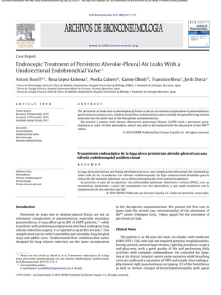
Fig. 1. Appearance of an unfolded IBV valve and endoscopic view after its placement. Alveolar-pleural fistulas are caused by lung resections, bul-
lous emphysema, advanced-stage sarcoidosis, post-radiotherapy
fibrotic changes or ablation by radiofrequency. An air leak is
hyperclarity. A pleural drain was placed, but given the persistence defined, by consensus, as being persistent if it does not stop in
of the air leak, the patient was transferred to the reference hos- 7 days. There is no physiological base to consider that this time
pital for elective surgical treatment. Two weeks later, we carried it is required in order to consider initiating therapeutic actions,
out apical bullectomy and talc pleurodesis using video-assisted while other variables such as the patient’s baseline disease, clinical
thoracoscopy, and the patient was discharged with lung reex- repercussions or magnitude of the air leak should also be consid-
pansion a week later. However, 48 h afterwards, the patient was ered. In this manner, if the debit of the leak is small or moderate,
re-hospitalized at his district hospital for right basal pneumo- a conservative approach with maintenance of the drainage tube
nia in bacterial filiation that became complicated three days later after the 7 days can be adequate. For important leaks or those
with hydropneumothorax. He was sent once again to the refer- with clinical repercussions in the patient, early surgical treatment
ence hospital, where pleuroscopy was performed with a second before the 7 days is the best option. According to current guide-
talc pleurodesis that was not effective, and at 15 days a third pleu- lines on the management of pneumothorax, when surgery is not
rodesis was done with autologous blood, with no success. Given the possible due to either advanced baseline disease, unstable clini-
persistence of the air leak, the patient was sent to our center. Upon cal situation or the refusal of the patient, alternative treatments
arrival, the conventional water-seal drainage system was with- can be contemplated, such as endoscopic interventions. However,
drawn and replaced with the ThopazTM electric aspiration system given the progressive evidence of its effectiveness, it is possible that
(Medela, Baar, Switzerland), with an observed debit of approxi- future guideline editions may even recommend endoscopic treat-
mately 330 ml/min under aspiration of −20 cm of water. The patient ment with unidirectional valves before surgery. The overall cost
was intubated with an orotracheal tube with an internal diameter of the procedure (approximately 2200 D per valve in Europe) also
of 9 mm to avoid a low tidal volume that could diminish the air favors their implantation. There are different types of endoscopic
leak. With an Olympus BF-1T180 therapeutic video-assisted bron- treatment for persistent air leak due to alveolar-pleural fistula.
choscope (Tokyo, Japan) and a number 4 Fogarty arterial balloon These include the application of adhesive substances, such as fib-
(Edwards Lifesciences LLC, Irvine, CA, USA) the lobar bronchi were rin, albumin or glutaraldehyde, other proinflammatory irritants,
sequentially occluded and, in the case of decline of the air leak, such as ethanol or antibiotics, and a third group made up of the
so were the pertaining segmental bronchi. LSD certified a signifi- implantation of endobronchial blockers, such as Watanabe spigots
cant fall in the debit during the occlusion of the anterior and apical (Novatech, Cedex, France) and more recently unidirectional flow
segments, which was mild (40 ml/min) in the posterior segment. It valves like Zephyr® by Pulmonx (Redwood City, CA, USA) and IBV®
was decided to implant valves only in the first two. After measur- by Olympus Corp (Tokyo, Japan).
ing the diameters with a previously calibrated balloon for the IBV® The Watanabe spigots are specifically designed for reducing air
system, a 7-mm diameter valve was placed in the anterior segment leaks by means of total occlusion of the affected bronchus. They
and a 6-mm valve in the apical segment (Fig. 1), to stop the air leak. are manufactured out of radiopaque silicone and are made in var-
However, a few hours later, the air leak re-initiated with a leak of ious diameters (5, 6, and 7 mm) with a length of 1 and 1.5 cm.
about 150 ml/min under continuous aspiration −20 cm of water. Watanabe et al. reached in 60 patients a resolution in 40% and a
Simple chest radiograph after the procedure with continuous aspi- reduction of the debit in 38% of the cases, with a mean of 4 spig-
ration showed a practical reexpansion of the right pneumothorax ots per patient.4 The Zephyr® and IBV® unidirectional valves allow
(Fig. 2). The patient was derived to his hospital of origin, where he for the centripetal flow of air and secretions, and were initially
was discharged with a drain tube connected to a Heimlich valve designed for lung volume reduction treatment in emphysema. After
incorporating sufficient bibliographic evidence, the Food and Drug
Administration (FDA) in the United States approved the indication
of IBV® as a treatment for air leaks in 2006. The Zephyr® valve has
an aperture in its center and works by means of a system similar
to the Heimlich valve. On the other hand, the IBV® valve allows
for the flow of air and secretions around its perimeter by a sys-
tem of flexible metallic umbrella-like rods. Travaline et al. used the
Zephyr® valves in the treatment of 40 subjects affected by persis-
tent alveolar-pleural air leak of varying etiologies (25 spontaneous,
7 post-surgery, 6 iatrogenic, 1 volume reduction surgery, 1 trau-
matic), reaching complete resolution in 47.5% of the cases and a
reduction of the leak in 45%.5 A mean of between 2 and 3 valves
were used per patient, ranging between 1 and 9 valves. Apart from
this publication, another 13 patients were reported in 6 different
articles with a high success rate. The only case published using the
IBV® valves corresponds with a patient who presented pneumoth-
24.03.10
orax and massive subcutaneous emphysema 8 days later due to
radiofrequency ablation of a local relapse after atypical resection
Fig. 2. Reexpansion of the right pneumothorax. due to squamous carcinoma three years before. The authors report
3. Document downloaded from http://www.archbronconeumol.org, day 23/10/2012. This copy is for personal use. Any transmission of this document by any media or format is strictly prohibited.
A. Rosell et al. / Arch Bronconeumol. 2011;47(7):371–373 373
a significant reduction of the leak with the implantation of two Conflict of interest
valves, measured 15 min after the procedure.6
All the endoscopic approaches require the localization of the The authors declare having no conflict of interests.
most distal bronchus possible on which the entering air flow
depends. The most widely used system is the consecutive occlu- Acknowledgement
sion of the bronchial openings from larger to smaller caliber during
a time ranging between 30 s and 3 min, depending on the authors. We would like to thank Dr. X. González for his comments.
The visualization of the variation in intensity of the bubbling of
the conventional water-seal aspiration systems is to date the pro-
cedure used to guide the pulmonologist during balloon occlusion. References
The incorporation of electrical suction systems with digital screens,
1. George RB, Herbert SJ, Shames JM, Ellithorpe DB, Weill H, Ziskind MM.
such as ThopazTM (Medela), enables the specialist to numerically Pneumothorax complicating pulmonary emphysema. JAMA. 1975;234:
check the real-time air debit, and therefore it is an advancement in 389–93.
the management of these situations. 2. Videm V, Pillgram-Larsen J, Ellingsen O, Andersen G, Ovrum E. Spontaneous
pneumothorax in chronic obstructive pulmonary disease: complications, treat-
The cases published verify two facts that indirectly reflect the ment and recurrences. Eur J Respir Dis. 1987;71:365–71.
complexity of persistent air leaks. First of all, the fact that com- 3. Ciccone AM, Meyers BF, Guthrie TJ, Davis GE, Yusen RD, Lefrak SS, et al. Long-
plete resolution of the air leak is not reached in all patients upon term outcome of bilateral lung volume reduction in 250 consecutive patients
with emphysema. J Thorac Cardiovasc Surg. 2003;125:513–25.
implantation and, second of all that the number of valves neces- 4. Watanabe Y, Matsuo K, Tamaoki A, Komoto R, Hiraki S. Bronchial occlu-
sary oscillates between 2 and 4 on an average. The model with sion with endobronchial with endobronchial Watanabe spigot. J Bronchol.
one fistulous orifice depending on one segmental bronchus would 2003;10:264–7.
5. Travaline JM, McKenna Jr RJ, De Giacomo T, Venuta F, Hazelrigg SR, Boomer M,
represent a simplistic model. The incomplete reduction of the et al. Treatment of persistent pulmonary air leaks using endobronchial valves.
air leak, either during balloon occlusion or after the implanta- Chest. 2009;136:355–60.
tion of the valves, could be explained by the intrapulmonary air 6. Abu-Hijleh M, Blundin M. Emergency use of an endobronchial one-way valve
in the management of severe air leak and massive subcutaneous emphysema.
circulation7 or the presence of multiple fistulous orifices. Com- Lung. 2010;188:253–7.
plementary explorations such as CT,8 bronchography by CT9 or 7. Fessler HE. Collateral ventilation, the bane of bronchoscopic volume reduction.
ventilation gammagraphy with Tc99m 10 can be useful for localiz- Am J Respir Crit Care Med. 2005;171:423–4.
8. Ricci ZJ, Haramati LB, Rosenbaum AT, Liebling MS. Role of computed tomogra-
ing and determining the number of fistulas. Recently, the Pulmonx
phy in guiding the management of peripheral bronchopleural fistula. J Thorac
company has made available the Chartis® system, a pneumota- Imaging. 2002;17:214–8.
chometer coupled to an endobronquial balloon, that could be a 9. Sarkar P, Patel N, Chusid J, Shah R, Talwar A. The role of computed tomogra-
useful tool for determining the existence of collateral circulation.11 phy bronchography in the management of bronchopleural fistulas. J Thorac
Imaging. 2010;25:W10–3.
The impossibility of complete occlusion of the fistula after the 10. Nielsen KR, Blake LM, Mark JB, DeCampli W, McDougall IR. Localization of
placement of the valves should not be initially considered a failure. bronchopleural fistula using ventilation scintigraphy. J Nucl Med. 1994;35:
The simple reduction of the air debit may transform an incontrol- 867–9.
11. Aljuri N, Freitag L. Validation and pilot clinical study of a new bronchoscopic
lable air leak into a situation for conservative management until it method to measure collateral ventilation prior to endobronchial lung volume
heals, as occurred in our patient as described. reduction. J Appl Physiol. 2009;106:774–83.