2. Overview of Anatomy and
Physiology
human anatomy is the scientific study of the body’s structures. Some of these
structures are very small and can only
be observed and analyzed with the assistance of a microscope. Other larger
structures can readily be seen, manipulated,
measured, and weighed. The word “anatomy” comes from a Greek root that
means “to cut apart.”
Whereas anatomy is about structure, physiology is about function. Human
physiology is the scientific study of the chemistry
and physics of the structures of the body and the ways in which they work
together to support the functions of life. Much
of the study of physiology centers on the body’s tendency toward homeostasis.
Homeostasis is the state of steady internal
conditions maintained by living things.
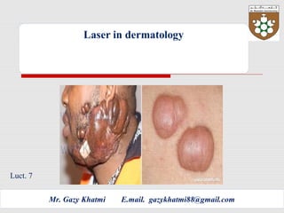
3. Dermatology is the branch of medicine dealing with the hair, nails, skin and its
diseases. It is a specialty with both medical and surgical
aspects. A dermatologist treats diseases, in the widest sense, and some cosmetic
problems of the skin, scalp, hair, and nails.
4. The Integumentary System
The integumentary system is an organ system consisting of the skin, hair, nails,
and exocrine glands. The skin is only a few millimeters thick yet is by far the
largest organ in the body. The average person’s skin weighs 10 pounds and has
a surface area of almost 20 square feet. Skin forms the body’s outer covering
and forms a barrier to protect the body from chemicals, disease, UV light, and
physical damage. Hair and nails extend from the skin to reinforce the skin and
protect it from environmental.
5. Anatomy of integumentary
system
Skin (Integument)
ƒ Consists of three major regions
ƒ Epidermis – outermost superficial
region
ƒ Dermis – middle region
ƒ Hypodermis (superficial fascia) –
deepest region
6.
7. EPIDERMIS
Epidermis
ƒThe epidermis is composed of keratinized. It is made of four or five layers of
epithelial cells, depending on its location in the body. It does not have any blood
vessels within it (i.e., it is avascular). Skin that
has four layers of cells is referred to as “thin skin.” From deep to superficial,
these layers are the stratum basale, stratum spinosum, stratum granulosum,
and stratum corneum. Most of the skin can be classified as thin skin. “Thick skin”
is found only on the palms of the hands and the soles of the feet. It has a fifth
layer, called the stratum lucidum, located between the stratum corneum and the
stratum granulosum.
The cells in all of the layers except the stratum basale are called keratinocytes. A
keratinocyte is a cell that manufactures and stores the protein keratin. Keratin is
an intracellular fibrous protein that gives hair, nails, and skin their hardness and
water-resistant properties. The keratinocytes in the stratum corneum are dead
and regularly slough away, being replaced by cells from the deeper layers
8.
9. Cells of the Epidermis
ƒ Keratinocytes – produce the fibrous protein keratin
ƒMelanocytes – produce the brown pigment melanin
Langerhans’ cells – epidermal macrophages that help
activate the immune system
ƒMerkel cells – function as touch receptors in
association with sensory nerve endings
10. Dermis
The dermis might be considered the “core” of the integumentary system (derma-
= “skin”), as distinct from the epidermis
(epi- = “upon” or “over”) and hypodermis (hypo- = “below”). It contains blood
and lymph vessels, nerves, and other
structures, such as hair follicles and sweat glands. The dermis is made of two
layers of connective tissue that compose an
interconnected mesh of elastin and collagenous fibers, produced by fibroblasts
11. Papillary Layer
The papillary layer is made of loose, areolar connective tissue, which means the
collagen and elastin fibers of this layer form a loose mesh. This superficial layer
of the dermis projects into the stratum basale of the epidermis to form finger-
like
dermal papillae (see Figure above). Within the papillary layer are fibroblasts, a
small number of fat cells (adipocytes), and an abundance of small blood vessels.
In addition, the papillary layer contains phagocytes, defensive cells that help
fight bacteria or other infections that have breached the skin. This layer also
contains lymphatic capillaries, nerve fibers, and touch receptors called the
Meissner corpuscles.
Reticular Layer
Underlying the papillary layer is the much thicker reticular layer, composed of
dense, irregular connective tissue
12. Hypodermis
Hypodermis
The hypodermis (also called the subcutaneous layer or superficial fascia) is a
layer directly below the dermis and serves
to connect the skin to the underlying fascia (fibrous tissue) of the bones and
muscles. It is not strictly a part of the skin,
although the border between the hypodermis and dermis can be difficult to
distinguish.
13. Pigmentation
The color of skin is influenced by a number of pigments, including
melanin, carotene, and hemoglobin. Recall that melanin
is produced by cells called melanocytes, which are found scattered
throughout the stratum basale of the epidermis. The
melanin is transferred into the keratinocytes via a cellular vesicle called
a melanosome (Figure 5.8).
14. Skin Color
ƒ Three pigments contribute to skin color
ƒ Melanin – yellow to reddish-brown to black
pigment, responsible for dark skin colors
ƒ Freckles and pigmented moles – result from local
accumulations of melanin
ƒ Carotene – yellow to orange pigment, most
obvious in the palms and soles of the feet
ƒ Hemoglobin – reddish pigment responsible for the
pinkish hue of the skin
15. Hair
Hair is a keratinous filament growing
out of the epidermis. It is primarily
made of dead, keratinized cells.
Strands of hair
originate in an epidermal penetration
of the dermis called the hair follicle.
16.
17.
18. Photobiological Effect
The overriding beneficial effect of laser energy is absorption of the light by the
target tissue and the transfer of laser energy, thus causing a tissue interaction
(Photobiological Effect). There are four basic interactions that can occur following
absorption of laser energy:
(1) Photochemical (Photochemolysis): certain wavelengths of laser light are
absorbed by naturally occurring chromophores or wavelength- specific light
absorbing substances that are able to induce certain biochemical reactions at
cellular level. Derivatives of naturally occurring chromophores or dyes have been
used as photosensitizers to induce biological reactions within tissues for both
diagnostic and therapeutic applications. Photochemical interactions include
photobiostimulation, photodynamic therapy, and tissue fluorescence. Certain
biological pigments, upon absorbing laser light, can fluoresce, which can be used
for detecting teeth caries. Lasers can also be used in a non- surgical mode for
biostimulation or more rapid wound healing, pain relief, increased collagen
growth and a general anti- inflammatory effect.
20. (2) Photothermal
(Photothermolysis):
light energy absorbed by the tissues is transformed into heat energy which then
produces tissue effects as follows:
Coagulation and haemostasis: from 60o
C to 70o
C, this is the secondary effects
through conduction of the heat generated.
Photopyrolysis: from 75o
C to 90o
C, target tissue proteins undergo permanent
morphological change (protein denaturation) as result of dissociation of covalent
bonds.
Photovaporolysis: at 100o
C +, inter- and intra-cellular water in soft tissue and
interstitial water in hard tissue is vaporised. This destructive phase transfer
results in expansive volume change, which can aid the ablative effect of the laser
by dissociating large tissue elements. This will be carried onto a further phase:
transfer to hydrocarbon gases and production of residual carbon (carbonization).4
The amount of laser energy absorbed by the tissue largely determines the
thermal interaction produced and is in turn dependant on the wavelength of the
laser light to a great degree, but also on other parameters such as spot size,
power density, pulse duration and frequency, and the optical properties and
composition of the tissue irradiated. The CO2(10600nm) is highly absorbed by the
water content of oral soft tissues, whereby 90% of the energy is absorbed within
the first 100 microns of penetrating the tissue surface5
. Hence, even at relatively
low power densities using a focused beam, there is rapid tissue vaporization of
the water with charring and burning of the organic content of the tissue.
21. Photothermal interaction causes the irradiated target tissue to absorb the laser
energy and converts it into heat, thereby producing a direct temperature rise in
the irradiated tissue volume. When this energy is applied for long enough, heat
conduction will cause a temperature rise in surrounding tissues as well. Hence,
thermal effects, such as coagulation necrosis, are produced indirectly in
collateral areas and are one of the mechanisms responsible for haemostasis
when cutting or vaporizing with a laser.
22. (4) Photomechanical and
photoelectrical:
These are non- thermal interactions produced by high energy, short pulsed laser
light, including: photodisruption, , photoplasmolysis and photoacoustic
interaction. Absorption of laser energy pulses results in rapid expansion or
generation of shock waves that are capable of rupturing intermolecular and
atomic bonds (photo-disruption ). Thus, the laser beam's energy is transformed
into vibration or kinetic energy. A pulse of laser energy on hard dentinal tissues
can produce a shock wave, which might explode the tissue. This is an example of
the photoacoustic effect of laserlight.12
23. Contact vs non-contact modes
Laser light will undergo some divergence on exit from a quartz fibre delivery
system and most non-fibre systems (hollow waveguide and articulated arm) use
a focusing lens. Consequently, the 'spot size' of the beam, relative to the target
tissue, will determine the concentration of laser energy – fluence and power
density – being delivered over an area.11
The spot size will change with distance
for any delivery system – it will increase with distance for a fibre-optic delivered
beam and change relative to the focal length of the lens in those delivery
systems where a focusing hand-piece is used
24. Acne
What is acne?
.
Acne is a skin condition that occurs when your hair follicles become plugged with oil
and dead skin cells. Acne usually appears on your face, neck, chest, back and
shoulders. Effective treatments are available, but acne can be persistent. The
pimples and bumps heal slowly, and when one begins to go away, others seem
to crop up.
Acne is most common among teenagers, with a reported prevalence of 70 to 87
percent. Increasingly, younger children are getting acne as well.
25.
26. What causes acne?
The sebaceous (oil-producing) glands of people who get acne are particularly sensitive to normal blood
levels of a hormone called testosterone, which is present in both men and women. This causes the
glands to produce an excess of oil. At the same time, the dead skin cells lining the pores are not shed
properly and clog up the follicles. These two effects result in a build-up of oil producing blackheads
(where a darkened plug of oil is visible) and whiteheads.
The acne bacterium (known as Propionibacterium acnes) lives on everyone’s skin, usually causing no
problems, but, in those prone to acne, the build up of oil creates an ideal environment in which these
bacteria can multiply. This triggers inflammation and the formation of red or pus-filled spots.
Is acne hereditary?
Acne can run in families, but this does not necessarily mean that if your parents had acne you will get
it too.
27. With respect to acne, Er:Yag lasers are used almost exclusively to treat acne
scars. Because the Er:YAG does not penetrate as deeply into the skin as a CO2
laser, they are used primarily for superficial skin resurfacing procedures. Er:YAG
laser therapy can be performed at low power, non-ablative and minimally
ablative settings to mildly improve the appearance of damaged skin. Low power
Er:YAG treatments generally have a relatively short recovery period and mild
side effects. However, they are less effective than ablative treatments for
significant acne scarring.
Erbium:YAG laser :
2940 nM
IR
Target chromophore: water
Superficial ablative resurfacing
28. keloid
What is a keloid?
When a wound heals, it leaves a scar. A keloid is a special type of scar: one that grows too much and
can even become larger than the original wound. It is not uncommon for surgical or injury scars to
become a little lumpy (hypertrophic). A keloid differs from these in several ways:
A keloid can come up after very minor skin damage, such as an acne spot, or even if there has been no
obvious damage to the skin at all.
It can spread outside the original area of skin damage.
It may last for many years.
Are keloids hereditary?
They can be - a tendency to get keloids certainly runs in some families.
29.
30. hirsutism
What is hirsutism?
Hirsutism is the term used when a woman grows too much body or facial hair in a pattern seen
normally occurring only in men.
What causes hirsutism?
Androgens are often thought of as 'male hormones' but, in fact, both men and women produce them -
men usually in greater amounts than women. Testosterone is the best-known androgen, but there are
several others too. Hirsutism can be caused either by abnormally high levels of androgens, or by the
hair follicles being more sensitive than usual to normal androgen levels.
31.
32. Psoriasis
What is psoriasis?
Psoriasis is a common skin problem
affecting about 2% of the population. It
occurs equally in men and women, at any
age, and tends to come and go
unpredictably. It is not infectious, and does
not scar the skin.
33.
34. What causes psoriasis?
The skin is a complex organ made up of several different layers.
The outer layer of skin (the epidermis) contains cells which are
formed at the bottom and then move up towards the surface,
gradually changing as they go, finally dying before they are shed
from the surface. This journey normally takes between 3 and 4
weeks. In psoriasis, the rate of turnover is dramatically increased
within the affected skin, so that cells are formed and shed in as
little as 3 or 4 days. The reasons for this are still not fully
understood.
Some people are more likely to develop psoriasis than others,
particularly if there is someone else in their family who has
psoriasis: in other words, it is a genetic or hereditary disease .
However, the trigger for psoriasis to appear is often an outside
event, such as a throat infection, stress or an injury to the skin.