2. Cells 1600’s: Robert Hooke The Cell Theory All living things are made up of cells The cell is the basic unit of structure & function of all living things All cells come from pre-existing cells
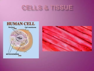
3. Review of Cells C, O, H, N Interstitial fluid Made from blood All exchgsbt cells & blood made thru this fluid Nucleus Plasma membrane Cytoplasm
5. Cell Diversity Cells that: Connect body parts Fibroblast Elongated Lies along the cable-like fibers that it secretes Has lots of RER & GA to make & secrete protein bldg blocks of these fibers Erthrocyte (RBC) Carries O2 Concave disc=↑ SA Streamlined = flows easily So much O-carrying pigment is packed in that all other organelles have been removed to make room
6. Cells that: Covers & lines organs Epithelial cells Hexagonal Pack together well Many intermediat filaments wc resist tearing when rubbed/pulled Move organs & body parts Skeletal muscle & smooth muscle cells Elongated Filled w/ contractile filaments Shorten Move bones Change size of organs
7. Cells that: Store nutrients Fat cells Large & round From lg lipid droplet in cytoplasm Fight disease Macrophage (phagocytic cell) Long pseudopods sent out to infection Has many lysosomeswc digest infectious microorgs
8. Cells that: Gather info & controls body functions Nerve cell Very long Receives & transmits to other parts of body Covered w/ lg plasma membr Many rER to make parts of membr Are for reproduction Oocyte (f) Lgst cell Has many copies of all organells to distribute to daughter cells Sperm (m) Long, streamlined for “swimming” Flagellum to propel
9. Body Tissues Epithelial Tissue (epithelium) lining, covering & glandular tissue Glandular epithelium forms glands Lining & covering epith covers all free body surfaces Contains versatile cells intestine
10. Can form outer layer of skin Almost all subs that body give/receives must go thru epith Can dip in to line cavities Functions: Protection Absoption Filtration Secretion kidney
11. Special Characteristics Fit close together, forming sheets Bound together at many pts by cell junctions (desmosomes & tight junctions) Has an apical surface (unattached) Slick, smooth, microvilli, cilia Has a basement membr (lower surface) Secreted by both epithel cells & connective tissue cells next to it Avascular (lack own blood supply) Depends on diffusion from capillaries for nutrients & O2 Easily self regenerates
12. Classification of Epithelium 2 names First: = number of cell layers it has Simple epith= one layer Stratified epith=more than one cell layer Second = describe the shape of its cells Squamous = flat Cuboidal = cube Columnar = columns Stratified epith named for apical surface
13. Simple Epithelia Thin layer ≠ protection =absorption, secretion, filtration Simple Squamous Rests on basement layer Air sacs Walls of capillaries Simple Cuboidal Rests on basement layer In glands and ducts Salivary glands, pancreas, walls of kidney tubules, surface of ovaries Simple Columnar Tall Goblet cells Product lubricating mucus Entire length of digestive tract (stomach to anus) Line body cavities open to body exterior = mucosae/mucus membranes
14. Pseudostratified columnar Rests on basement membr BUT: some cells shorter than others Nuclei at diff heights False impression that it is stratified Absorption/secretion Ciliated variety = lines resp tract Goblet cells traps dust/debris
15. Stratified Epithelia More durable = protection Stratified Squamous Most common stratepith Apical = squamous Basement memb = cub or col Subject to abuse/friction Esophagus, mouth, outer skin
16. Stratified Cuboidal Usually just 2 layers At least apical cells cub Stratified Columnar Apical cells col Basal cells vary in size & shape Both fairly rare Mainly in ducts of lg glands
17. Transitional Epithelium Highly modified stratified squamousepith Forms lining of only few organs Urinary bladder, ureters & parts of urethra Subject to considerable stretching Basal layer cub or col Apical cells vary Not Stretched = membr many layered, surface cells rounded/domed Stretched = (w/urine) epith thins, surface cells flatten & become squamous-like Allows cells to slip past ea other & chg shape (=transition)
18. Glandular Epithelium Gland=make & secrete a specific product = secretion 2 Major Types of Glands Endocrine glands Lose connection to surface (duct) = ductless All hormones Secretions diffuse directly into blood vessels Thyroid, adrenals & pituitary
19. Exocrine Glands Have ducts where secretion occurs to surface Sweat & oil glands, liver & pancreas Both internal & external
20. Connective Tissue Connects body parts Everywhere, most abundant & distributed tissue Protecting, supporting & binding together other body tissues
21. Characteristics Variations in blood supply Most well vascularized Tendons & ligaments = poor blood supply Cartilages = avascular = slow to heal Would you rather break a bone or tear a ligament?
22. Extracellular Matrix made of many diff types of cells & varying amts of nonliving substance outside the cells = extracellular matrix
23. Extracellular Matrix Made by CT then secreted to outside 2 main elements Structureless ground substance fibers Ground substance = mostly water plus adhesion proteins (“glue”) and lg charged polysaccharide molecule (will trap water as they intertwine) Matrix can be very fluid, gel-like, firm, rock hard
24. Fibers = various types & amts Include: collagen = white, hi tensile strength Elastic = yellow Reticular = fine collagen fibers wc form internal “skeleton” of soft organs (spleen)
25. Due to the extracellular matrix, connective tissue is able to form soft packaging tissue around organs bear weight w/stand stretching w/stand abuse/abrasion w/c other tissues couldn’t
26. Variations of connective tissue One extreme to another Fat tissue = many cells, matrix is soft Bone & cartilage = few cells, very hard matrix (strong)
27. Types of Connective Tissue Bone (osseous) = bone cells in lacunae (pits) surrounded by very hard matrix matrix contains Ca salts & lg amts of collagen fibers Excellent protection & support
28. Cartilage =less hard, more flexible only in a few places most common = hyaline cartilage (glassy) Many collagen fibers in rubbery matrix Forms supporting structures Larynx Ribs to breastbone Ends of bones at joints Fetus = mostly hyaline, replaced by bone by birth Fibrocartilage Highly compressible Disks bt vertebrae Elastic cartilage ear
29. Dense Connective Tissue Main matrix element = collagen fibers (in rows w/fibroblasts) Forms strong rope-like structures Tendons (m2b) Ligaments (b2b) More elastic/stretchy Lower layer of skin (dermis) = in sheets
30. Loose CT Softer More cells, fewer fibers (except blood) Areolar tissue Soft/cobwebby Cushions/protects organs ‘glue’ wc holds organs in their positions Fluid matrix w/all types of fibers Appears to be empty space A resevoir of water & salts for all other cells to get nutrients and dispose of “wastes” Adipose tissue Reticular CT
31. Adipose tissue Fat Mostly oil… pushes nucleus to one side Beneath skin Insulates/protects Kidneys surrounded Cushions eye in socket For fuel if needed Reticular CT Interwoven reticular fibers Limited sites: stroma to support free blood cells in lymph nodes, spleen, bone marrow
32. Loose connective tissue types Reticular connective tissue Delicate network of interwoven fibers Forms stroma (internal supporting network) of lymphoid organs Lymph nodes Spleen Bone marrow
33. Blood (vascular tissue) Blood cells surrounded by fluid matrix called blood plasma Fibers are visible during clotting Functions as the transport vehicle for materials
34. Muscle Tissue Function is to produce movement Three types Skeletal muscle Cardiac muscle Smooth muscle
35. Muscle Tissue Types Skeletal muscle Under voluntary control Contracts to pull on bones or skin Produces gross body movements or facial expressions Characteristics of skeletal muscle cells Striated Multinucleate (more than one nucleus) Long, cylindrical
36. Cardiac muscle Under involuntary control Found only in the heart Function is to pump blood Characteristics of cardiac muscle cells Cells are attached to other cardiac muscle cells at intercalated disks Striated One nucleus per cell
37. Smooth muscle Under involuntary muscle Found in walls of hollow organs such as stomach, uterus, and blood vessels Characteristics of smooth muscle cells No visible striations One nucleus per cell Spindle-shaped cells
38. Nervous Tissue Composed of neurons and nerve support cells Function is to send impulses to other areas of the body Irritability Conductivity
39. Tissue Repair (Wound Healing) Regeneration Replacement of destroyed tissue by the same kind of cells Fibrosis Repair by dense (fibrous) connective tissue (scar tissue) Determination of method Type of tissue damaged Severity of the injury
40. Events in Tissue Repair Capillaries become very permeable Introduce clotting proteins A clot walls off the injured area Formation of granulation tissue Growth of new capillaries Rebuild collagen fibers Regeneration of surface epithelium Scab detaches
41. Regeneration of Tissues Tissues that regenerate easily Epithelial tissue (skin and mucous membranes) Fibrous connective tissues and bone Tissues that regenerate poorly Skeletal muscle Tissues that are replaced largely with scar tissue Cardiac muscle Nervous tissue within the brain and spinal cord
42. Developmental Aspects of Tissue Epithelial tissue arises from all three primary germ layers Muscle and connective tissue arise from the mesoderm Nervous tissue arises from the ectoderm With old age, there is a decrease in mass and viability in most tissues