SlideShare une entreprise Scribd logo
1  sur  181
STAYING HEALTHY
1.    Describe the function of blood including transport of oxygen, carbon dioxide, digested food, waste
      products, hormones, antibodies and heat.
2.    Name the parts of the blood and the function of each part
3.    Describe the structure of each blood cell type
4.    Describe the function of the heart as a “muscular pump that maintains the flow of blood around the
      body.
5.    Identify and label the parts of the heart and describe the function of each part.
6.    Draw a diagram to show the movement of blood through the heart. Describe reason for double
      system
7.    Name the major blood vessels entering and leaving the heart and describe where they come from/
      go to
8.    Describe the structure and function of the three types of blood vessels found in the body
9.    Define your pulse as “a wave of blood passing along an artery” and explain where it can be felt in
      the body
10.   Describe factors that affect heart/pulse rate
11.   Describe the causes and symptoms of some heart and circulatory diseases - examples include
      atherosclerosis, coronary heart disease, high blood pressure and varicose veins.
12.   Label the parts of the breathing system and explain the function of each part.
13.   Describe the role of the diaphragm and intercostal muscles in breathing.
14.   Explain the effect of exercise on breathing rate.
15.   Describe and define the difference between breathing, gas exchange and respiration
16.   Explain how to measure lung capacity and relate this to good respiratory function.


Tuesday, 27 April 2010
Living World (level 5)
• Life processes LW pro: Identify the key structural features and functions involved in the life processes of plants and animals. Describe
   the organisation of life at a cellular level.
• Ecology LW eco: Investigate the interdependence of living things (including humans) in an ecosystem.
• Evolution LW evo: Describe the basic processes by which genetic information is passed from one generation to the next.


      Specific Learning Outcome                                   Suggested Learning Activities                                  References

1.   Name several                 • Read about the function of blood and discuss the function of blood                 Year 10 Study Guide
     functions of blood               emphasising the need for cells to be supplied with nutrients and oxygen and
                                      have the wastes and carbon dioxide removed.


2.   List the components          •   Students view cows blood under the microscope and draw the red blood cells.      Slide 9 - 4 Sci2 p67
     of blood and their           •   Students view Platelets, red and white blood cells on the projector screen and
     functions                        describe their appearance. As an exercise they can compare and contrast the      Slides 10 to 12
                                      appearance of these structures


3.   Compare the                  • Students dissect and view cross-sections of veins using the binocular
     differences in                   microscopes. They compare and contrast these vessels
     structure and                •   Cut and paste diagrams and complete close exercise on slide.                     Hipkins and Connor
     function of blood                                                                                                 Slide 16
     vessels


4.   Define ‘pulse’ and           •    Listen to a normal heart beat on the internet                                   http://www.med.ucla.edu/
     give examples of                                                                                                  wilkes/intro.html
     where it can be
     located
                                  •    Take both the radial and the carotid pulse and compare the pulse rate of
                                       boys and girls.
                                  •    Discuss the difference between pulse rates of boys and girls and other
                                       features of heart rate.


5.   Label a simple               •   Cut and paste a simple diagram of the human circulatory system into books. Year 10 Study Guide
     circulatory system           •   Use slide 32 to describe the function of the circulatory system in terms of the
                                      major structures.                                                               Slide 32


6.   Describe the function        •   Students work through the worksheet, “The heart of the matter”
     of the parts of the          •   Complete the cloze exercise summary
     heart


Tuesday, 27 April 2010
7.   Show the               •   With reference to a detailed OHT heart structure, students label the-se Slide 24 and 27 (OHT)
     circulation of blood       cross ction that is projected on the whiteboard
     through the heart      •   Students dissect a sheep’s heart and identify the relevant structures
     and name the
     major blood vessels
     and their function


8.   Define                 •   Brainstorm factors that students think will lead to coronary artery     Reading Sheet: “What
     artherosclerosis           damage.                                                                 is atherosclerosis”
                            •   Read the handout sheet “What is atherosclerosis” and copy and
                                complete the notes as a cloze exercise.


9.   Investigate factors    • Read the sheet “Risk factors for heart disease”                           Reading sheet “Risk
     that affect coronary   • Complete the mind map on slide 35                                         factors for heart
     arteries of the        • Answer the questions on slide 36                                          disease”
     heart and lead to                                                                                  Slides 35 & 36
     heart disease


10. Identify factors that   •   Students read the sheet on training and pulse rate. They form buzz      refer to sheet from
    affect resting heart        groups and discuss the questions handed out. Each group nominates a     “Training, lactate &
    rate                        recorder and a reporter and after the discussion their answers are      pulse rate” from Peter
                                reported back to the class one group at a time.                         Janssen
                            •   Use a datalogger to monitor resting heart rate. Students volunteer to
                                be subjects.
                            •   Complete the cloze summary on resting heart rate.                       Slide 48


11. Describe how            •   Students use a heart rate monitor to record heart rate with time as
    exercise affects the        they exercise. This is plotted on a set of axes.
    heart rate and          •   Exercise on Peter’s pulse                                               Slides 49 & 50
    explain the purpose     •   Compare fitness of two individuals                                      Slide 51
    of a raised heart
    rate during exercise


Tuesday, 27 April 2010
12. Label parts of the     •   Choose from a list of labels to complete the labelled diagram of the      Slide 53 to be re-
    breathing system           breathing system. Labels match structure and function                     developed.
    and identify the
    function of each
    part


13. Describe the           •   Teacher demonstration: Bell jar breathing model                           Slide 53 to be re-
    breathing process      •   Students explain why the balloons inflate and deflate                     developed.
                           •   Cut and paste diagrams which summarise the breathing process.             Bell jar
                           •   Discuss the difference between breathing, gas exchange and                breathing model.
                               respiration.


14. Describe how           • Record breathing rate before and after exercise.                            Slide 57
    exercise affects the   • Discuss reasons for breathing rate needing to increase
    breathing rate and
    explain the purpose
    of an elevated
    breathing rate
    during exercise

15. Determine a profile    • Students work their way around a circus of activities that involves them
    of health and              taking measurements of each others resting ECG, Repiration rate
    fitness that has an        (before and after exercise), Heart rate (before, after and during
    emphasis on                exercise), lung capacity plus peak flow and blood pressure.
    cardiovascular
    fitness.

16. Define respiration     •   Discuss respiration. Stress that it is a cellular process and using simple Slide 58
    and write an               ideas about cell structure explain the chemical change that takes
    equation for this          place.
    reaction


Tuesday, 27 April 2010
INTRO
Tuesday, 27 April 2010
Word search                          KEY WORDS

                         Y   N   I   P   N   O   T   T   S   Y   E   R   C   W   N
     plasma              R   B   O   H   O   C   R   U   R   Z   R   W   I   O   G
     platelets
     pulse               A   R   K   I   I   L   K   A   N   A   Z   E   I   E   P
     aorta
     atrium              L   O   R   E   T   S   E   L   O   H   C   T   T   U   R
     artery              L   N   R   P   A   A   P   A   C   Q   A   H   L   R   C
     ventricle
     pulmonary           I   C   L   T   L   L   R   A   I   L   C   M   E   X   A
     vena cava
     cholesterol
                         P   H   Y   O   A   A   V   I   A   C   O   J   V   A   J
     inhalation          A   I   R   S   H   A   T   H   P   N   X   A   E   R   R
     exhalation
     trachea             C   K   M   Y   N   G   X   E   A   S   U   N   N   O   K
     bronchi
     bronchiole
                         V   A   Q   N   I   E   V   R   L   Y   E   S   T   H   M
     alveolus            M   U   I   R   T   A   Y   L   J   E   C   R   R   T   J
     diaphragm
     thorax              M   G   A   R   H   P   A   I   D   V   T   S   I   W   T
     vein                B   R   O   N   C   H   I   O   L   E   E   S   C   F   W
     capillary
     respiration         A   L   V   E   O   L   U   S   S   N   P   U   L   S   E
                         U   C   H   K   C   O   E   D   Z   A   P   E   E   F   W
Tuesday, 27 April 2010
AEROBIC RESPIRATION (air-o-bick res-purr-ay-shun) – respiration that use oxygen.
  ALVEOLI (al-vee-oh-lie) – air sacs in the lungs where gas exchange occurs.
  ANAEROBIC RESPIRATION (an-air-o-bick res-purr-ay-shun) – respiration that does not use oxygen.
  AORTA (ay-ort-ar) – major artery that carries blood from the heart to the body.
  ARTERIOSCLEROSIS (are-tear-ee-oh-skleer-oh-sis) – hardening of the arteries.
  ARTERY (are-ter-ee) – blood vessels that carry blood away from the heart.
  ATRIUM (ay-tree-um) – one of two chambers in the heart that collect blood.
  BREATHING (bree-thing) – the physical movement of air in and out of the lungs.
  BRONCHI (bronk-eye)– two tubes that connect the trachea to the lungs.
  BRONCHIOLES (bronk-ee-oles) – narrow tubes in the lungs that branch off from the bronchi.
  CAPILLARY (ka-pill-are-ee) – the smallest blood vessel with walls only one cell thick.
  CILIA (sill-ee-a) – tiny hairs present on the surface of cells lining the airways.
  CIRCULATORY SYSTEM (sir-kew-lar-tory sis-tim) – the blood, blood vessels and heart.
  DEOXYGENATED (dee-ox-I-gin-ay-ted) – without oxygen.
  DIAPHRAGM (dye-a-fram) – sheet of muscle separating the chest area from the abdomen that has an important role in breathing.
  EXHALE (ex-hail) – to breathe air out of the body.
  GAS EXCHANGE (gass ex-change) – the exchange that occurs in the lungs of oxygen for carbon dioxide.
  HAEMOGLOBIN(he-mow-glow-bin) – a protein in red blood cells that carries oxygen.
  HEART (hart) – a muscle that pumps blood around the body.
  INHALE (in-hay-l) – to breathe air into the body.
  LARYNX (lar-inx) – voice box.
  OXYGENATED (ox-i-gin) – containing oxygen.
  PLASMA (plaz-ma) – pale yellow liquid part of blood.
  PLATELETS (plate-lets) – a cell in blood involved in blood clotting.
  PULMONARY (pull-mun-airy) – refers to the lungs.
  PULSE (pole-z) – expanding and contracting of arteries as a wave of blood passes.
  RED BLOOD CELLS (red blud cells) – cells that carry oxygen around the body.
  RESPIRATION (res-purr-ay-shun) – a chemical process that occurs in cells and releases energy.
  RESPIRATORY SYSTEM (res-purr-a-tory sis-tim) – the organs involved in getting air into the body and exchanging oxygen for carbon dioxide.
  TRACHEA (tray-key-a) – the wind pipe.
  VALVES (valve-z) – flaps in a blood vessel that only allow blood to go one way.
  VEIN (vay-n) – blood vessels that return blood to the heart.
  VENA CAVA (vee-na car-va) – vein that carries blood that is returning from the body to the heart
  VENTRICLE (ven-trick-kill) – one of two chambers in the heart that pumps blood.
  WHITE BLOOD CELLS (why-te blud cells)– cells involved in defence against disease-causing organisms.
Tuesday, 27 April 2010
Write the answers only in the back of your book

            Term         Definition           GLOSSARY 1      Match the term with its definition

A. Aerobic respiration 1.    two tubes that connect the trachea to the lungs
B. Alveoli               2. hardening of the arteries
C. Anaerobic
                         3. without oxygen
   respiration
D. Aorta                 4. narrow tubes in the lungs that branch off from the bronchi.
E. Arteriosclerosis      5. to breathe air out of the body
F. Artery                6. the physical movement of air in and out of the lungs
G. Atrium                7. major artery that carries blood from the heart to the body.
                         8. sheet of muscle separating the chest area from the
H. Breathing
                            abdomen that has an important role in breathing
I. Bronchi               9. respiration that use oxygen.
J. Bronchioles           10. one of two chambers in the heart that collect blood
K. Capillary             11. blood vessels that carry blood away from the heart.
L. Cilia                 12. air sacs in the lungs where gas exchange occurs
M. Circulatory system 13. respiration that does not use oxygen
N. Deoxygenated          14. the smallest blood vessel with walls only one cell thick
O. Diaphragm             15. the blood, blood vessels and heart
P. Exhale                16. tiny hairs present on the surface of cells lining the airways
Tuesday, 27 April 2010
Term          Definition               GLOSSARY 1
Aerobic respiration respiration that use oxygen.
Alveoli                  air sacs in the lungs where gas exchange occurs
Anaerobic
                         respiration that does not use oxygen
respiration
Aorta                    major artery that carries blood from the heart to the body.
Arteriosclerosis         hardening of the arteries
Artery                   blood vessels that carry blood away from the heart.
Atrium                   one of two chambers in the heart that collect blood
Breathing                the physical movement of air in and out of the lungs
Bronchi                  two tubes that connect the trachea to the lungs
Bronchioles              narrow tubes in the lungs that branch off from the bronchi.
Capillary                the smallest blood vessel with walls only one cell thick
Cilia                    tiny hairs present on the surface of cells lining the airways
Circulatory system the blood, blood vessels and heart
Deoxygenated             without oxygen
                         sheet of muscle separating the chest area from the abdomen
Diaphragm
                         that has an important role in breathing
Exhale                   to breathe air out of the body

Tuesday, 27 April 2010
Term          Definition                 GLOSSARY 2             Match the term with its definition

A.    Gas exchange         1.   containing oxygen

B.    Haemoglobin          2.   a cell in blood involved in blood clotting

C.    Heart                3.   the exchange that occurs in the lungs of oxygen for carbon dioxide

D.    Inhale               4.   cells that carry oxygen around the body.

E.    Larynx               5.   pale yellow liquid part of blood

F.    Oxygenated           6.   voice box

G.    Plasma               7.   a muscle that pumps blood around the body

H.    Platelets            8.   a protein in red blood cells that carries oxygen

I.    Pulmonary            9.   to breathe air into the body

J.    Pulse                10. flaps in a blood vessel that only allow blood to go one way.

K.    Red blood cells      11. refers to the lungs

L.    Respiration          12. blood vessels that return blood to the heart
M.    Respiratory system   13. expanding and contracting of arteries as a wave of blood passes
                           14. the organs involved in getting air into the body and exchanging oxygen for
N.    trachea
                               carbon dioxide
O.    valves               15. the wind pipe

P.    vein                 16. one of two chambers in the heart that pumps blood

Q.    vena cava            17. cells involved in defence against disease-causing organisms

R.    ventricle            18. a chemical process that occurs in cells and releases energy

S.    white blood cells    19. vein that carries blood that is returning from the body to the heart

Tuesday, 27 April 2010
Term          Definition                         GLOSSARY 2
Gas exchange             the exchange that occurs in the lungs of oxygen for carbon dioxide

Haemoglobin              a protein in red blood cells that carries oxygen

Heart                    a muscle that pumps blood around the body

Inhale                   to breathe air into the body

Larynx                   voice box

Oxygenated               containing oxygen

Plasma                   pale yellow liquid part of blood

Platelets                a cell in blood involved in blood clotting

Pulmonary                refers to the lungs

Pulse                    expanding and contracting of arteries as a wave of blood passes

Red blood cells          cells that carry oxygen around the body.

Respiration              a chemical process that occurs in cells and releases energy
                         the organs involved in getting air into the body and exchanging oxygen for carbon
Respiratory system       dioxide
trachea                  the wind pipe

valves                   flaps in a blood vessel that only allow blood to go one way.

vein                     blood vessels that return blood to the heart

vena cava                vein that carries blood that is returning from the body to the heart

ventricle                one of two chambers in the heart that pumps blood

white blood cells        cells involved in defence against disease-causing organisms
Tuesday, 27 April 2010
Back of book                   WHAT DO I ALREADY KNOW?               Label the diagrams

         (a)                        (b)                  (c)               (d)


                                                               (g)
                                             (f)


                         (e)


   GUT, HEART, STOMACH, LIVER, LUNG
   PANCREAS, KIDNEY

Tuesday, 27 April 2010
LOCATION OF THE BODY’S ORGANS


     1. Draw the outline
     2. Draw in where you think the organs (from the previous slide) are.
     (Use the pictures on the previous slide to show you the shape of each organ


     Check your work with the teacher when you think you have finished


Tuesday, 27 April 2010
BLOOD -
FUNCTION
Tuesday, 27 April 2010
1. DESCRIBE THE FUNCTION OF BLOOD ...
                                    INTERESTING FACTS

            COPY & ANSWER THE QUESTIONS (BELOW) INTO YOUR EXERCISE
            BOOKS
            USE A COMPUTER TO RESEARCH ANSWERS
            Start in class ---> Finish for homework
1. What volume of blood is contained in the body of an average adult male?    5L
2. At what rate is this blood circulated through the heart? 5 L min-1

3. What effect does smoking have on the way our blood Narrows the blood vessels
                                                          in legs and arms (mainly)
   circulates through our body?
                                                            High blood pressure.
4. Name one blood-related disease and describe it briefly.
                                                            Leukemia, Clotting
                                                            disorders (haemophilia),
 Copy and complete the notes
                                                            Anaemia.
  Blood is important because it carries substances around the body.
   • Heat is carried to the skin.
   • Wastes to the        kidneys

   • Carbon dioxide to the     lungs

   • Food from the       gut & stomach   to the        cells

   • Hormones from the       glands       to the   body organs
Tuesday, 27 April 2010
BLOOD -
          BITS
Tuesday, 27 April 2010
Copy the notes (below) and use computers to help you to complete them


                                 Mainly water (50%)

                                                                      Straw
                                 4%                                   colored


                                 1% other


                                 White blood cells - fight


                                  Platelets -


                                 Red blood cells - carry
                                                   around the body

Tuesday, 27 April 2010
LOOKING AT BLOOD CELLS
       Practical                                                        Do not copy this

        Copy the aim and draw the method then follow this instruction


                                                                          Practical: 4Sci2 p67


     Also, look at the OHT of red blood cells set up on the projector
Tuesday, 27 April 2010
IMAGES
Tuesday, 27 April 2010
WHAT YOU MIGHT SEE, 500 X MAGNIFIED

                                      1000 x MAGNIFIED


Tuesday, 27 April 2010
SICKLE CELL            NORMAL CELL


Tuesday, 27 April 2010
Red blood cell


                         White blood cell


Tuesday, 27 April 2010
WHERE DID I COME FROM?


Tuesday, 27 April 2010
Extra for Experts   HOW A CLOT FORMS

                                     1. Study the diagram (left and below)
                                     2. For homework find out what fibrin is
                                        and write the definition in the space
                                        below.
                                     3. Research how aspirin works and
                                        what it is used for and write a brief
                                        paragraph to summarize your
                                        findings

                                     Fibrin is
                                     ____________________________
                                     ____________________________
                                     ____________________________
                                     ____________________________
                                     ____________________________
                                     Aspirin is used for people who have
                                     had a __________ attack. It works
                                     by __________________________
                                     ____________________________
                                     ____________________________
                                     ____________________________
                                     ____________________________
                                     ____________________________
                                     ____________________________
                                     ____________________________
                                     ____________________________
Tuesday, 27 April 2010
BLOOD BITS - MIX & MATCH


                                                    Platelets

                                                    Cut out the pictures,
 Red blood cells          White blood cells         descriptions & functions


Tuesday, 27 April 2010
BLOOD BITS - MIX & MATCH


                                                    Platelets

                                                    Cut out the pictures,
 Red blood cells          White blood cells         descriptions & functions

 Donut shaped
 (discs which have a hollow on each
 side)
 - this increases surface area


Tuesday, 27 April 2010
BLOOD BITS - MIX & MATCH


                                                        Platelets

                                                        Cut out the pictures,
 Red blood cells               White blood cells        descriptions & functions

 Donut shaped
 (discs which have a hollow on each
 side)
 - this increases surface area


                         Have no nucleus


Tuesday, 27 April 2010
BLOOD BITS - MIX & MATCH


                                                         Platelets

                                                         Cut out the pictures,
 Red blood cells                White blood cells        descriptions & functions

 Donut shaped
 (discs which have a hollow on each
 side)
 - this increases surface area


                         Red in colour

                         Have no nucleus


Tuesday, 27 April 2010
BLOOD BITS - MIX & MATCH


                                                          Platelets

                                                         Cut out the pictures,
 Red blood cells                White blood cells        descriptions & functions

 Donut shaped
                                                Granular (rough) in appearance
 (discs which have a hollow on each
 side)
 - this increases surface area


                         Red in colour

                         Have no nucleus


Tuesday, 27 April 2010
BLOOD BITS - MIX & MATCH


                                                              Platelets

                                                              Cut out the pictures,
 Red blood cells                White blood cells             descriptions & functions

 Donut shaped
                                                Granular (rough) in appearance
 (discs which have a hollow on each
 side)
 - this increases surface area

                                                    Larger than red blood cells
                         Red in colour

                         Have no nucleus


Tuesday, 27 April 2010
BLOOD BITS - MIX & MATCH


                                                              Platelets

                                                              Cut out the pictures,
 Red blood cells                White blood cells             descriptions & functions

 Donut shaped
                                                Granular (rough) in appearance
 (discs which have a hollow on each
 side)
 - this increases surface area
 Fewer in number than red blood cells
                                                    Larger than red blood cells
 Have a nucleus          Red in colour
                                                                                  Flat
                         Have no nucleus


Tuesday, 27 April 2010
BLOOD BITS - MIX & MATCH


                                                              Platelets

                                                              Cut out the pictures,
 Red blood cells                White blood cells             descriptions & functions

 Donut shaped
                                                Granular (rough) in appearance
 (discs which have a hollow on each
 side)
 - this increases surface area
 Fewer in number than red blood cells
                                                    Larger than red blood cells
 Have a nucleus          Red in colour
                                             Irregularly shaped discs             Flat
                         Have no nucleus
                                              Cell fragments
Tuesday, 27 April 2010
BLOOD BITS - MIX & MATCH


                                                              Platelets

                                                              Cut out the pictures,
 Red blood cells                White blood cells             descriptions & functions

 Donut shaped
                                                Granular (rough) in appearance
 (discs which have a hollow on each
 side)
 - this increases surface area
 Fewer in number than red blood cells
                                                    Larger than red blood cells
 Have a nucleus          Red in colour
                                             Irregularly shaped discs             Flat
 Very small in size Have no nucleus
                          Carry oxygen        Cell fragments
Tuesday, 27 April 2010
BLOOD BITS - MIX & MATCH


                                                              Platelets

                                                              Cut out the pictures,
 Red blood cells                White blood cells             descriptions & functions

 Donut shaped
                                                Granular (rough) in appearance
 (discs which have a hollow on each
 side)                                          Can cause bacteria to clump so
 - this increases surface area                  that antibodies can attack them
 Fewer in number than red blood cells
                                                    Larger than red blood cells
 Have a nucleus          Red in colour
                                             Irregularly shaped discs             Flat
 Very small in size Have no nucleus
  Can eat bacteria        Carry oxygen        Cell fragments
Tuesday, 27 April 2010
BLOOD BITS - MIX & MATCH


                                                              Platelets

                                                              Cut out the pictures,
 Red blood cells                White blood cells             descriptions & functions

 Donut shaped
                                                Granular (rough) in appearance
 (discs which have a hollow on each
 side)                                          Can cause bacteria to clump so
 - this increases surface area                  that antibodies can attack them
 Fewer in number than red blood cells
                                                    Larger than red blood cells
 Have a nucleus          Red in colour
                                             Irregularly shaped discs             Flat
 Very small in size Have no nucleus
  Can eat bacteria        Carry oxygen        Cell fragments       Help blood to clot
Tuesday, 27 April 2010
Match these pictures, descriptions & functions
 in a table such as the one shown (below)

                 Picture             Descriptions   Functions


          Red blood cells


         White blood cells


               Platelets


Tuesday, 27 April 2010
HAEMOGLOBIN

                            Haemoglobin is a very large molecule (a big
                            group of atoms joined together). In the
                            centre of the molecule there are iron atoms
                            which allow the molecule to carry oxygen
                            atoms (and some carbon dioxide).

                            Haemoglobin gives blood its red colour. When
                            someone is low in haemoglobin we say that
                            they are anaemic.


                            Oxygen is collected from the lungs and
                            delivered to the body tissues:


Tuesday, 27 April 2010
BLOOD -
      VESSELS
Tuesday, 27 April 2010
Copy the notes & COMPLETE them


Arteries           -     have              walls
                   -     carry blood                          the heart
                   -     have elastic fibres that withstand                 .
Veins          -    have               walls
              -    carry blood                 to the heart
              -    do not need elastic fibres because the                 in veins is at a low
                    pressure
              -    rely on                to make sure that blood flows back to the heart in
                   one direction only.                also help blood return to the heart by
                   squeezing on veins
              -    Veins are found in the veins of calves, hands and arms as these
                   vessels carry blood against the force of gravity

Capillaries              - are           blood vessels that ensure that blood reaches every cell
                         - their walls are one cell thick so blood can pass through them easily
                         - they supply           &            and remove
                            and              .
                         - have walls that are one cell thick
Tuesday, 27 April 2010
Answers                  I SEE RED, I SEE RED, I SEE RED!


1. Dash needs concentrated red cells because his body is not making red
   cells.

2. Marcus needs chemicals in the plasma because he needs more factor
   VIII.

3. Ignatius needs concentrated blood cells to replace the blood cells that
   have been lost and concentrated plasma to maintain fluid balance.
   After the operation he needs immunity proteins from plasma to help
   fight infection.

4. Joan needs concentrated plasma as this will replace the fluids lost in
   the burn blisters.


Tuesday, 27 April 2010
Interactive exercise     ARTERY OR VEIN - WHICH IS WHICH?


A                                      B


                    C


Tuesday, 27 April 2010
CIRCULATION

Tuesday, 27 April 2010
WHAT IS CIRCULATION??


 • Circulation is the flow of blood around the
   body.

 • This is required to supply cells with nutrients
   and oxygen.

 • Nutrients are absorbed into the bloodstream
   through the small intestine and become
   dissolved in the plasma.

 • Oxygen is carried on haemoglobin.

 • Circulation is required to remove wastes and
   carbon dioxide from the cells. Carbon dioxide
   is carried on haemoglobin.

 • Wastes are dissolved in the plasma.


Tuesday, 27 April 2010
ARTERIES CARRY BLOOD AWAY FROM THE HEART. VEINS CARRY BLOOD
                 BACK TO THE HEART.


Tuesday, 27 April 2010
Artery

                              Vein

 ARTERIES & VEINS ARE
 CONNECTED BY A CAPILLARY
 NETWORK
Tuesday, 27 April 2010
ARTERIES & VEINS ARE FOUND TOGETHER AROUND ORGANS


Tuesday, 27 April 2010
HEART AS
     A PUMP
Tuesday, 27 April 2010
THE HEART AND THE TWO LOOP SYSTEM


                                    The function of the heart is to ________
                                    blood around the body.


                                    Blood circulates through two loops.

                                    •   One loop involves the _________.

                                    •   The other loop involves the flow of
                                        blood through body’s tissues and
                                        _________ (these include the brain,
                                        _____________ kidneys, and
                                        ________________ .


Tuesday, 27 April 2010
Blood pressure results from the
       back pressure of the capillaries
       on the arteries due to the
       hearts contraction


Tuesday, 27 April 2010
Blood pressure results from the
       back pressure of the capillaries
       on the arteries due to the
       hearts contraction


    A healthy blood
    pressure for an
    adult is 120
    over 80.


Tuesday, 27 April 2010
Blood pressure results from the
       back pressure of the capillaries
       on the arteries due to the
       hearts contraction


    A healthy blood
    pressure for an
    adult is 120
    over 80.


Tuesday, 27 April 2010
Blood pressure results from the
       back pressure of the capillaries
       on the arteries due to the
       hearts contraction


    A healthy blood
    pressure for an
    adult is 120
    over 80.


Tuesday, 27 April 2010
Blood pressure results from the
       back pressure of the capillaries
       on the arteries due to the
       hearts contraction


    A healthy blood
    pressure for an
    adult is 120
    over 80.


                              120 mm Hg - max contraction
Tuesday, 27 April 2010
Blood pressure results from the
       back pressure of the capillaries
       on the arteries due to the
       hearts contraction


    A healthy blood
    pressure for an
    adult is 120
    over 80.


                              120 mm Hg - max contraction
Tuesday, 27 April 2010
Blood pressure results from the
       back pressure of the capillaries
       on the arteries due to the
       hearts contraction


                                          80 mm Hg - heart
    A healthy blood                       relaxes as blood
    pressure for an                       flows into it.
    adult is 120
    over 80.


                              120 mm Hg - max contraction
Tuesday, 27 April 2010
Typical Blood
    Pressures include ...


Tuesday, 27 April 2010
Typical Blood
    Pressures include ...


Tuesday, 27 April 2010
Typical Blood
    Pressures include ...   80/45 in infants


Tuesday, 27 April 2010
Typical Blood
    Pressures include ...   80/45 in infants


Tuesday, 27 April 2010
Typical Blood
    Pressures include ...   80/45 in infants


                                               120/80 at age 30


Tuesday, 27 April 2010
Typical Blood
    Pressures include ...              80/45 in infants


                                                          120/80 at age 30


                         140/85 at age 40 and over
Tuesday, 27 April 2010
Typical Blood
    Pressures include ...              80/45 in infants


                                                          120/80 at age 30


                         140/85 at age 40 and over
Tuesday, 27 April 2010
Blood Pressure can
                identify any disease
                that...


Tuesday, 27 April 2010
Blood Pressure can
                identify any disease
                that...


Tuesday, 27 April 2010
Blood Pressure can
                identify any disease
                that...
                                       1. widens blood
                                       vessels


Tuesday, 27 April 2010
Blood Pressure can
                identify any disease
                that...
                                       1. widens blood
                                       vessels


Tuesday, 27 April 2010
Blood Pressure can
                identify any disease
                that...
                                          1. widens blood
                                          vessels


                                       2. narrows blood
                                       vessels
      3. makes blood vessels
      less stretchy


Tuesday, 27 April 2010
Blood Pressure can
                identify any disease
                that...
                                             1. widens blood
                                             vessels


                                         2. narrows blood
                                         vessels
      3. makes blood vessels
      less stretchy
                                       4. effects the pumping
                                       power of the heart

Tuesday, 27 April 2010
Video worksheet          PUMPING LIFE - THE BLOOD & CIRCULATORY SYSTEM

                                            continually
In order for blood to do its job it must ______________ flow throughout the body.

                        pump
The heart acts as a ____________ to drive blood through the circulatory system. It
                                          muscle
can do this because it is nearly all _____________.

                 four                                            atria
The heart has _________ chambers. The top chambers are called _________ and the
                             ventricles             atrium
bottom chambers are called __________. The right _________ and the right
     ventricle                               atrium            ventricle
_____________ are connected and the left ___________ and left __________ are
                                                        right       left
also connected but there is no connection between the _______ and ________ side

of the heart.

                                                left
The right side of the heart appears on the ____________ from our point of view.

The right atrium accepts blood (that is low in ____________) from the body tissues
                                                  oxygen

and organs. It then passes into the right ____________. The right __________
                                              ventricle             ventricle

pumps blood to the capillaries of the lungs. Capillaries allow ___________ _________
                                                                     carbon dioxide

to enter the lungs and pick up _____________ from the lungs.
                                    oxygen
Tuesday, 27 April 2010
HEART,
 VESSELS &
 FUNCTIONS
Tuesday, 27 April 2010
___________________
            carries blood to body
            tissues & organs


                                    _______________
                                    carries blood back
                                    to the heart from
_______________                     the lungs
Carries blood to the
lungs                                 Word list
                                      Valves
_______________                       Left atrium
carries blood back                    Right atrium
to the heart from                     Left ventricle
organs & tissues
                                      Right ventricle
                                      Septum
                                      Pulmonary artery
                                      Pulmonary vein
BLOOD VESSELS
& CHAMBERS OF                         Aorta
THE HEART                             Vena cavae


Tuesday, 27 April 2010
THE CUT AND PASTE HEART


                                 PART 4


Tuesday, 27 April 2010
THE CUT AND PASTE HEART instructions

Cut out Part 1

Colour the white areas on the side labeled “right side” BLUE. The grey shaded areas
are the muscular wall. Paste Part 1 in the centre of a new page in your exercise book.

Cut out parts 2 and 3

Colour these RED. Glue the dotted edges (not the whole thing) and stick edges on top
of the numbers 2 and 3 on Part one.

Cut out parts 4, 5 and 6.

Colour these parts BLUE. Glue the arrowed edges (not the whole thing) and stick
edges on top of the numbers 4, 5 and 6 on Part one. Lift part 3 so part 6 lies
underneath it.

Cut out parts 7 and 8

Colour these parts YELLOW. Glue the arrow edges (not the whole thing) and stick
edges on top of the numbers 7 and 8 on Part one.

Cut out Part 9.

Colour this PINK. Glue the back of the tab and stick in your book so it covers all of
Part 1.

Once you have completed this exercise, label the blood vessels.
Tuesday, 27 April 2010
Practical                   HEART DISSECTION
1. The left side will feel thicker and more muscular. Using this knowledge, place the heart on the
   dissection tray so that the left side is where you would expect to see it if you were looking at
   a picture of your heart.
2. Draw and label your heart showing the blood vessels that enter and leave the heart. Also
   draw and label the coronary arteries.

                                    B


                                                                   3. Make cuts A, B and C (in
                               A                                      that order. Use the 3D
                                                                      picture on the next slide to
                                                            C
                                                                      help you to identify the
                                                                      highlighted structures
Tuesday, 27 April 2010
Theory                   MY HEART FROM THE OUTSIDE   H/O
                                Now label this


Tuesday, 27 April 2010
BLOOD
                         FLOW
                         (THROUGH THE HEART)


Tuesday, 27 April 2010
Use the diagram to
                         MY CIRCULATORY SYSTEM
                                                    complete these notes
                         • The right side of the heart receives blood from
                           all body ______ and ______. This blood is
                           dark in colour because it does not contain
                           much oxygen.
                         • Blood enters the right side of the heart through
                           the _____ ______ and into the ______
                           ______ .It then passes into the right ventricle
                           which squeezes blood into the pulmonary
                           arteries.
                         • ___________ ________ carry blood to the
                           lungs.
                         • The lungs oxygenate the blood.
                         • The ________ ______ carry blood from the
                           lungs back to the heart where it enters the left
                           atrium. This blood is bright red in colour
                           because it is rich in oxygen.
                         • From the left atrium, blood passes into the
                           _______ _____________.
                         • The left ventricle squeezes blood into the
                           _________ which branches into smaller blood
                           vessels. These supply body tissues and organs
                           with oxygenated blood.
Tuesday, 27 April 2010
BLOOD VESSELS & FLOW THROUGH THE HEART

         1. Cut and Paste the diagram of the human heart (below) into your book.
         2. Use the “3D DIAGRAM” on OHT to help you to label this diagram using
            the word list below. Use every word once only.


 Word list                                                      Labels for arrows
 Aorta
                                                                From body
 Right ventricle
                                                                From lungs
 Left ventricle
                                                                to lungs
 Pulmonary artery
 Vena Cava                                                      to body

 Pulmonary vein
 Valve tendon
 bicuspid valve
 tricuspid valve
 Right atrium
 Left atrium
 semilunar valve

Tuesday, 27 April 2010
DISEASE
Tuesday, 27 April 2010
Staying Health                   Research Assignment          Date due

A disease is a condition in which a tissue, organ or organ system does not function properly.
Atherosclerosis is one form of the disease arteriosclerosis and it affects part of the
circulatory system.
In your report you must answer the following questions: -
  •	

 Name the part of the circulatory system this disease affects and how the disease affects
       it
  •	

 Describe the causes of the disease
  •	

 Describe the symptoms a person with this disease might show
  •	

 Discuss how the disease can be prevented and how it is treated.

You can present your report as either an A3 sized poster or a written/typed report of no
more than 2 A4 pages.

Your written work must be in your own words (no cut and paste from internet sites).
You must also include at least one diagram/picture that helps support your written
answers. Make sure it has either labels or a caption that links to your written work.

Include a bibliography at end of the written report or on the back of poster.
This will include:-
  •	

 the title and author(s) of any books you use in alphabetical order
  •	

 the web address of any world wide web sites used

Hint - start by looking up arteriosclerosis and then look for Atherosclerosis
Tuesday, 27 April 2010
ATHEROSCLEROSIS               http://www.torrancememorial.org/carrisk.htm


    1. Read the handout sheet entitled “What is Atherosclerosis?”
    2. Copy and Complete the following notes:
• Also called “.......................... ..... ......... ..................... “ .
• Caused by a build up of ............ and ...................... on the inside walls of arteries.
• This can cause ...................... of the arteries and a ..................... in blood flow.
• Permanent damage to tissues and organs can occur if .............. supply is disrupted.
• If the coronary arteries are affected by atherosclerosis, they could become further
  narrowed by substances in the blood and block altogether. THIS RESULTS IN A HEART
  ATTACK.


Tuesday, 27 April 2010
Mind map                          RISK FACTORS FOR HEART DISEASE

    1. Read the handout sheet entitled “Risk factors and Coronary Heart Disease?”
    2. Copy & Complete the mind map below:

                                              All Risk factors


                                  Major


              can be managed or controlled                 ( --> obesity)


                                                                                  Contributing


      can be managed or controlled                               can not be managed or controlled
ACTIVITY: With these factors up on the white board; Students all stand. Go thru each factor. Students who think they
have it sit. By the end of the lesson...no one is standing ---> no one is safe from heart disease)
Tuesday, 27 April 2010
Mind map                          RISK FACTORS FOR HEART DISEASE

    1. Read the handout sheet entitled “Risk factors and Coronary Heart Disease?”
    2. Copy & Complete the mind map below:

                                              All Risk factors


                                  Major


    smoking

              can be managed or controlled                 ( --> obesity)


                                                                                  Contributing


      can be managed or controlled                               can not be managed or controlled
ACTIVITY: With these factors up on the white board; Students all stand. Go thru each factor. Students who think they
have it sit. By the end of the lesson...no one is standing ---> no one is safe from heart disease)
Tuesday, 27 April 2010
Mind map                           RISK FACTORS FOR HEART DISEASE

    1. Read the handout sheet entitled “Risk factors and Coronary Heart Disease?”
    2. Copy & Complete the mind map below:

                                              All Risk factors


                                   Major


    smoking              Cholesterol

              can be managed or controlled                 ( --> obesity)


                                                                                  Contributing


      can be managed or controlled                               can not be managed or controlled
ACTIVITY: With these factors up on the white board; Students all stand. Go thru each factor. Students who think they
have it sit. By the end of the lesson...no one is standing ---> no one is safe from heart disease)
Tuesday, 27 April 2010
Mind map                           RISK FACTORS FOR HEART DISEASE

    1. Read the handout sheet entitled “Risk factors and Coronary Heart Disease?”
    2. Copy & Complete the mind map below:

                                              All Risk factors


                                   Major


    smoking              Cholesterol     diabetes

              can be managed or controlled                 ( --> obesity)


                                                                                  Contributing


      can be managed or controlled                               can not be managed or controlled
ACTIVITY: With these factors up on the white board; Students all stand. Go thru each factor. Students who think they
have it sit. By the end of the lesson...no one is standing ---> no one is safe from heart disease)
Tuesday, 27 April 2010
Mind map                           RISK FACTORS FOR HEART DISEASE

    1. Read the handout sheet entitled “Risk factors and Coronary Heart Disease?”
    2. Copy & Complete the mind map below:

                                              All Risk factors


                                   Major


    smoking              Cholesterol     diabetes           inactivity

              can be managed or controlled                 ( --> obesity)


                                                                                  Contributing


             stress

      can be managed or controlled                               can not be managed or controlled
ACTIVITY: With these factors up on the white board; Students all stand. Go thru each factor. Students who think they
have it sit. By the end of the lesson...no one is standing ---> no one is safe from heart disease)
Tuesday, 27 April 2010
Mind map                           RISK FACTORS FOR HEART DISEASE

    1. Read the handout sheet entitled “Risk factors and Coronary Heart Disease?”
    2. Copy & Complete the mind map below:

                                              All Risk factors


                                   Major


    smoking              Cholesterol      diabetes          inactivity

              can be managed or controlled                 ( --> obesity)


                                                                                  Contributing


             stress             alcohol

      can be managed or controlled                               can not be managed or controlled
ACTIVITY: With these factors up on the white board; Students all stand. Go thru each factor. Students who think they
have it sit. By the end of the lesson...no one is standing ---> no one is safe from heart disease)
Tuesday, 27 April 2010
Mind map                           RISK FACTORS FOR HEART DISEASE

    1. Read the handout sheet entitled “Risk factors and Coronary Heart Disease?”
    2. Copy & Complete the mind map below:

                                              All Risk factors


                                   Major


    smoking              Cholesterol      diabetes          inactivity

              can be managed or controlled                 ( --> obesity)


                                                                                  Contributing


             stress             alcohol                                age

      can be managed or controlled                               can not be managed or controlled
ACTIVITY: With these factors up on the white board; Students all stand. Go thru each factor. Students who think they
have it sit. By the end of the lesson...no one is standing ---> no one is safe from heart disease)
Tuesday, 27 April 2010
Mind map                           RISK FACTORS FOR HEART DISEASE

    1. Read the handout sheet entitled “Risk factors and Coronary Heart Disease?”
    2. Copy & Complete the mind map below:

                                              All Risk factors


                                   Major


    smoking              Cholesterol      diabetes          inactivity

              can be managed or controlled                 ( --> obesity)


                                                                                  Contributing


             stress             alcohol                                age            gender

      can be managed or controlled                               can not be managed or controlled
ACTIVITY: With these factors up on the white board; Students all stand. Go thru each factor. Students who think they
have it sit. By the end of the lesson...no one is standing ---> no one is safe from heart disease)
Tuesday, 27 April 2010
Mind map                           RISK FACTORS FOR HEART DISEASE

    1. Read the handout sheet entitled “Risk factors and Coronary Heart Disease?”
    2. Copy & Complete the mind map below:

                                              All Risk factors


                                   Major


    smoking              Cholesterol      diabetes          inactivity

              can be managed or controlled                 ( --> obesity)


                                                                                  Contributing


             stress             alcohol                                age            gender             heredity

      can be managed or controlled                               can not be managed or controlled
ACTIVITY: With these factors up on the white board; Students all stand. Go thru each factor. Students who think they
have it sit. By the end of the lesson...no one is standing ---> no one is safe from heart disease)
Tuesday, 27 April 2010
PULSE  DEFINITION & FACTORS AFFECTING IT


Tuesday, 27 April 2010
WHAT’S IN A PULSE?
         http://www.blaufuss.org/

         http://www.med.ucla.edu/wilkes/intro.html
When the doctor listens to my heart using a stethoscope a “lud doog” sound is heard.
This is the sound of one beat. These sounds are from the valves in the heart
snapping shut.
Definition
The pulse is a wave of blood passing through an artery and it is the pressure
caused by the compression of blood that is felt when someone feels for a pulse.

Things you need to know
1. My pulse rate is the number of beats that my heart makes per minute.
2. The unit of pulse rate is __________ _____ _____________ (____) .
3. The average adult resting pulse rate is between 60 and 80 bpm
4. Newborns have a pulse rate of about __________.
5. Women have a ___________ pulse rate than men.
6. Recovery rate is the time it takes for a person’s heart rate to return to its resting
    value once exercise has stopped. The lower the recovery rate, the greater the level
    of fitness
7. A fit person also has a low resting pulse and takes a long time to reach maximum
    heart rate
Tuesday, 27 April 2010
RECOVERY TIME


                         recovery time


Tuesday, 27 April 2010
Practical                         TAKING MY PULSE

There are at least 2 places where my pulse can be taken:
(a) at my wrist (radial pulse)
(b) at my neck (carotid pulse)


1. Take your pulse using both methods, each time counting the number of beats in 15
   seconds and multiplying by 4. (What are the units of your answer? Are both of
   your answers the same?)
2. Calculate your average pulse and write it into your book.
3. Record your class results in the following table


Class results

                                                                          Average
    Boys

                                                                          Average
    Girls


Tuesday, 27 April 2010
Practical               FACTORS AFFECTING PULSE RATE


For each of the activities shown in the table (below) measure your pulse rate and
record this in your books in a table (as shown below)

          Activity       Pulse rate (bpm)    Copy and answer the questions:

  Sitting
  Standing
  Jogging
  Step-ups/stairs

  Questions

  (a) Which activity that would give a lower pulse rate than standing
      ______________________________________________________________

(b) Trial your suggestion and record your result in the space in the table

(c) Explain why your pulse rate changes when you change from sitting down to each
    of the other activities
    _________________________________________________________________
    _________________________________________________________________
    _________________________________________________________________
Tuesday, 27 April 2010
HOW LONG-TERM EXERCISE AFFECTS THE RESTING PULSE RATE


Tuesday, 27 April 2010
OTHER FACTORS


Tuesday, 27 April 2010
WHO IS FITTER?


Pulse rate
                             Heart rate vs time graph for two individuals
(bpm)

                                                                                  Key:
                                                                                  Paul
                                                                                  Peter
      200


      150


      100


      50


                  2      4      6    8    10   12   14   16   18   20   22   24   26      time
                                                                                          (min)
Tuesday, 27 April 2010
Cut &
                         Copy & answer the following questions into your book   Paste


Questions
1. What is Paul’s resting heart rate? ____________

2. At what time does the graph suggest that exercise started? __________

3. How long does it take Paul’s heart rate to reach a maximum? __________

4. What is Peter’s maximum heart rate? __________

5. For how long does Peter maintain his maximum heart rate? __________

6. At what time does the graph suggest that exercise stops? __________

7. How long does it take Paul’s heart rate to return to its resting value? __________

8. Explain why Peter is fitter than Paul.
   ___________________________________________________________________
   ___________________________________________________________________
   ___________________________________________________________________
   ___________________________________________________________________


Tuesday, 27 April 2010
SUMMARY - MY HEART AT REST

                            Copy and Complete the following notes:

• Resting heart rate is a measure of the overall .................... of an athlete. The

    lower the better.

• Resting heart rate is best measured in the ................... so that

   accurate comparisons can be made.

• Over several months of exercise resting heart rate can be ........................... .

• Resting heart rate can remain elevated a day or two after

   a heavy workout or race as increased blood flow is required

   to compensate for the low .................. level of recovering muscle cells.

• During a fever the resting heart rate ............. to compensate for

   a ..................... in blood pressure caused by illness.


Tuesday, 27 April 2010
BREATHING
 SYSTEM                  PARTS & FUNCTION


Tuesday, 27 April 2010
THE FLOW OF AIR                         Cut & Paste


1. Cut and paste the diagram (of the breathing system) into your
   book.
2. Copy a flow diagram into your book that correctly represents the
   pathway that air flows through as it enters the body (the format
   of this diagram is shown below and the words needed to
   complete it are in the box)


 bronchiole, mouth, alveoli
 bronchus, trachea


 1. Cut & paste the picture of the trachea into your book.
 2. The cartilage rings are like the ribs of a vacuum cleaner hose.
    Copy and complete the sentence below next to your pasted
    diagram

 The trachea
Another name for the trachea is the __________________ .                                     Cartilage
                                                                                             rings
Its walls are reinforced with cartilage rings. Cartilage is a soft yet stiff
material. The cartilage prevents the wind pipe from ____________
when we breathe in.
Tuesday, 27 April 2010
Alveoli

 Use the pictures on the right to complete the
 following sentence, describing the function of
 the alveoli:

 The ____________ is an air sac at the end of a
 ______________ . It is surrounded by a
 ________________ which carries blood from the
 heart. ___________ ___________ passes
 into the alveolus through the ____________
 _____________. _______________ from the alveolus
 passes into the capillary and this blood is carried back
 to the _______________


 The surface of the alveolar wall must be moist
 so that gases can ___________ . This must
 happen before they can pass into the blood
 stream.


Tuesday, 27 April 2010
DID YOU KNOW?                                  “Not having enough
                                               pulmonary surfactant
                                               causes respiratory
                                               distress syndrome in
                                               infants.”

A moist alveolar surface is important
but ... the water creates a lot of
surface tension
There is a special type of alveolar cell
called a type II cell which secretes a
substance called pulmonary surfactant.
This is a chemical which reduces the
surface tension of the water that lines the
alveolus.
Without this chemical it would be very
difficult to expand the alveolus so that air
could be dragged into them. The effort
required would be tremendous.
Taking deep breaths stimulates the
release of this chemical and allows
breathing to be more effective.
Tuesday, 27 April 2010
BREATHING
 PROCESS
Tuesday, 27 April 2010
“Check out the bell jar”     DIAPHRAGM


                                      Glass tube
                                                                                 Ribs
                                      Rubber bung


                                       Balloon


                                                                            Diaphragm


                                       Rubber sheet (represents the diaphragm)


                PULL DOWN


Observation


Tuesday, 27 April 2010
The diaphragm is a sheet of _______.
                                              When the diaphragm __________ it
                                              flattens out and __________ the
                                              space inside the chest cavity. Air
                                              rushes in to fill this space (you
                                              breathe in). When the diaphragm
                                              relaxes it arches up and __________
                                              the space so air must leave the chest
                                              cavity (you breathe out)


                                    INTERCOSTALS

                         These are the muscles between the ribs. When you breathe in
                         the diaphragm contracts and flattens while these muscles
                         contract to ______ the rib cage.
                         When you breathe out the intercostals _________.


Tuesday, 27 April 2010
PEAK EXPIRATORY FLOW                       Copy

                         • Peak expiratory flow (PEF) is a measure of the maximum
                           speed that air can be blown out from your lungs.

                         • It is useful for helping doctors to find out how serious
                           someone’s asthma symptoms are.


                         • The doctor has a record of the patient’s PEF when that person
                           is well.

                         • If the person arrives at the surgery complaining of problems
                           with breathing the doctor measures the PEF.

                         • A 15 to 20% drop in the person’s PEF shows that the problem
                           is significant.


Tuesday, 27 April 2010
HOW TO USE A PEAK FLOW METER


 Step 3: “Make sure the indicator
 is at the bottom of the meter

 1. Stand up straight.
 2. Make sure the indicator is at the bottom of the meter.
 3. Take a deep breath filling your lungs completely.
 4. Place the mouthpiece in your mouth; lightly bite with your teeth, and close your
    lips on it.
 5. Blast the air out as hard and as fast as possible in a single blow.
 6. Record the number that appears on the meter.
 7. Repeat these steps three times.
 8. Record the highest of the three readings

 My results:


Tuesday, 27 April 2010
BREATHING
   RATE                  & FACTORS AFFECTING IT


Tuesday, 27 April 2010
Practical        THE EFFECT OF EXERCISE ON BREATHING RATE                         Copy

                     My breathing rate is the number of times I inhale or exhale
                     in 1 minute.


 My breathing rate

                         before exercise =        breaths per minute

                         after exercise   =       breaths per minute


 Notes                                                “Copy and complete the following notes”


 1. My breathing rate increases during exercise because ...


 2. The process which uses the extra oxygen to make chemical
      energy in the cells is called

 3. During exercise this process occurs more


Tuesday, 27 April 2010
RESPIRATION

Tuesday, 27 April 2010
BREATHING, GAS EXCHANGE and RESPIRATION              Copy

               Breathing          How air gets into and out of the lungs
               Gas exchange How gases get into and out of the blood stream
               Respiration        How oxygen is used to make energy

    Respiration
 • Respiration is the process which occurs inside cells that uses glucose and oxygen
   to make chemical potential energy. This energy can be used for growth and
   movement.
 • Carbon dioxide and water are products of respiration.


The equation which summarises respiration is:

Carried to cells by the blood                      Carried away from cells by the blood


          Glucose        +    Oxygen          Carbon dioxide   +   Water


                             Breathed in               Breathed out


Tuesday, 27 April 2010
STARTERS
Tuesday, 27 April 2010
MICROSCOPE PARTS


Your teacher will show you a microscope
and explain how it works
1. Cut and Paste the microscope picture into
   your book
2. Label the picture using the list of parts
   shown below.


                                                      Arm


List of microscope parts:
base, objective lenses, stage,
course focusing knob,
fine focusing knob, eyepiece lens
light source, body tube,
stage clips.
Tuesday, 27 April 2010
Focus Knob, Mirror, Eyepiece lens, Condenser,
                          Objective lens, Stage

                         Use the words given to label the
                         diagram:

                                                                       (a)
 LABEL THE PARTS OF A MICROSCOPE


                         (b)                                                    (c)


                         (e)                         •
                         (f)


                         Use some of the same words to complete the sentences
                         below:

                         The (g)....................... is used to get a clear picture.

                         The (h)....................... is used to support the object you

                         are looking at.

                         The (i)...................... is used to reflect the light.

                         The (j)...................... is what you look through.
Tuesday, 27 April 2010
USING THE MICROSCOPE


A        Change to the                 E   Focus the microscope
         big lens                          using the large knob

B        Put a drop of water           F   Turn the lenses till
         between the two                   you are on the
         sheets of glass                   smallest.
                                       G   Put the sheets of
C        Focus using the                   glass on the stage
         small knob

                                       H
                                           Put a cover slip over
                                           the top
D
          Adjust the light
          source
Tuesday, 27 April 2010
STEPS IN USING A MICROSCOPE


IN THE BACK OF YOUR BOOKS. Write out the steps that you would follow when you
use a microscope in the correct order. (the steps below are jumbled)

1. Change to the medium power objective and adjust using the fine
   focus knob

2. Turn the light on.

3. Place the specimen on the stage

4. Check that the microscope is set to low power.

5. Plug the electrical lead of the microscope into the wall socket.

6. Adjust using the course focus knob

7. Adjust the amount of light entering the microscope by adjusting the
   condenser.

8. Carry the microscope to the bench with one hand on the arm and the
   other hand under the base of the microscope.
Tuesday, 27 April 2010
BLOOD BITS REVISION


     Name three substances transported by the blood.
     1. _____________________________________
     2. _____________________________________
     3. _____________________________________
     Use the following terms to complete the sentences below:

             plasma,     haemoglobin,   red blood cells,
             antibodies,   white blood cells,   platelets,   oxygen

  (a) There are 600 ……………………. to every one ……………………. in the blood

  (b) The clear liquid in blood is called …………………………..

  (c) The red colour of blood is caused by ………………………………

  (d) White blood cells make ……………………… which destroy ‘germs’ in the blood

  (e) Haemoglobin carries ………………………. around the body

  (f) The cell fragments that make blood clots to heal wounds are called
         ………………………….
Tuesday, 27 April 2010
LABEL THE BLOOD VESSELS


Name of vessel
                                                   Word list
                                                   (you may use some words
                                                   more than once)

                                                   Valve
                                                   Vein
                                                   Strong muscular layer
                                                   Weak muscular layer
                                                   Thin wall
                                                   Outer layer
                                                   Lining of vessel
                                                   Artery
                                                   Capillary


Tuesday, 27 April 2010
QUESTIONS


1. Explain why capillaries have thin walls?


2. Explain why arteries have elastic fibres in their walls?


3. Explain why veins have valves?


Tuesday, 27 April 2010
THE HEART AND CIRCULATION


• The heart is labelled from the point of view of the
   animal from which the heart is taken so the left side
   of the heart is on the ............. side of the animal’s body.
• The left ventricle has thicker walls because ...........
• The human heart acts as a ....................
• It pumps blood to the lungs where blood is ......................
• Oxygenated blood returns to the heart and from
   there it is pumped to the rest of the body.
   (............. and ....................)
• Blood flows away from the heart through ....................
• Blood flows back to the heart through ....................
• The heart never stops pumping. It is made up of
      a special muscle that never fatigues called
   ............... .................. .


Tuesday, 27 April 2010
AN INCREDIBLE JOURNEY
http://subscription.echalk.co.uk/index.htm


Tuesday, 27 April 2010
A MIXED BAG OF WORDS                    Use after teaching SLO’s 1 and 2

                         Match the term with its correct definition
 Term                                 Definition
 1. oxygen                             A. blood proteins that act against foreign substances in the blood

 2. carbon dioxide                     B. the speed with which something moves or changes

 3. hormones                           C. the rectangular sheet of glass upon which a sample is placed in
                                          preparation for it to be viewed microscopically
 4. antibodies                         D. a colourless odourless gas produced by burning and respiration

 5. circulates                         E. an optical instrument used for viewing very small objects

 6. rate                               F. moves continuously around a closed circuit in the body

 7. plasma                             G. a small thin slither of glass used to cover a sample that is being
                                          viewed under a microscope
 8. platelets                          H. the colourless part of the blood

 9. cells                              I. disc - shaped fragments which have no nucleus. They are found
                                          in blood and are involved in clotting
 10. microscope                        J. the building blocks of living things

 11. cover slip                        K. a colourless odourless gas that is essential for life

 12. slide                             L. substances that stimulate cells into action

Tuesday, 27 April 2010
BLOOD BITS


Tuesday, 27 April 2010
THE BIG HEART CROSSWORD CHALLENGE


Tuesday, 27 April 2010
CLUES


Tuesday, 27 April 2010
ANSWERS


Tuesday, 27 April 2010
HEART PARTS


1. The heart is a hollow ball of muscle which sits to the left side of the chest
   _____________.

2. The heart is connected to the lungs by the ____________ arteries and veins.

3. Arteries carry blood ______ from the heart and veins carry blood back to the
   heart.

4. The pulmonary arteries supply the ________ with blood and the pulmonary veins
   return blood from the ________ to the heart.

5. The heart has ________ chambers.

6. These are the left and right _________ and the left and right ___________.

7. The left and right sides of the heart are separated by a thick muscular wall.

8. _________ separate each atrium from each ventricle and ensure that blood flows
   in the right direction as it flows through the heart.

9. Blood flow is powered by the ____________ of the atria and ventricles.

10. Atria contract to force blood into the __________. Valves close to prevent blood
    flowing ________ ______ ___ _________.

11. The left ventricle consists of muscular walls that are ___________ than the right
    ventricle because _ _ _ _ _ _ _ _ _ _ _ _ _ _ _
Tuesday, 27 April 2010
EXERCISES

Tuesday, 27 April 2010
Hwk 1


Tuesday, 27 April 2010
Hwk 2


Tuesday, 27 April 2010
OTHER
                         STUFF
Tuesday, 27 April 2010
THE HEART OF THE MATTER   H/O: 	

“How the heart
                                                      	

     pumps blood”


          1.	

Research the following
          	

 questions.
          2.	

Answer them as full
          	

 sentences on a sheet
          	

 of A4
          3.	

Hand your work in for
          	

 marking


Tuesday, 27 April 2010
QUESTIONS

(a) What is cholesterol, LDL and HDL?

(b) Describe what is meant by heredity?

(c) Explain how smoking makes it difficult for the heart
     to get enough oxygen?

(d) Explain how hypertension increases the workload on the
    heart.

(e) Discuss the difference between a major risk factor and a
    contributing risk factor.


Tuesday, 27 April 2010
QUESTIONS


                         Answer the following questions as full sentences.


    1. Explain in words how the breathing tubes branch. (Hint:
       start from the trachea)

    2. The trachea has rings of cartilage as part of its structure.
       Explain the function of these rings of cartilage. (Hint: think
       of a vacuum cleaner tube)

    3. Alveoli have a large, moist surface area. Explain why this
       is necessary. (Hint: use the words “sticking” and “diffuses”)


Tuesday, 27 April 2010
CRITERIA           EVIDENCE                                                             Judgement


                                                                                                     √ or x
Names the part of the           can affect all large and medium-sized arteries, including the                    Need 2     A
circulatory system that is      coronary, carotid, and cerebral arteries, the aorta, its branches,               out of 3
                                and major arteries of the extremities
affected                                                                                                         A grades

States how this is affected     Atherosclerosis is patchy intimal plaques (atheromas) in                                    A
                                medium-sized and large arteries; the plaques contain lipids,
                                inflammatory cells, smooth muscle cells, and connective
                                tissue.

Describes the cause of the      Causal/Risk factors include dyslipidemia, diabetes, cigarette                               A
disease                         smoking, family history, sedentary lifestyle, obesity, and
                                hypertension.

Describes symptoms              Symptoms develop when growth or rupture of the plaque                            Must       M
                                reduces or obstructs blood flow; symptoms vary by artery                         have
                                affected. (angina if coronary arteries affected)

Discusses how the disease       Reduction of Causal/Risk factors: ie. correction of                              Must       M
might be prevented              dyslipidemia with sensible diet low in saturated fats and high in                have
                                polyunsaturated fatty acids, controlling diabetes, stop cigarette
                                smoking, exercise, eating moderately, and controlling
                                hypertension.

Work is in the students own                                                                                      Must       E
words                                                                                                            have

There is at least one picture                                                                                    Need 2     A
or diagram                                                                                                       out of 3
                                                                                                                 A grades
A bibliography is included                                                                                                  A
The work is well presented                                                                                                  A
Tuesday, 27 April 2010
Teacher resource   READING ABOUT ATHEROSCLEROSIS


Atherosclerosis is patchy intimal plaques (atheromas) in medium-sized and large arteries; the
plaques contain lipids, inflammatory cells, smooth muscle cells, and connective tissue. Risk factors
include dyslipidemia, diabetes, cigarette smoking, family history, sedentary lifestyle, obesity, and
hypertension. Symptoms develop when growth or rupture of the plaque reduces or obstructs blood
flow; symptoms vary by artery affected. Diagnosis is clinical and confirmed by angiography,
ultrasonography, or other imaging tests. Treatment includes risk factor and dietary modification,
physical activity, antiplatelet drugs, and antiatherogenic drugs.

Atherosclerosis can affect all large and medium-sized arteries, including the coronary, carotid, and
cerebral arteries, the aorta, its branches, and major arteries of the extremities. It is the leading
cause of morbidity and mortality in the US and in most developed countries. In recent years, age-
related mortality attributable to atherosclerosis has been decreasing, but in 2005, cardiovascular
disease, primarily coronary and cerebrovascular atherosclerosis still caused almost 870,000 deaths
in the US (more than cancer and almost 9 times more than injuries). Atherosclerosis is rapidly
increasing in prevalence in developing countries, and as people in developed countries live longer,
incidence will increase. By 2020, atherosclerosis is expected to be the leading cause of death
worldwide.


Tuesday, 27 April 2010
DEVELOP INTO
                  A CLOSE
                  EXERCISE

Tuesday, 27 April 2010
DEVELOP INTO
                  A CLOSE
                  EXERCISE

Tuesday, 27 April 2010
DEVELOP INTO
                  A CLOSE
                  EXERCISE

Tuesday, 27 April 2010
DEVELOP INTO
                         Red Blood
                         cells are made

                  A CLOSE
                         inside bones
                         ( the bone
                         marrow).
                  EXERCISE

Tuesday, 27 April 2010
Blood & circulation
Blood & circulation
Blood & circulation
Blood & circulation

Contenu connexe

Similaire à Blood & circulation

5. PHYSIOLOGY.pdf2. PREFACES - MBBS COURSE CURRICULAM DOCS
5. PHYSIOLOGY.pdf2. PREFACES - MBBS COURSE CURRICULAM DOCS5. PHYSIOLOGY.pdf2. PREFACES - MBBS COURSE CURRICULAM DOCS
5. PHYSIOLOGY.pdf2. PREFACES - MBBS COURSE CURRICULAM DOCSLits IT
 
Innovative lesson plan Structure of Heart
Innovative lesson plan Structure of HeartInnovative lesson plan Structure of Heart
Innovative lesson plan Structure of Heartroshni17890
 
Study group bn ap1 l1 pptx(1)
Study group bn ap1 l1 pptx(1)Study group bn ap1 l1 pptx(1)
Study group bn ap1 l1 pptx(1)Cheuk Yin
 
PEShare.co.uk Shared Resource
PEShare.co.uk Shared ResourcePEShare.co.uk Shared Resource
PEShare.co.uk Shared Resourcepeshare.co.uk
 
Rpt 2013 science f4
Rpt 2013 science f4Rpt 2013 science f4
Rpt 2013 science f4MOHD AZLEE
 
Study guide year 8
Study guide year 8Study guide year 8
Study guide year 8Ed
 
Tissues_1_ppt.pptx
Tissues_1_ppt.pptxTissues_1_ppt.pptx
Tissues_1_ppt.pptxssusere91cce
 
life processes new.pptx
life processes new.pptxlife processes new.pptx
life processes new.pptxaswin678
 
The Transport System (Core)
The Transport System (Core)The Transport System (Core)
The Transport System (Core)Stephen Taylor
 
Lp2 peredaran darah
Lp2  peredaran darahLp2  peredaran darah
Lp2 peredaran darahJeny Hardiah
 
HAP-I, Syllabus for Theory and Practical
HAP-I, Syllabus for Theory and PracticalHAP-I, Syllabus for Theory and Practical
HAP-I, Syllabus for Theory and PracticalAudumbar Mali
 
PEShare.co.uk Shared Resource
PEShare.co.uk Shared ResourcePEShare.co.uk Shared Resource
PEShare.co.uk Shared Resourcepeshare.co.uk
 
PEShare.co.uk Shared Resource
PEShare.co.uk Shared ResourcePEShare.co.uk Shared Resource
PEShare.co.uk Shared Resourcepeshare.co.uk
 
Essential Biology 11.3 The Kidney (AHL)
Essential Biology 11.3 The Kidney (AHL)Essential Biology 11.3 The Kidney (AHL)
Essential Biology 11.3 The Kidney (AHL)Stephen Taylor
 

Similaire à Blood & circulation (20)

Circulatory
CirculatoryCirculatory
Circulatory
 
5. PHYSIOLOGY.pdf2. PREFACES - MBBS COURSE CURRICULAM DOCS
5. PHYSIOLOGY.pdf2. PREFACES - MBBS COURSE CURRICULAM DOCS5. PHYSIOLOGY.pdf2. PREFACES - MBBS COURSE CURRICULAM DOCS
5. PHYSIOLOGY.pdf2. PREFACES - MBBS COURSE CURRICULAM DOCS
 
Hp qb 28 3-2020
Hp qb 28 3-2020Hp qb 28 3-2020
Hp qb 28 3-2020
 
Sowsf4 smkcb
Sowsf4 smkcbSowsf4 smkcb
Sowsf4 smkcb
 
Innovative lesson plan Structure of Heart
Innovative lesson plan Structure of HeartInnovative lesson plan Structure of Heart
Innovative lesson plan Structure of Heart
 
Study group bn ap1 l1 pptx(1)
Study group bn ap1 l1 pptx(1)Study group bn ap1 l1 pptx(1)
Study group bn ap1 l1 pptx(1)
 
PEShare.co.uk Shared Resource
PEShare.co.uk Shared ResourcePEShare.co.uk Shared Resource
PEShare.co.uk Shared Resource
 
Rpt 2013 science f4
Rpt 2013 science f4Rpt 2013 science f4
Rpt 2013 science f4
 
Study guide year 8
Study guide year 8Study guide year 8
Study guide year 8
 
Tissues_1_ppt.pptx
Tissues_1_ppt.pptxTissues_1_ppt.pptx
Tissues_1_ppt.pptx
 
life processes new.pptx
life processes new.pptxlife processes new.pptx
life processes new.pptx
 
The Transport System (Core)
The Transport System (Core)The Transport System (Core)
The Transport System (Core)
 
Lesson template
Lesson templateLesson template
Lesson template
 
Lp2 peredaran darah
Lp2  peredaran darahLp2  peredaran darah
Lp2 peredaran darah
 
HAP-I, Syllabus for Theory and Practical
HAP-I, Syllabus for Theory and PracticalHAP-I, Syllabus for Theory and Practical
HAP-I, Syllabus for Theory and Practical
 
PEShare.co.uk Shared Resource
PEShare.co.uk Shared ResourcePEShare.co.uk Shared Resource
PEShare.co.uk Shared Resource
 
PEShare.co.uk Shared Resource
PEShare.co.uk Shared ResourcePEShare.co.uk Shared Resource
PEShare.co.uk Shared Resource
 
Unit 9 transport in animals
Unit 9   transport in animalsUnit 9   transport in animals
Unit 9 transport in animals
 
ebook
ebookebook
ebook
 
Essential Biology 11.3 The Kidney (AHL)
Essential Biology 11.3 The Kidney (AHL)Essential Biology 11.3 The Kidney (AHL)
Essential Biology 11.3 The Kidney (AHL)
 

Plus de gueste5efd8

Circular Motion Assnt Answers
Circular Motion Assnt AnswersCircular Motion Assnt Answers
Circular Motion Assnt Answersgueste5efd8
 
Atomic Structure Student Copy
Atomic Structure   Student CopyAtomic Structure   Student Copy
Atomic Structure Student Copygueste5efd8
 
Atomic Structure
Atomic StructureAtomic Structure
Atomic Structuregueste5efd8
 
Assnt Answers Linear M
Assnt Answers   Linear MAssnt Answers   Linear M
Assnt Answers Linear Mgueste5efd8
 
Student Notes Y10
Student Notes   Y10Student Notes   Y10
Student Notes Y10gueste5efd8
 

Plus de gueste5efd8 (6)

Circular Motion Assnt Answers
Circular Motion Assnt AnswersCircular Motion Assnt Answers
Circular Motion Assnt Answers
 
Atomic Structure Student Copy
Atomic Structure   Student CopyAtomic Structure   Student Copy
Atomic Structure Student Copy
 
Projectiles
ProjectilesProjectiles
Projectiles
 
Atomic Structure
Atomic StructureAtomic Structure
Atomic Structure
 
Assnt Answers Linear M
Assnt Answers   Linear MAssnt Answers   Linear M
Assnt Answers Linear M
 
Student Notes Y10
Student Notes   Y10Student Notes   Y10
Student Notes Y10
 

Blood & circulation

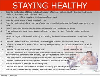
  • 1. STAYING HEALTHY 1. Describe the function of blood including transport of oxygen, carbon dioxide, digested food, waste products, hormones, antibodies and heat. 2. Name the parts of the blood and the function of each part 3. Describe the structure of each blood cell type 4. Describe the function of the heart as a “muscular pump that maintains the flow of blood around the body. 5. Identify and label the parts of the heart and describe the function of each part. 6. Draw a diagram to show the movement of blood through the heart. Describe reason for double system 7. Name the major blood vessels entering and leaving the heart and describe where they come from/ go to 8. Describe the structure and function of the three types of blood vessels found in the body 9. Define your pulse as “a wave of blood passing along an artery” and explain where it can be felt in the body 10. Describe factors that affect heart/pulse rate 11. Describe the causes and symptoms of some heart and circulatory diseases - examples include atherosclerosis, coronary heart disease, high blood pressure and varicose veins. 12. Label the parts of the breathing system and explain the function of each part. 13. Describe the role of the diaphragm and intercostal muscles in breathing. 14. Explain the effect of exercise on breathing rate. 15. Describe and define the difference between breathing, gas exchange and respiration 16. Explain how to measure lung capacity and relate this to good respiratory function. Tuesday, 27 April 2010
  • 2. Living World (level 5) • Life processes LW pro: Identify the key structural features and functions involved in the life processes of plants and animals. Describe the organisation of life at a cellular level. • Ecology LW eco: Investigate the interdependence of living things (including humans) in an ecosystem. • Evolution LW evo: Describe the basic processes by which genetic information is passed from one generation to the next. Specific Learning Outcome Suggested Learning Activities References 1. Name several • Read about the function of blood and discuss the function of blood Year 10 Study Guide functions of blood emphasising the need for cells to be supplied with nutrients and oxygen and have the wastes and carbon dioxide removed. 2. List the components • Students view cows blood under the microscope and draw the red blood cells. Slide 9 - 4 Sci2 p67 of blood and their • Students view Platelets, red and white blood cells on the projector screen and functions describe their appearance. As an exercise they can compare and contrast the Slides 10 to 12 appearance of these structures 3. Compare the • Students dissect and view cross-sections of veins using the binocular differences in microscopes. They compare and contrast these vessels structure and • Cut and paste diagrams and complete close exercise on slide. Hipkins and Connor function of blood Slide 16 vessels 4. Define ‘pulse’ and • Listen to a normal heart beat on the internet http://www.med.ucla.edu/ give examples of wilkes/intro.html where it can be located • Take both the radial and the carotid pulse and compare the pulse rate of boys and girls. • Discuss the difference between pulse rates of boys and girls and other features of heart rate. 5. Label a simple • Cut and paste a simple diagram of the human circulatory system into books. Year 10 Study Guide circulatory system • Use slide 32 to describe the function of the circulatory system in terms of the major structures. Slide 32 6. Describe the function • Students work through the worksheet, “The heart of the matter” of the parts of the • Complete the cloze exercise summary heart Tuesday, 27 April 2010
  • 3. 7. Show the • With reference to a detailed OHT heart structure, students label the-se Slide 24 and 27 (OHT) circulation of blood cross ction that is projected on the whiteboard through the heart • Students dissect a sheep’s heart and identify the relevant structures and name the major blood vessels and their function 8. Define • Brainstorm factors that students think will lead to coronary artery Reading Sheet: “What artherosclerosis damage. is atherosclerosis” • Read the handout sheet “What is atherosclerosis” and copy and complete the notes as a cloze exercise. 9. Investigate factors • Read the sheet “Risk factors for heart disease” Reading sheet “Risk that affect coronary • Complete the mind map on slide 35 factors for heart arteries of the • Answer the questions on slide 36 disease” heart and lead to Slides 35 & 36 heart disease 10. Identify factors that • Students read the sheet on training and pulse rate. They form buzz refer to sheet from affect resting heart groups and discuss the questions handed out. Each group nominates a “Training, lactate & rate recorder and a reporter and after the discussion their answers are pulse rate” from Peter reported back to the class one group at a time. Janssen • Use a datalogger to monitor resting heart rate. Students volunteer to be subjects. • Complete the cloze summary on resting heart rate. Slide 48 11. Describe how • Students use a heart rate monitor to record heart rate with time as exercise affects the they exercise. This is plotted on a set of axes. heart rate and • Exercise on Peter’s pulse Slides 49 & 50 explain the purpose • Compare fitness of two individuals Slide 51 of a raised heart rate during exercise Tuesday, 27 April 2010
  • 4. 12. Label parts of the • Choose from a list of labels to complete the labelled diagram of the Slide 53 to be re- breathing system breathing system. Labels match structure and function developed. and identify the function of each part 13. Describe the • Teacher demonstration: Bell jar breathing model Slide 53 to be re- breathing process • Students explain why the balloons inflate and deflate developed. • Cut and paste diagrams which summarise the breathing process. Bell jar • Discuss the difference between breathing, gas exchange and breathing model. respiration. 14. Describe how • Record breathing rate before and after exercise. Slide 57 exercise affects the • Discuss reasons for breathing rate needing to increase breathing rate and explain the purpose of an elevated breathing rate during exercise 15. Determine a profile • Students work their way around a circus of activities that involves them of health and taking measurements of each others resting ECG, Repiration rate fitness that has an (before and after exercise), Heart rate (before, after and during emphasis on exercise), lung capacity plus peak flow and blood pressure. cardiovascular fitness. 16. Define respiration • Discuss respiration. Stress that it is a cellular process and using simple Slide 58 and write an ideas about cell structure explain the chemical change that takes equation for this place. reaction Tuesday, 27 April 2010
  • 6. Word search KEY WORDS Y N I P N O T T S Y E R C W N plasma R B O H O C R U R Z R W I O G platelets pulse A R K I I L K A N A Z E I E P aorta atrium L O R E T S E L O H C T T U R artery L N R P A A P A C Q A H L R C ventricle pulmonary I C L T L L R A I L C M E X A vena cava cholesterol P H Y O A A V I A C O J V A J inhalation A I R S H A T H P N X A E R R exhalation trachea C K M Y N G X E A S U N N O K bronchi bronchiole V A Q N I E V R L Y E S T H M alveolus M U I R T A Y L J E C R R T J diaphragm thorax M G A R H P A I D V T S I W T vein B R O N C H I O L E E S C F W capillary respiration A L V E O L U S S N P U L S E U C H K C O E D Z A P E E F W Tuesday, 27 April 2010
  • 7. AEROBIC RESPIRATION (air-o-bick res-purr-ay-shun) – respiration that use oxygen. ALVEOLI (al-vee-oh-lie) – air sacs in the lungs where gas exchange occurs. ANAEROBIC RESPIRATION (an-air-o-bick res-purr-ay-shun) – respiration that does not use oxygen. AORTA (ay-ort-ar) – major artery that carries blood from the heart to the body. ARTERIOSCLEROSIS (are-tear-ee-oh-skleer-oh-sis) – hardening of the arteries. ARTERY (are-ter-ee) – blood vessels that carry blood away from the heart. ATRIUM (ay-tree-um) – one of two chambers in the heart that collect blood. BREATHING (bree-thing) – the physical movement of air in and out of the lungs. BRONCHI (bronk-eye)– two tubes that connect the trachea to the lungs. BRONCHIOLES (bronk-ee-oles) – narrow tubes in the lungs that branch off from the bronchi. CAPILLARY (ka-pill-are-ee) – the smallest blood vessel with walls only one cell thick. CILIA (sill-ee-a) – tiny hairs present on the surface of cells lining the airways. CIRCULATORY SYSTEM (sir-kew-lar-tory sis-tim) – the blood, blood vessels and heart. DEOXYGENATED (dee-ox-I-gin-ay-ted) – without oxygen. DIAPHRAGM (dye-a-fram) – sheet of muscle separating the chest area from the abdomen that has an important role in breathing. EXHALE (ex-hail) – to breathe air out of the body. GAS EXCHANGE (gass ex-change) – the exchange that occurs in the lungs of oxygen for carbon dioxide. HAEMOGLOBIN(he-mow-glow-bin) – a protein in red blood cells that carries oxygen. HEART (hart) – a muscle that pumps blood around the body. INHALE (in-hay-l) – to breathe air into the body. LARYNX (lar-inx) – voice box. OXYGENATED (ox-i-gin) – containing oxygen. PLASMA (plaz-ma) – pale yellow liquid part of blood. PLATELETS (plate-lets) – a cell in blood involved in blood clotting. PULMONARY (pull-mun-airy) – refers to the lungs. PULSE (pole-z) – expanding and contracting of arteries as a wave of blood passes. RED BLOOD CELLS (red blud cells) – cells that carry oxygen around the body. RESPIRATION (res-purr-ay-shun) – a chemical process that occurs in cells and releases energy. RESPIRATORY SYSTEM (res-purr-a-tory sis-tim) – the organs involved in getting air into the body and exchanging oxygen for carbon dioxide. TRACHEA (tray-key-a) – the wind pipe. VALVES (valve-z) – flaps in a blood vessel that only allow blood to go one way. VEIN (vay-n) – blood vessels that return blood to the heart. VENA CAVA (vee-na car-va) – vein that carries blood that is returning from the body to the heart VENTRICLE (ven-trick-kill) – one of two chambers in the heart that pumps blood. WHITE BLOOD CELLS (why-te blud cells)– cells involved in defence against disease-causing organisms. Tuesday, 27 April 2010
  • 8. Write the answers only in the back of your book Term Definition GLOSSARY 1 Match the term with its definition A. Aerobic respiration 1. two tubes that connect the trachea to the lungs B. Alveoli 2. hardening of the arteries C. Anaerobic 3. without oxygen respiration D. Aorta 4. narrow tubes in the lungs that branch off from the bronchi. E. Arteriosclerosis 5. to breathe air out of the body F. Artery 6. the physical movement of air in and out of the lungs G. Atrium 7. major artery that carries blood from the heart to the body. 8. sheet of muscle separating the chest area from the H. Breathing abdomen that has an important role in breathing I. Bronchi 9. respiration that use oxygen. J. Bronchioles 10. one of two chambers in the heart that collect blood K. Capillary 11. blood vessels that carry blood away from the heart. L. Cilia 12. air sacs in the lungs where gas exchange occurs M. Circulatory system 13. respiration that does not use oxygen N. Deoxygenated 14. the smallest blood vessel with walls only one cell thick O. Diaphragm 15. the blood, blood vessels and heart P. Exhale 16. tiny hairs present on the surface of cells lining the airways Tuesday, 27 April 2010
  • 9. Term Definition GLOSSARY 1 Aerobic respiration respiration that use oxygen. Alveoli air sacs in the lungs where gas exchange occurs Anaerobic respiration that does not use oxygen respiration Aorta major artery that carries blood from the heart to the body. Arteriosclerosis hardening of the arteries Artery blood vessels that carry blood away from the heart. Atrium one of two chambers in the heart that collect blood Breathing the physical movement of air in and out of the lungs Bronchi two tubes that connect the trachea to the lungs Bronchioles narrow tubes in the lungs that branch off from the bronchi. Capillary the smallest blood vessel with walls only one cell thick Cilia tiny hairs present on the surface of cells lining the airways Circulatory system the blood, blood vessels and heart Deoxygenated without oxygen sheet of muscle separating the chest area from the abdomen Diaphragm that has an important role in breathing Exhale to breathe air out of the body Tuesday, 27 April 2010
  • 10. Term Definition GLOSSARY 2 Match the term with its definition A. Gas exchange 1. containing oxygen B. Haemoglobin 2. a cell in blood involved in blood clotting C. Heart 3. the exchange that occurs in the lungs of oxygen for carbon dioxide D. Inhale 4. cells that carry oxygen around the body. E. Larynx 5. pale yellow liquid part of blood F. Oxygenated 6. voice box G. Plasma 7. a muscle that pumps blood around the body H. Platelets 8. a protein in red blood cells that carries oxygen I. Pulmonary 9. to breathe air into the body J. Pulse 10. flaps in a blood vessel that only allow blood to go one way. K. Red blood cells 11. refers to the lungs L. Respiration 12. blood vessels that return blood to the heart M. Respiratory system 13. expanding and contracting of arteries as a wave of blood passes 14. the organs involved in getting air into the body and exchanging oxygen for N. trachea carbon dioxide O. valves 15. the wind pipe P. vein 16. one of two chambers in the heart that pumps blood Q. vena cava 17. cells involved in defence against disease-causing organisms R. ventricle 18. a chemical process that occurs in cells and releases energy S. white blood cells 19. vein that carries blood that is returning from the body to the heart Tuesday, 27 April 2010
  • 11. Term Definition GLOSSARY 2 Gas exchange the exchange that occurs in the lungs of oxygen for carbon dioxide Haemoglobin a protein in red blood cells that carries oxygen Heart a muscle that pumps blood around the body Inhale to breathe air into the body Larynx voice box Oxygenated containing oxygen Plasma pale yellow liquid part of blood Platelets a cell in blood involved in blood clotting Pulmonary refers to the lungs Pulse expanding and contracting of arteries as a wave of blood passes Red blood cells cells that carry oxygen around the body. Respiration a chemical process that occurs in cells and releases energy the organs involved in getting air into the body and exchanging oxygen for carbon Respiratory system dioxide trachea the wind pipe valves flaps in a blood vessel that only allow blood to go one way. vein blood vessels that return blood to the heart vena cava vein that carries blood that is returning from the body to the heart ventricle one of two chambers in the heart that pumps blood white blood cells cells involved in defence against disease-causing organisms Tuesday, 27 April 2010
  • 12. Back of book WHAT DO I ALREADY KNOW? Label the diagrams (a) (b) (c) (d) (g) (f) (e) GUT, HEART, STOMACH, LIVER, LUNG PANCREAS, KIDNEY Tuesday, 27 April 2010
  • 13. LOCATION OF THE BODY’S ORGANS 1. Draw the outline 2. Draw in where you think the organs (from the previous slide) are. (Use the pictures on the previous slide to show you the shape of each organ Check your work with the teacher when you think you have finished Tuesday, 27 April 2010
  • 15. 1. DESCRIBE THE FUNCTION OF BLOOD ... INTERESTING FACTS COPY & ANSWER THE QUESTIONS (BELOW) INTO YOUR EXERCISE BOOKS USE A COMPUTER TO RESEARCH ANSWERS Start in class ---> Finish for homework 1. What volume of blood is contained in the body of an average adult male? 5L 2. At what rate is this blood circulated through the heart? 5 L min-1 3. What effect does smoking have on the way our blood Narrows the blood vessels in legs and arms (mainly) circulates through our body? High blood pressure. 4. Name one blood-related disease and describe it briefly. Leukemia, Clotting disorders (haemophilia), Copy and complete the notes Anaemia. Blood is important because it carries substances around the body. • Heat is carried to the skin. • Wastes to the kidneys • Carbon dioxide to the lungs • Food from the gut & stomach to the cells • Hormones from the glands to the body organs Tuesday, 27 April 2010
  • 16. BLOOD - BITS Tuesday, 27 April 2010
  • 17. Copy the notes (below) and use computers to help you to complete them Mainly water (50%) Straw 4% colored 1% other White blood cells - fight Platelets - Red blood cells - carry around the body Tuesday, 27 April 2010
  • 20. LOOKING AT BLOOD CELLS Practical Do not copy this Copy the aim and draw the method then follow this instruction Practical: 4Sci2 p67 Also, look at the OHT of red blood cells set up on the projector Tuesday, 27 April 2010
  • 22. WHAT YOU MIGHT SEE, 500 X MAGNIFIED 1000 x MAGNIFIED Tuesday, 27 April 2010
  • 25. SICKLE CELL NORMAL CELL Tuesday, 27 April 2010
  • 27. Red blood cell White blood cell Tuesday, 27 April 2010
  • 28. WHERE DID I COME FROM? Tuesday, 27 April 2010
  • 30. Extra for Experts HOW A CLOT FORMS 1. Study the diagram (left and below) 2. For homework find out what fibrin is and write the definition in the space below. 3. Research how aspirin works and what it is used for and write a brief paragraph to summarize your findings Fibrin is ____________________________ ____________________________ ____________________________ ____________________________ ____________________________ Aspirin is used for people who have had a __________ attack. It works by __________________________ ____________________________ ____________________________ ____________________________ ____________________________ ____________________________ ____________________________ ____________________________ ____________________________ Tuesday, 27 April 2010
  • 32. BLOOD BITS - MIX & MATCH Platelets Cut out the pictures, Red blood cells White blood cells descriptions & functions Tuesday, 27 April 2010
  • 33. BLOOD BITS - MIX & MATCH Platelets Cut out the pictures, Red blood cells White blood cells descriptions & functions Donut shaped (discs which have a hollow on each side) - this increases surface area Tuesday, 27 April 2010
  • 34. BLOOD BITS - MIX & MATCH Platelets Cut out the pictures, Red blood cells White blood cells descriptions & functions Donut shaped (discs which have a hollow on each side) - this increases surface area Have no nucleus Tuesday, 27 April 2010
  • 35. BLOOD BITS - MIX & MATCH Platelets Cut out the pictures, Red blood cells White blood cells descriptions & functions Donut shaped (discs which have a hollow on each side) - this increases surface area Red in colour Have no nucleus Tuesday, 27 April 2010
  • 36. BLOOD BITS - MIX & MATCH Platelets Cut out the pictures, Red blood cells White blood cells descriptions & functions Donut shaped Granular (rough) in appearance (discs which have a hollow on each side) - this increases surface area Red in colour Have no nucleus Tuesday, 27 April 2010
  • 37. BLOOD BITS - MIX & MATCH Platelets Cut out the pictures, Red blood cells White blood cells descriptions & functions Donut shaped Granular (rough) in appearance (discs which have a hollow on each side) - this increases surface area Larger than red blood cells Red in colour Have no nucleus Tuesday, 27 April 2010
  • 38. BLOOD BITS - MIX & MATCH Platelets Cut out the pictures, Red blood cells White blood cells descriptions & functions Donut shaped Granular (rough) in appearance (discs which have a hollow on each side) - this increases surface area Fewer in number than red blood cells Larger than red blood cells Red in colour Have no nucleus Tuesday, 27 April 2010
  • 39. BLOOD BITS - MIX & MATCH Platelets Cut out the pictures, Red blood cells White blood cells descriptions & functions Donut shaped Granular (rough) in appearance (discs which have a hollow on each side) - this increases surface area Fewer in number than red blood cells Larger than red blood cells Have a nucleus Red in colour Have no nucleus Tuesday, 27 April 2010
  • 40. BLOOD BITS - MIX & MATCH Platelets Cut out the pictures, Red blood cells White blood cells descriptions & functions Donut shaped Granular (rough) in appearance (discs which have a hollow on each side) - this increases surface area Fewer in number than red blood cells Larger than red blood cells Have a nucleus Red in colour Flat Have no nucleus Tuesday, 27 April 2010
  • 41. BLOOD BITS - MIX & MATCH Platelets Cut out the pictures, Red blood cells White blood cells descriptions & functions Donut shaped Granular (rough) in appearance (discs which have a hollow on each side) - this increases surface area Fewer in number than red blood cells Larger than red blood cells Have a nucleus Red in colour Irregularly shaped discs Flat Have no nucleus Tuesday, 27 April 2010
  • 42. BLOOD BITS - MIX & MATCH Platelets Cut out the pictures, Red blood cells White blood cells descriptions & functions Donut shaped Granular (rough) in appearance (discs which have a hollow on each side) - this increases surface area Fewer in number than red blood cells Larger than red blood cells Have a nucleus Red in colour Irregularly shaped discs Flat Have no nucleus Cell fragments Tuesday, 27 April 2010
  • 43. BLOOD BITS - MIX & MATCH Platelets Cut out the pictures, Red blood cells White blood cells descriptions & functions Donut shaped Granular (rough) in appearance (discs which have a hollow on each side) - this increases surface area Fewer in number than red blood cells Larger than red blood cells Have a nucleus Red in colour Irregularly shaped discs Flat Very small in size Have no nucleus Cell fragments Tuesday, 27 April 2010
  • 44. BLOOD BITS - MIX & MATCH Platelets Cut out the pictures, Red blood cells White blood cells descriptions & functions Donut shaped Granular (rough) in appearance (discs which have a hollow on each side) - this increases surface area Fewer in number than red blood cells Larger than red blood cells Have a nucleus Red in colour Irregularly shaped discs Flat Very small in size Have no nucleus Carry oxygen Cell fragments Tuesday, 27 April 2010
  • 45. BLOOD BITS - MIX & MATCH Platelets Cut out the pictures, Red blood cells White blood cells descriptions & functions Donut shaped Granular (rough) in appearance (discs which have a hollow on each side) Can cause bacteria to clump so - this increases surface area that antibodies can attack them Fewer in number than red blood cells Larger than red blood cells Have a nucleus Red in colour Irregularly shaped discs Flat Very small in size Have no nucleus Carry oxygen Cell fragments Tuesday, 27 April 2010
  • 46. BLOOD BITS - MIX & MATCH Platelets Cut out the pictures, Red blood cells White blood cells descriptions & functions Donut shaped Granular (rough) in appearance (discs which have a hollow on each side) Can cause bacteria to clump so - this increases surface area that antibodies can attack them Fewer in number than red blood cells Larger than red blood cells Have a nucleus Red in colour Irregularly shaped discs Flat Very small in size Have no nucleus Can eat bacteria Carry oxygen Cell fragments Tuesday, 27 April 2010
  • 47. BLOOD BITS - MIX & MATCH Platelets Cut out the pictures, Red blood cells White blood cells descriptions & functions Donut shaped Granular (rough) in appearance (discs which have a hollow on each side) Can cause bacteria to clump so - this increases surface area that antibodies can attack them Fewer in number than red blood cells Larger than red blood cells Have a nucleus Red in colour Irregularly shaped discs Flat Very small in size Have no nucleus Can eat bacteria Carry oxygen Cell fragments Help blood to clot Tuesday, 27 April 2010
  • 48. Match these pictures, descriptions & functions in a table such as the one shown (below) Picture Descriptions Functions Red blood cells White blood cells Platelets Tuesday, 27 April 2010
  • 49. HAEMOGLOBIN Haemoglobin is a very large molecule (a big group of atoms joined together). In the centre of the molecule there are iron atoms which allow the molecule to carry oxygen atoms (and some carbon dioxide). Haemoglobin gives blood its red colour. When someone is low in haemoglobin we say that they are anaemic. Oxygen is collected from the lungs and delivered to the body tissues: Tuesday, 27 April 2010
  • 50. BLOOD - VESSELS Tuesday, 27 April 2010
  • 53. Copy the notes & COMPLETE them Arteries - have walls - carry blood the heart - have elastic fibres that withstand . Veins - have walls - carry blood to the heart - do not need elastic fibres because the in veins is at a low pressure - rely on to make sure that blood flows back to the heart in one direction only. also help blood return to the heart by squeezing on veins - Veins are found in the veins of calves, hands and arms as these vessels carry blood against the force of gravity Capillaries - are blood vessels that ensure that blood reaches every cell - their walls are one cell thick so blood can pass through them easily - they supply & and remove and . - have walls that are one cell thick Tuesday, 27 April 2010
  • 54. Answers I SEE RED, I SEE RED, I SEE RED! 1. Dash needs concentrated red cells because his body is not making red cells. 2. Marcus needs chemicals in the plasma because he needs more factor VIII. 3. Ignatius needs concentrated blood cells to replace the blood cells that have been lost and concentrated plasma to maintain fluid balance. After the operation he needs immunity proteins from plasma to help fight infection. 4. Joan needs concentrated plasma as this will replace the fluids lost in the burn blisters. Tuesday, 27 April 2010
  • 55. Interactive exercise ARTERY OR VEIN - WHICH IS WHICH? A B C Tuesday, 27 April 2010
  • 57. WHAT IS CIRCULATION?? • Circulation is the flow of blood around the body. • This is required to supply cells with nutrients and oxygen. • Nutrients are absorbed into the bloodstream through the small intestine and become dissolved in the plasma. • Oxygen is carried on haemoglobin. • Circulation is required to remove wastes and carbon dioxide from the cells. Carbon dioxide is carried on haemoglobin. • Wastes are dissolved in the plasma. Tuesday, 27 April 2010
  • 58. WHAT IS CIRCULATION?? • Circulation is the flow of blood around the body. • This is required to supply cells with nutrients and oxygen. • Nutrients are absorbed into the bloodstream through the small intestine and become dissolved in the plasma. • Oxygen is carried on haemoglobin. • Circulation is required to remove wastes and carbon dioxide from the cells. Carbon dioxide is carried on haemoglobin. • Wastes are dissolved in the plasma. Tuesday, 27 April 2010
  • 59. ARTERIES CARRY BLOOD AWAY FROM THE HEART. VEINS CARRY BLOOD BACK TO THE HEART. Tuesday, 27 April 2010
  • 60. Artery Vein ARTERIES & VEINS ARE CONNECTED BY A CAPILLARY NETWORK Tuesday, 27 April 2010
  • 61. ARTERIES & VEINS ARE FOUND TOGETHER AROUND ORGANS Tuesday, 27 April 2010
  • 62. HEART AS A PUMP Tuesday, 27 April 2010
  • 63. THE HEART AND THE TWO LOOP SYSTEM The function of the heart is to ________ blood around the body. Blood circulates through two loops. • One loop involves the _________. • The other loop involves the flow of blood through body’s tissues and _________ (these include the brain, _____________ kidneys, and ________________ . Tuesday, 27 April 2010
  • 66. Blood pressure results from the back pressure of the capillaries on the arteries due to the hearts contraction Tuesday, 27 April 2010
  • 67. Blood pressure results from the back pressure of the capillaries on the arteries due to the hearts contraction A healthy blood pressure for an adult is 120 over 80. Tuesday, 27 April 2010
  • 68. Blood pressure results from the back pressure of the capillaries on the arteries due to the hearts contraction A healthy blood pressure for an adult is 120 over 80. Tuesday, 27 April 2010
  • 69. Blood pressure results from the back pressure of the capillaries on the arteries due to the hearts contraction A healthy blood pressure for an adult is 120 over 80. Tuesday, 27 April 2010
  • 70. Blood pressure results from the back pressure of the capillaries on the arteries due to the hearts contraction A healthy blood pressure for an adult is 120 over 80. 120 mm Hg - max contraction Tuesday, 27 April 2010
  • 71. Blood pressure results from the back pressure of the capillaries on the arteries due to the hearts contraction A healthy blood pressure for an adult is 120 over 80. 120 mm Hg - max contraction Tuesday, 27 April 2010
  • 72. Blood pressure results from the back pressure of the capillaries on the arteries due to the hearts contraction 80 mm Hg - heart A healthy blood relaxes as blood pressure for an flows into it. adult is 120 over 80. 120 mm Hg - max contraction Tuesday, 27 April 2010
  • 73. Blood pressure results from the back pressure of the capillaries on the arteries due to the hearts contraction 80 mm Hg - heart A healthy blood relaxes as blood pressure for an flows into it. adult is 120 over 80. 120 mm Hg - max contraction Tuesday, 27 April 2010
  • 75. Typical Blood Pressures include ... Tuesday, 27 April 2010
  • 76. Typical Blood Pressures include ... Tuesday, 27 April 2010
  • 77. Typical Blood Pressures include ... 80/45 in infants Tuesday, 27 April 2010
  • 78. Typical Blood Pressures include ... 80/45 in infants Tuesday, 27 April 2010
  • 79. Typical Blood Pressures include ... 80/45 in infants 120/80 at age 30 Tuesday, 27 April 2010
  • 80. Typical Blood Pressures include ... 80/45 in infants 120/80 at age 30 140/85 at age 40 and over Tuesday, 27 April 2010
  • 81. Typical Blood Pressures include ... 80/45 in infants 120/80 at age 30 140/85 at age 40 and over Tuesday, 27 April 2010
  • 85. Blood Pressure can identify any disease that... Tuesday, 27 April 2010
  • 86. Blood Pressure can identify any disease that... Tuesday, 27 April 2010
  • 87. Blood Pressure can identify any disease that... 1. widens blood vessels Tuesday, 27 April 2010
  • 88. Blood Pressure can identify any disease that... 1. widens blood vessels Tuesday, 27 April 2010
  • 89. Blood Pressure can identify any disease that... 1. widens blood vessels 2. narrows blood vessels Tuesday, 27 April 2010
  • 90. Blood Pressure can identify any disease that... 1. widens blood vessels 2. narrows blood vessels Tuesday, 27 April 2010
  • 91. Blood Pressure can identify any disease that... 1. widens blood vessels 2. narrows blood vessels 3. makes blood vessels less stretchy Tuesday, 27 April 2010
  • 92. Blood Pressure can identify any disease that... 1. widens blood vessels 2. narrows blood vessels 3. makes blood vessels less stretchy Tuesday, 27 April 2010
  • 93. Blood Pressure can identify any disease that... 1. widens blood vessels 2. narrows blood vessels 3. makes blood vessels less stretchy 4. effects the pumping power of the heart Tuesday, 27 April 2010
  • 94. Video worksheet PUMPING LIFE - THE BLOOD & CIRCULATORY SYSTEM continually In order for blood to do its job it must ______________ flow throughout the body. pump The heart acts as a ____________ to drive blood through the circulatory system. It muscle can do this because it is nearly all _____________. four atria The heart has _________ chambers. The top chambers are called _________ and the ventricles atrium bottom chambers are called __________. The right _________ and the right ventricle atrium ventricle _____________ are connected and the left ___________ and left __________ are right left also connected but there is no connection between the _______ and ________ side of the heart. left The right side of the heart appears on the ____________ from our point of view. The right atrium accepts blood (that is low in ____________) from the body tissues oxygen and organs. It then passes into the right ____________. The right __________ ventricle ventricle pumps blood to the capillaries of the lungs. Capillaries allow ___________ _________ carbon dioxide to enter the lungs and pick up _____________ from the lungs. oxygen Tuesday, 27 April 2010
  • 95. HEART, VESSELS & FUNCTIONS Tuesday, 27 April 2010
  • 96. ___________________ carries blood to body tissues & organs _______________ carries blood back to the heart from _______________ the lungs Carries blood to the lungs Word list Valves _______________ Left atrium carries blood back Right atrium to the heart from Left ventricle organs & tissues Right ventricle Septum Pulmonary artery Pulmonary vein BLOOD VESSELS & CHAMBERS OF Aorta THE HEART Vena cavae Tuesday, 27 April 2010
  • 97. THE CUT AND PASTE HEART PART 4 Tuesday, 27 April 2010
  • 98. THE CUT AND PASTE HEART instructions Cut out Part 1 Colour the white areas on the side labeled “right side” BLUE. The grey shaded areas are the muscular wall. Paste Part 1 in the centre of a new page in your exercise book. Cut out parts 2 and 3 Colour these RED. Glue the dotted edges (not the whole thing) and stick edges on top of the numbers 2 and 3 on Part one. Cut out parts 4, 5 and 6. Colour these parts BLUE. Glue the arrowed edges (not the whole thing) and stick edges on top of the numbers 4, 5 and 6 on Part one. Lift part 3 so part 6 lies underneath it. Cut out parts 7 and 8 Colour these parts YELLOW. Glue the arrow edges (not the whole thing) and stick edges on top of the numbers 7 and 8 on Part one. Cut out Part 9. Colour this PINK. Glue the back of the tab and stick in your book so it covers all of Part 1. Once you have completed this exercise, label the blood vessels. Tuesday, 27 April 2010
  • 99. Practical HEART DISSECTION 1. The left side will feel thicker and more muscular. Using this knowledge, place the heart on the dissection tray so that the left side is where you would expect to see it if you were looking at a picture of your heart. 2. Draw and label your heart showing the blood vessels that enter and leave the heart. Also draw and label the coronary arteries. B 3. Make cuts A, B and C (in A that order. Use the 3D picture on the next slide to C help you to identify the highlighted structures Tuesday, 27 April 2010
  • 101. Theory MY HEART FROM THE OUTSIDE H/O Now label this Tuesday, 27 April 2010
  • 102. BLOOD FLOW (THROUGH THE HEART) Tuesday, 27 April 2010
  • 103. Use the diagram to MY CIRCULATORY SYSTEM complete these notes • The right side of the heart receives blood from all body ______ and ______. This blood is dark in colour because it does not contain much oxygen. • Blood enters the right side of the heart through the _____ ______ and into the ______ ______ .It then passes into the right ventricle which squeezes blood into the pulmonary arteries. • ___________ ________ carry blood to the lungs. • The lungs oxygenate the blood. • The ________ ______ carry blood from the lungs back to the heart where it enters the left atrium. This blood is bright red in colour because it is rich in oxygen. • From the left atrium, blood passes into the _______ _____________. • The left ventricle squeezes blood into the _________ which branches into smaller blood vessels. These supply body tissues and organs with oxygenated blood. Tuesday, 27 April 2010
  • 104. BLOOD VESSELS & FLOW THROUGH THE HEART 1. Cut and Paste the diagram of the human heart (below) into your book. 2. Use the “3D DIAGRAM” on OHT to help you to label this diagram using the word list below. Use every word once only. Word list Labels for arrows Aorta From body Right ventricle From lungs Left ventricle to lungs Pulmonary artery Vena Cava to body Pulmonary vein Valve tendon bicuspid valve tricuspid valve Right atrium Left atrium semilunar valve Tuesday, 27 April 2010
  • 106. Staying Health Research Assignment Date due A disease is a condition in which a tissue, organ or organ system does not function properly. Atherosclerosis is one form of the disease arteriosclerosis and it affects part of the circulatory system. In your report you must answer the following questions: - • Name the part of the circulatory system this disease affects and how the disease affects it • Describe the causes of the disease • Describe the symptoms a person with this disease might show • Discuss how the disease can be prevented and how it is treated. You can present your report as either an A3 sized poster or a written/typed report of no more than 2 A4 pages. Your written work must be in your own words (no cut and paste from internet sites). You must also include at least one diagram/picture that helps support your written answers. Make sure it has either labels or a caption that links to your written work. Include a bibliography at end of the written report or on the back of poster. This will include:- • the title and author(s) of any books you use in alphabetical order • the web address of any world wide web sites used Hint - start by looking up arteriosclerosis and then look for Atherosclerosis Tuesday, 27 April 2010
  • 107. ATHEROSCLEROSIS http://www.torrancememorial.org/carrisk.htm 1. Read the handout sheet entitled “What is Atherosclerosis?” 2. Copy and Complete the following notes: • Also called “.......................... ..... ......... ..................... “ . • Caused by a build up of ............ and ...................... on the inside walls of arteries. • This can cause ...................... of the arteries and a ..................... in blood flow. • Permanent damage to tissues and organs can occur if .............. supply is disrupted. • If the coronary arteries are affected by atherosclerosis, they could become further narrowed by substances in the blood and block altogether. THIS RESULTS IN A HEART ATTACK. Tuesday, 27 April 2010
  • 108. Mind map RISK FACTORS FOR HEART DISEASE 1. Read the handout sheet entitled “Risk factors and Coronary Heart Disease?” 2. Copy & Complete the mind map below: All Risk factors Major can be managed or controlled ( --> obesity) Contributing can be managed or controlled can not be managed or controlled ACTIVITY: With these factors up on the white board; Students all stand. Go thru each factor. Students who think they have it sit. By the end of the lesson...no one is standing ---> no one is safe from heart disease) Tuesday, 27 April 2010
  • 109. Mind map RISK FACTORS FOR HEART DISEASE 1. Read the handout sheet entitled “Risk factors and Coronary Heart Disease?” 2. Copy & Complete the mind map below: All Risk factors Major smoking can be managed or controlled ( --> obesity) Contributing can be managed or controlled can not be managed or controlled ACTIVITY: With these factors up on the white board; Students all stand. Go thru each factor. Students who think they have it sit. By the end of the lesson...no one is standing ---> no one is safe from heart disease) Tuesday, 27 April 2010
  • 110. Mind map RISK FACTORS FOR HEART DISEASE 1. Read the handout sheet entitled “Risk factors and Coronary Heart Disease?” 2. Copy & Complete the mind map below: All Risk factors Major smoking Cholesterol can be managed or controlled ( --> obesity) Contributing can be managed or controlled can not be managed or controlled ACTIVITY: With these factors up on the white board; Students all stand. Go thru each factor. Students who think they have it sit. By the end of the lesson...no one is standing ---> no one is safe from heart disease) Tuesday, 27 April 2010
  • 111. Mind map RISK FACTORS FOR HEART DISEASE 1. Read the handout sheet entitled “Risk factors and Coronary Heart Disease?” 2. Copy & Complete the mind map below: All Risk factors Major smoking Cholesterol diabetes can be managed or controlled ( --> obesity) Contributing can be managed or controlled can not be managed or controlled ACTIVITY: With these factors up on the white board; Students all stand. Go thru each factor. Students who think they have it sit. By the end of the lesson...no one is standing ---> no one is safe from heart disease) Tuesday, 27 April 2010
  • 112. Mind map RISK FACTORS FOR HEART DISEASE 1. Read the handout sheet entitled “Risk factors and Coronary Heart Disease?” 2. Copy & Complete the mind map below: All Risk factors Major smoking Cholesterol diabetes inactivity can be managed or controlled ( --> obesity) Contributing can be managed or controlled can not be managed or controlled ACTIVITY: With these factors up on the white board; Students all stand. Go thru each factor. Students who think they have it sit. By the end of the lesson...no one is standing ---> no one is safe from heart disease) Tuesday, 27 April 2010
  • 113. Mind map RISK FACTORS FOR HEART DISEASE 1. Read the handout sheet entitled “Risk factors and Coronary Heart Disease?” 2. Copy & Complete the mind map below: All Risk factors Major smoking Cholesterol diabetes inactivity can be managed or controlled ( --> obesity) Contributing stress can be managed or controlled can not be managed or controlled ACTIVITY: With these factors up on the white board; Students all stand. Go thru each factor. Students who think they have it sit. By the end of the lesson...no one is standing ---> no one is safe from heart disease) Tuesday, 27 April 2010
  • 114. Mind map RISK FACTORS FOR HEART DISEASE 1. Read the handout sheet entitled “Risk factors and Coronary Heart Disease?” 2. Copy & Complete the mind map below: All Risk factors Major smoking Cholesterol diabetes inactivity can be managed or controlled ( --> obesity) Contributing stress alcohol can be managed or controlled can not be managed or controlled ACTIVITY: With these factors up on the white board; Students all stand. Go thru each factor. Students who think they have it sit. By the end of the lesson...no one is standing ---> no one is safe from heart disease) Tuesday, 27 April 2010
  • 115. Mind map RISK FACTORS FOR HEART DISEASE 1. Read the handout sheet entitled “Risk factors and Coronary Heart Disease?” 2. Copy & Complete the mind map below: All Risk factors Major smoking Cholesterol diabetes inactivity can be managed or controlled ( --> obesity) Contributing stress alcohol age can be managed or controlled can not be managed or controlled ACTIVITY: With these factors up on the white board; Students all stand. Go thru each factor. Students who think they have it sit. By the end of the lesson...no one is standing ---> no one is safe from heart disease) Tuesday, 27 April 2010
  • 116. Mind map RISK FACTORS FOR HEART DISEASE 1. Read the handout sheet entitled “Risk factors and Coronary Heart Disease?” 2. Copy & Complete the mind map below: All Risk factors Major smoking Cholesterol diabetes inactivity can be managed or controlled ( --> obesity) Contributing stress alcohol age gender can be managed or controlled can not be managed or controlled ACTIVITY: With these factors up on the white board; Students all stand. Go thru each factor. Students who think they have it sit. By the end of the lesson...no one is standing ---> no one is safe from heart disease) Tuesday, 27 April 2010
  • 117. Mind map RISK FACTORS FOR HEART DISEASE 1. Read the handout sheet entitled “Risk factors and Coronary Heart Disease?” 2. Copy & Complete the mind map below: All Risk factors Major smoking Cholesterol diabetes inactivity can be managed or controlled ( --> obesity) Contributing stress alcohol age gender heredity can be managed or controlled can not be managed or controlled ACTIVITY: With these factors up on the white board; Students all stand. Go thru each factor. Students who think they have it sit. By the end of the lesson...no one is standing ---> no one is safe from heart disease) Tuesday, 27 April 2010
  • 118. PULSE DEFINITION & FACTORS AFFECTING IT Tuesday, 27 April 2010
  • 119. WHAT’S IN A PULSE? http://www.blaufuss.org/ http://www.med.ucla.edu/wilkes/intro.html When the doctor listens to my heart using a stethoscope a “lud doog” sound is heard. This is the sound of one beat. These sounds are from the valves in the heart snapping shut. Definition The pulse is a wave of blood passing through an artery and it is the pressure caused by the compression of blood that is felt when someone feels for a pulse. Things you need to know 1. My pulse rate is the number of beats that my heart makes per minute. 2. The unit of pulse rate is __________ _____ _____________ (____) . 3. The average adult resting pulse rate is between 60 and 80 bpm 4. Newborns have a pulse rate of about __________. 5. Women have a ___________ pulse rate than men. 6. Recovery rate is the time it takes for a person’s heart rate to return to its resting value once exercise has stopped. The lower the recovery rate, the greater the level of fitness 7. A fit person also has a low resting pulse and takes a long time to reach maximum heart rate Tuesday, 27 April 2010
  • 120. RECOVERY TIME recovery time Tuesday, 27 April 2010
  • 121. Practical TAKING MY PULSE There are at least 2 places where my pulse can be taken: (a) at my wrist (radial pulse) (b) at my neck (carotid pulse) 1. Take your pulse using both methods, each time counting the number of beats in 15 seconds and multiplying by 4. (What are the units of your answer? Are both of your answers the same?) 2. Calculate your average pulse and write it into your book. 3. Record your class results in the following table Class results Average Boys Average Girls Tuesday, 27 April 2010
  • 122. Practical FACTORS AFFECTING PULSE RATE For each of the activities shown in the table (below) measure your pulse rate and record this in your books in a table (as shown below) Activity Pulse rate (bpm) Copy and answer the questions: Sitting Standing Jogging Step-ups/stairs Questions (a) Which activity that would give a lower pulse rate than standing ______________________________________________________________ (b) Trial your suggestion and record your result in the space in the table (c) Explain why your pulse rate changes when you change from sitting down to each of the other activities _________________________________________________________________ _________________________________________________________________ _________________________________________________________________ Tuesday, 27 April 2010
  • 123. HOW LONG-TERM EXERCISE AFFECTS THE RESTING PULSE RATE Tuesday, 27 April 2010
  • 125. WHO IS FITTER? Pulse rate Heart rate vs time graph for two individuals (bpm) Key: Paul Peter 200 150 100 50 2 4 6 8 10 12 14 16 18 20 22 24 26 time (min) Tuesday, 27 April 2010
  • 126. Cut & Copy & answer the following questions into your book Paste Questions 1. What is Paul’s resting heart rate? ____________ 2. At what time does the graph suggest that exercise started? __________ 3. How long does it take Paul’s heart rate to reach a maximum? __________ 4. What is Peter’s maximum heart rate? __________ 5. For how long does Peter maintain his maximum heart rate? __________ 6. At what time does the graph suggest that exercise stops? __________ 7. How long does it take Paul’s heart rate to return to its resting value? __________ 8. Explain why Peter is fitter than Paul. ___________________________________________________________________ ___________________________________________________________________ ___________________________________________________________________ ___________________________________________________________________ Tuesday, 27 April 2010
  • 127. SUMMARY - MY HEART AT REST Copy and Complete the following notes: • Resting heart rate is a measure of the overall .................... of an athlete. The lower the better. • Resting heart rate is best measured in the ................... so that accurate comparisons can be made. • Over several months of exercise resting heart rate can be ........................... . • Resting heart rate can remain elevated a day or two after a heavy workout or race as increased blood flow is required to compensate for the low .................. level of recovering muscle cells. • During a fever the resting heart rate ............. to compensate for a ..................... in blood pressure caused by illness. Tuesday, 27 April 2010
  • 128. BREATHING SYSTEM PARTS & FUNCTION Tuesday, 27 April 2010
  • 129. THE FLOW OF AIR Cut & Paste 1. Cut and paste the diagram (of the breathing system) into your book. 2. Copy a flow diagram into your book that correctly represents the pathway that air flows through as it enters the body (the format of this diagram is shown below and the words needed to complete it are in the box) bronchiole, mouth, alveoli bronchus, trachea 1. Cut & paste the picture of the trachea into your book. 2. The cartilage rings are like the ribs of a vacuum cleaner hose. Copy and complete the sentence below next to your pasted diagram The trachea Another name for the trachea is the __________________ . Cartilage rings Its walls are reinforced with cartilage rings. Cartilage is a soft yet stiff material. The cartilage prevents the wind pipe from ____________ when we breathe in. Tuesday, 27 April 2010
  • 130. Alveoli Use the pictures on the right to complete the following sentence, describing the function of the alveoli: The ____________ is an air sac at the end of a ______________ . It is surrounded by a ________________ which carries blood from the heart. ___________ ___________ passes into the alveolus through the ____________ _____________. _______________ from the alveolus passes into the capillary and this blood is carried back to the _______________ The surface of the alveolar wall must be moist so that gases can ___________ . This must happen before they can pass into the blood stream. Tuesday, 27 April 2010
  • 131. DID YOU KNOW? “Not having enough pulmonary surfactant causes respiratory distress syndrome in infants.” A moist alveolar surface is important but ... the water creates a lot of surface tension There is a special type of alveolar cell called a type II cell which secretes a substance called pulmonary surfactant. This is a chemical which reduces the surface tension of the water that lines the alveolus. Without this chemical it would be very difficult to expand the alveolus so that air could be dragged into them. The effort required would be tremendous. Taking deep breaths stimulates the release of this chemical and allows breathing to be more effective. Tuesday, 27 April 2010
  • 135. “Check out the bell jar” DIAPHRAGM Glass tube Ribs Rubber bung Balloon Diaphragm Rubber sheet (represents the diaphragm) PULL DOWN Observation Tuesday, 27 April 2010
  • 136. The diaphragm is a sheet of _______. When the diaphragm __________ it flattens out and __________ the space inside the chest cavity. Air rushes in to fill this space (you breathe in). When the diaphragm relaxes it arches up and __________ the space so air must leave the chest cavity (you breathe out) INTERCOSTALS These are the muscles between the ribs. When you breathe in the diaphragm contracts and flattens while these muscles contract to ______ the rib cage. When you breathe out the intercostals _________. Tuesday, 27 April 2010
  • 137. PEAK EXPIRATORY FLOW Copy • Peak expiratory flow (PEF) is a measure of the maximum speed that air can be blown out from your lungs. • It is useful for helping doctors to find out how serious someone’s asthma symptoms are. • The doctor has a record of the patient’s PEF when that person is well. • If the person arrives at the surgery complaining of problems with breathing the doctor measures the PEF. • A 15 to 20% drop in the person’s PEF shows that the problem is significant. Tuesday, 27 April 2010
  • 138. HOW TO USE A PEAK FLOW METER Step 3: “Make sure the indicator is at the bottom of the meter 1. Stand up straight. 2. Make sure the indicator is at the bottom of the meter. 3. Take a deep breath filling your lungs completely. 4. Place the mouthpiece in your mouth; lightly bite with your teeth, and close your lips on it. 5. Blast the air out as hard and as fast as possible in a single blow. 6. Record the number that appears on the meter. 7. Repeat these steps three times. 8. Record the highest of the three readings My results: Tuesday, 27 April 2010
  • 139. BREATHING RATE & FACTORS AFFECTING IT Tuesday, 27 April 2010
  • 140. Practical THE EFFECT OF EXERCISE ON BREATHING RATE Copy My breathing rate is the number of times I inhale or exhale in 1 minute. My breathing rate before exercise = breaths per minute after exercise = breaths per minute Notes “Copy and complete the following notes” 1. My breathing rate increases during exercise because ... 2. The process which uses the extra oxygen to make chemical energy in the cells is called 3. During exercise this process occurs more Tuesday, 27 April 2010
  • 142. BREATHING, GAS EXCHANGE and RESPIRATION Copy Breathing How air gets into and out of the lungs Gas exchange How gases get into and out of the blood stream Respiration How oxygen is used to make energy Respiration • Respiration is the process which occurs inside cells that uses glucose and oxygen to make chemical potential energy. This energy can be used for growth and movement. • Carbon dioxide and water are products of respiration. The equation which summarises respiration is: Carried to cells by the blood Carried away from cells by the blood Glucose + Oxygen Carbon dioxide + Water Breathed in Breathed out Tuesday, 27 April 2010
  • 145. MICROSCOPE PARTS Your teacher will show you a microscope and explain how it works 1. Cut and Paste the microscope picture into your book 2. Label the picture using the list of parts shown below. Arm List of microscope parts: base, objective lenses, stage, course focusing knob, fine focusing knob, eyepiece lens light source, body tube, stage clips. Tuesday, 27 April 2010
  • 146. Focus Knob, Mirror, Eyepiece lens, Condenser, Objective lens, Stage Use the words given to label the diagram: (a) LABEL THE PARTS OF A MICROSCOPE (b) (c) (e) • (f) Use some of the same words to complete the sentences below: The (g)....................... is used to get a clear picture. The (h)....................... is used to support the object you are looking at. The (i)...................... is used to reflect the light. The (j)...................... is what you look through. Tuesday, 27 April 2010
  • 147. USING THE MICROSCOPE A Change to the E Focus the microscope big lens using the large knob B Put a drop of water F Turn the lenses till between the two you are on the sheets of glass smallest. G Put the sheets of C Focus using the glass on the stage small knob H Put a cover slip over the top D Adjust the light source Tuesday, 27 April 2010
  • 148. STEPS IN USING A MICROSCOPE IN THE BACK OF YOUR BOOKS. Write out the steps that you would follow when you use a microscope in the correct order. (the steps below are jumbled) 1. Change to the medium power objective and adjust using the fine focus knob 2. Turn the light on. 3. Place the specimen on the stage 4. Check that the microscope is set to low power. 5. Plug the electrical lead of the microscope into the wall socket. 6. Adjust using the course focus knob 7. Adjust the amount of light entering the microscope by adjusting the condenser. 8. Carry the microscope to the bench with one hand on the arm and the other hand under the base of the microscope. Tuesday, 27 April 2010
  • 149. BLOOD BITS REVISION Name three substances transported by the blood. 1. _____________________________________ 2. _____________________________________ 3. _____________________________________ Use the following terms to complete the sentences below: plasma, haemoglobin, red blood cells, antibodies, white blood cells, platelets, oxygen (a) There are 600 ……………………. to every one ……………………. in the blood (b) The clear liquid in blood is called ………………………….. (c) The red colour of blood is caused by ……………………………… (d) White blood cells make ……………………… which destroy ‘germs’ in the blood (e) Haemoglobin carries ………………………. around the body (f) The cell fragments that make blood clots to heal wounds are called …………………………. Tuesday, 27 April 2010
  • 150. LABEL THE BLOOD VESSELS Name of vessel Word list (you may use some words more than once) Valve Vein Strong muscular layer Weak muscular layer Thin wall Outer layer Lining of vessel Artery Capillary Tuesday, 27 April 2010
  • 151. QUESTIONS 1. Explain why capillaries have thin walls? 2. Explain why arteries have elastic fibres in their walls? 3. Explain why veins have valves? Tuesday, 27 April 2010
  • 152. THE HEART AND CIRCULATION • The heart is labelled from the point of view of the animal from which the heart is taken so the left side of the heart is on the ............. side of the animal’s body. • The left ventricle has thicker walls because ........... • The human heart acts as a .................... • It pumps blood to the lungs where blood is ...................... • Oxygenated blood returns to the heart and from there it is pumped to the rest of the body. (............. and ....................) • Blood flows away from the heart through .................... • Blood flows back to the heart through .................... • The heart never stops pumping. It is made up of a special muscle that never fatigues called ............... .................. . Tuesday, 27 April 2010
  • 154. A MIXED BAG OF WORDS Use after teaching SLO’s 1 and 2 Match the term with its correct definition Term Definition 1. oxygen A. blood proteins that act against foreign substances in the blood 2. carbon dioxide B. the speed with which something moves or changes 3. hormones C. the rectangular sheet of glass upon which a sample is placed in preparation for it to be viewed microscopically 4. antibodies D. a colourless odourless gas produced by burning and respiration 5. circulates E. an optical instrument used for viewing very small objects 6. rate F. moves continuously around a closed circuit in the body 7. plasma G. a small thin slither of glass used to cover a sample that is being viewed under a microscope 8. platelets H. the colourless part of the blood 9. cells I. disc - shaped fragments which have no nucleus. They are found in blood and are involved in clotting 10. microscope J. the building blocks of living things 11. cover slip K. a colourless odourless gas that is essential for life 12. slide L. substances that stimulate cells into action Tuesday, 27 April 2010
  • 155. BLOOD BITS Tuesday, 27 April 2010
  • 156. THE BIG HEART CROSSWORD CHALLENGE Tuesday, 27 April 2010
  • 159. HEART PARTS 1. The heart is a hollow ball of muscle which sits to the left side of the chest _____________. 2. The heart is connected to the lungs by the ____________ arteries and veins. 3. Arteries carry blood ______ from the heart and veins carry blood back to the heart. 4. The pulmonary arteries supply the ________ with blood and the pulmonary veins return blood from the ________ to the heart. 5. The heart has ________ chambers. 6. These are the left and right _________ and the left and right ___________. 7. The left and right sides of the heart are separated by a thick muscular wall. 8. _________ separate each atrium from each ventricle and ensure that blood flows in the right direction as it flows through the heart. 9. Blood flow is powered by the ____________ of the atria and ventricles. 10. Atria contract to force blood into the __________. Valves close to prevent blood flowing ________ ______ ___ _________. 11. The left ventricle consists of muscular walls that are ___________ than the right ventricle because _ _ _ _ _ _ _ _ _ _ _ _ _ _ _ Tuesday, 27 April 2010
  • 161. Hwk 1 Tuesday, 27 April 2010
  • 163. Hwk 2 Tuesday, 27 April 2010
  • 167. OTHER STUFF Tuesday, 27 April 2010
  • 168. THE HEART OF THE MATTER H/O: “How the heart pumps blood” 1. Research the following questions. 2. Answer them as full sentences on a sheet of A4 3. Hand your work in for marking Tuesday, 27 April 2010
  • 169. QUESTIONS (a) What is cholesterol, LDL and HDL? (b) Describe what is meant by heredity? (c) Explain how smoking makes it difficult for the heart to get enough oxygen? (d) Explain how hypertension increases the workload on the heart. (e) Discuss the difference between a major risk factor and a contributing risk factor. Tuesday, 27 April 2010
  • 170. QUESTIONS Answer the following questions as full sentences. 1. Explain in words how the breathing tubes branch. (Hint: start from the trachea) 2. The trachea has rings of cartilage as part of its structure. Explain the function of these rings of cartilage. (Hint: think of a vacuum cleaner tube) 3. Alveoli have a large, moist surface area. Explain why this is necessary. (Hint: use the words “sticking” and “diffuses”) Tuesday, 27 April 2010
  • 171. CRITERIA EVIDENCE Judgement √ or x Names the part of the can affect all large and medium-sized arteries, including the Need 2 A circulatory system that is coronary, carotid, and cerebral arteries, the aorta, its branches, out of 3 and major arteries of the extremities affected A grades States how this is affected Atherosclerosis is patchy intimal plaques (atheromas) in A medium-sized and large arteries; the plaques contain lipids, inflammatory cells, smooth muscle cells, and connective tissue. Describes the cause of the Causal/Risk factors include dyslipidemia, diabetes, cigarette A disease smoking, family history, sedentary lifestyle, obesity, and hypertension. Describes symptoms Symptoms develop when growth or rupture of the plaque Must M reduces or obstructs blood flow; symptoms vary by artery have affected. (angina if coronary arteries affected) Discusses how the disease Reduction of Causal/Risk factors: ie. correction of Must M might be prevented dyslipidemia with sensible diet low in saturated fats and high in have polyunsaturated fatty acids, controlling diabetes, stop cigarette smoking, exercise, eating moderately, and controlling hypertension. Work is in the students own Must E words have There is at least one picture Need 2 A or diagram out of 3 A grades A bibliography is included A The work is well presented A Tuesday, 27 April 2010
  • 172. Teacher resource READING ABOUT ATHEROSCLEROSIS Atherosclerosis is patchy intimal plaques (atheromas) in medium-sized and large arteries; the plaques contain lipids, inflammatory cells, smooth muscle cells, and connective tissue. Risk factors include dyslipidemia, diabetes, cigarette smoking, family history, sedentary lifestyle, obesity, and hypertension. Symptoms develop when growth or rupture of the plaque reduces or obstructs blood flow; symptoms vary by artery affected. Diagnosis is clinical and confirmed by angiography, ultrasonography, or other imaging tests. Treatment includes risk factor and dietary modification, physical activity, antiplatelet drugs, and antiatherogenic drugs. Atherosclerosis can affect all large and medium-sized arteries, including the coronary, carotid, and cerebral arteries, the aorta, its branches, and major arteries of the extremities. It is the leading cause of morbidity and mortality in the US and in most developed countries. In recent years, age- related mortality attributable to atherosclerosis has been decreasing, but in 2005, cardiovascular disease, primarily coronary and cerebrovascular atherosclerosis still caused almost 870,000 deaths in the US (more than cancer and almost 9 times more than injuries). Atherosclerosis is rapidly increasing in prevalence in developing countries, and as people in developed countries live longer, incidence will increase. By 2020, atherosclerosis is expected to be the leading cause of death worldwide. Tuesday, 27 April 2010
  • 174. DEVELOP INTO A CLOSE EXERCISE Tuesday, 27 April 2010
  • 175. DEVELOP INTO A CLOSE EXERCISE Tuesday, 27 April 2010
  • 176. DEVELOP INTO A CLOSE EXERCISE Tuesday, 27 April 2010
  • 177. DEVELOP INTO Red Blood cells are made A CLOSE inside bones ( the bone marrow). EXERCISE Tuesday, 27 April 2010