

Recommandé
Contenu connexe
Tendances
Tendances (20)
En vedette
En vedette (20)
Similaire à Nervous tissue2k1
Similaire à Nervous tissue2k1 (20)
Dernier
Dernier (20)
Nervous tissue2k1
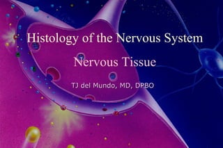
- 1. Histology of the Nervous System Nervous Tissue TJ del Mundo, MD, DPBO
- 2. environment animal organism Nervous system sensory neuron stimulus reaction effector Inter- neuron receptor Motor neuron Nervous System stimulus and reaction
- 4. Nervous Tissue Cellular Elements - Neuron (Nerve Cell) - Neuroglial Cells central neurglia astrocyte, oligodendrocyte, microglia and ependymal cell peripheral neuroglia Schwann cell in nerve and ganglion satellite (capsular) cell in ganglion Intercellular Substance : extremly scarse
- 5. Neuron Neuronal Morphology Neuronal Cell Body (Soma) - Nucleus - Perikaryon Neuronal Processes Axon Dendrites cf. Diversity of Neuronal Size and Morphology
- 7. Diversity of Neuronal Morphology
- 8. Morphology of Typical Motor Neuron 1. nucleus 2. perikaryon 3. cell body 4. axon 5. dendrite 6. Nissl body 7. axon hillock 8. myelin sheath 9. oligodendrocyte 10. Schwann cell 11. skeletal muscle cell 12. neuromuscular junction
- 9. Neuron Neuronal Function Communication Receptor - Neuron - Effector - Excitability (Irritability) - Conductivity through membrane in intraneuronal conduction via synapse in interneuronal conduction neurotransmitters
- 10. Neuronal Cell Body (Soma) NUCLEUS Ÿ Chromatin Pattern: Euchromatic euchromatin >>> heterochromatin active mRNA transcription cf. Barr body (facultative heterochromatin) inactive X chromosome in female Ÿ Conspicuous Nucleolus NOR (nucleolar organizing region) active rRNA transcription -- ribosome
- 11. NB: neuronal cell body NS: Nissl substance NL: nucleolus BB: Barr body
- 12. Murray Llewellyn Barr 1908-1995
- 14. Neuronal Cell Body (Soma) Cytoplasmic Organelles Ÿ Nissl substance: rER abundant, parallally arranged rER cisterna active site of protein (polypeptide) synthesis - transport vesicle Ÿ Golgi complex complex of perinuclear Golgi substance glycosylation, sulfation & phosphorylation packing and condensation protein to transport
- 17. Camilio Golgi (1843-1926) Golgi apparatus Golgi method (Golgi’s metallic impregnation) Golgi type I & II cell Golgi tendon organ Golgi-Mazzoni corpuscle
- 18. Neuronal Cell Body (Soma) Protein Synthesizing Assembly Ÿ Euchromatic Nucleus ------- mRNA transcription Ÿ Prominent Nucleolus ------- rRNA (ribosome) synthesis Ÿ Nissl substance: rER ------- polypeptide chain Ÿ Golgi complex ----- destined to transport - no protein synthesizing assembly in axon - neurotransmitters, enzymes, membrane proteins - transport via axonal (axoplasmic) transport
- 19. Neuronal Cell Body (Soma) Other Cytoplasmic Organelles Ÿ Mitochondria - energy source for ion exchange (Na + -K + exchange pump) and axonal transport Ÿ Lysosome - hydrolytic enzymes --- waste products - lysosomal storage disease (mental retardation, seizure) Pigment Granules Lipofuscin granule , Melanin granule
- 23. Neuronal Cell Body (Soma)
- 24. Neuronal Cell Body (Soma) Cytoskeleton Ÿ Neurofilament: Intermediate Filament - supporting - neurofibril ------- reduced silver staining Fink-Heimer method Ÿ Microtubule - tubulin - fast anterograde axonal transport and retrograde axonal transport cf. microfilament: actin, postsynaptic membrane
- 26. Neurofibril - neurofilament
- 27. Distinction between Axon and Dendrites Axon Dendrite Dimension number 1 multiple (0, 1, or more) length 200 m – 1 m less than 700 m diameter constant throughout gradually tapered Branching pattern angle almost right angle acute angle site distant from cell body near cell body Structural components Nissl substance absent could be present dendritic spine absent could be present myelin sheath could be associated not associated
- 30. Dendrite Nissl
- 32. dendritic spine
- 34. Distinction between Axon and Dendrites Axon Dendrite Staining property Golgi hard to impregnate well delineated impregnation (except rapid Golgi method) reduced silver more darkly stained less darkly stained Functional direction of efferent afferent conduction (soma -> axon terminal) (dendrite -> soma) exception: pseudounipolar neuron
- 35. Golgi method
- 37. SYNAPSE Ÿ Presynaptic Portion: Synaptic Button - synaptic vesicle - mitochondria - presynaptic membrane: tubulin Ÿ Synaptic Cleft - 20-30 nm Ÿ Postsynaptic Portion - postsynaptic membrane: actin, fodrin, spectrin - mitochondria
- 38. SYNAPSE
- 39. SYNAPSE
- 40. Components of Axonal (Axoplasmic) Transport Components Velocity (mm/day) Transporting Substances Anterograde Axonal Transport Fast Transport 200-400 synaptic vesicle, enzymes neurotransmitters Mitochondrial Transport 50-100 mitochondria Slow Transport Slow Components a (SCa) 0.1 - 1.0 tubulin, neurofilament protein Slow Comnponent b (SCb) 2 - 6 actin, clathrine, calmodulins spectrin, cytoplasmic enzymes Retrograde Axonal Transport 100-200 prelysosomal vesicles, recycled proteins, HRP, WGA neurotrophic viruses Axonal (Axoplasmic) Transport
- 41. Mechanism of Axonal Transport Fast Anterograde Axonal transport and Retrograde Axonal transport
- 42. Degeneration and Regeneration of Nervous System 1. Wallerian Degeneration - changes distal to the injury site - myelin breakdown - von B ü ngner’s band - Schwann cell - neuroma formation 2. Axon Reaction (Chromatolysis, Nissl Reaction) - displacement of nucleus - chromatolysis - regenerative processes
- 43. Augustus Waller (1816-1870) Wallerian degeneration
- 44. Axon (Nissl) Reaction - chromatolysis
- 46. Tract Tracing Methods 1) retrograde tracing methods axon, axon terminal cell body 2) anterograde tracing methods cell body axon, axon terminal cell body ---- axon terminal based on nerve degeneration and axonal transport
- 47. 1. Nissl Reaction methylene blue, toluidine blue, thionin, cresyl violet 2. Retrograde Tracer Horseradish Peroxidase (HRP) Wheat-germ Agglutinin (WGA) Fluorescence Tracer - Lucifer Yellow, Fast Blue, Nuclear Yellow Viruses and Toxoids Retrograde Tracing Methods
- 48. Retrograde Labeling of HRP
- 49. 1. Marchi Method OsO4 after tract of nerve lesion 2. Nauta Method - Fink Heimer method Reduced Silver Method after tract lesion 3. Autoradiograhy with Radiolabelled Amino Acid Tritiated Glycine 4. Anterograde Tracer Phageolus Vulgaris Leucoagglutinin (PHA-L) Anterograde Tracing Methods
- 50. Marchi method - Wallerian degeneration
- 51. Nauta method Reduced silver method after axon transection neurofilament transport continues after axon transection Nauta-Gygax method Fink-Heimer method
- 52. 3 H-labeled amino acid Autoradiography Amino acid is incorporated to protein in neuronal cell bodies Proteins are slowly transported to axonal endings
- 53. Classification of Neurons (1) by the Number of Processes 1. unipolar neuron 2. pseudounipolar neuron 3. bipolar neuron 4. multipolar neuron (2) by the Length of Axon 1. Golgi type I neuron 2. Golgi type II neuron (3) by the Morphology of Dendrites (Topognostic Value) 1. isodendritic neuron 2. allodendritic neuron 3. idiodendritic neuron
- 54. 1. unipolar neuron 2. bipolar neuron 3. pseudounipolar neuron 4. multipolar neuron a. axon d. dendrite
- 55. Pseudounipolar Neuron DRG (dorsal root ganglion) neuron pseudounipolar cell peripheral process central process telodendron cell body in DRG
- 56. Neuroglia (Neuroglial Cells) Central Neuroglia Astrocyte protoplasmic astrocyte fibrous astrocyte Oligodendrocyte perineuronal satellite cell interfascicular cell Microglia Ependymal Cell Peripheral Neuroglia Schwann Cell in peripheral nerve and ganglion Capsular (Satellite) Cell in ganglion
- 57. Astrocyte Oligodendrocyte Microglia Central Neuroglia
- 60. Protoplasmic Fibrous Synaptic Astrocyte Astrocyte Glomerulus
- 62. Oligodendrocyte 1. nucleus of oligodendrocyte 2. process of oligodendrocyte 3. myelin sheath 4. axon
- 63. Microglia Cell Body slender, indented, heterochromatic nucleus dark cytopasm - prominent secondary lysosome Processes short, highly branched Macrophage (Mononuclear Phagocytic) System Mesenchymal Origin - Blood Monocyte Increased in Inflammation
- 64. Microglia 1. nucleus of microglia 2. process of microglia 3. lysosome 4. capillary 5. pericyte
- 65. Ependymal Cell Epithelial Cell lining ventricular surface cilia and microvilli on luminal surface simple cuboidal cell with round nucleus Tanicyte basal process, numerous in 3rd ventricle most ependymal cell has basal process (Chung & Lee, 1988) Choroid Plexus Epithelial Cells ion transporting cell: numerous mitochondria
- 67. Schwann Cell Slender Cell with small heterochromatic nuclei Myelin forming cell in PNS - Myelin Sheath: Myelinated Fiber A Schwann cell constitutes a internodal Segment Surround Unmyelinated Axons A schwann cell surround many unmyelinated axons
- 68. Schwann Cell
- 69. Satellite (Capsular) Cell Squamous Cell encircles neuronal cell body in Ganglion - completely encircles pseudounipolar neuron in spinal and cranial ganglion - neurons of autonomic ganglia were less completely surrounded by satellite cell
- 72. Myelin Node of Ranvier - Internodal segment Schmidt-Lantermann’s cleft
- 75. Osmium tetroxide (OsO 4 ) stain for Myelin
- 76. Myelin Structure of fast nerve conduction
- 77. Giant squid axon 0.5-1 mm in diameter conduction velocity 25 m/s
- 78. Myelin Conduction velocity is proportional to 1. The Length of Internodal Segment 2. Thickness of Myelin 3. Diameter of Nerve Fiber
- 79. Conduction velocity of mammalian nerve fiber Group I A 10 - 20 70 -120 Group II A 5 - 12 30 - 70 A γ 3 - 6 15 - 30 Group III A δ 2 - 5 12 - 30 B < 3 3 - 15 Group IV C 0.1 - 1.5 0.5 - 2 Myelinated fiber Unmyelinated fiber
- 80. Myelin Formation
- 81. Myelin Myelin formation Schwann cell oligodendrocyte
- 82. MYELIN - Fusion of Plasma Membrane Major Dense Line - fusion of inner leaflet ---- Myelin Basic Protein (MBP) Intraperiod Line - fusion of outer leaflet ---- Proteolipid Protein (PLP) in oligodendrocyte ---- Protein Zero (P 0 ) in Schwann Cell Myelin
- 83. Myelin 1. trilaminar unit membrane 2. major dense line 3. intraperiod line 4. Cytoplasm of Schwann cell
- 84. Multiple Slerosis – disease of the myelin Jacqueline Du Pre oligodendrocyte
- 85. LEPROSY: Mycobactrium leprae infection of Schwann cell Rembrandt. The King Uzziah Stricken with Leprosy.
- 86. Organization of Nervous System Central Nervous System Gray Matter Nucleus and Cortex White Matter Tracts Peripheral Nervous System Nerve (Peripheral Nerve) Ganglion
- 87. Peripheral Nerve
- 89. Composition of Peripheral Nerve
- 90. Peripheral Nerve Endings: Afferent Endings R eceptor Neurons of Craniospinal Ganglion Ÿ pseudounipolar neurons of dorsal root ganglia Ÿ trigeminal (semilunar, Gasserian ganglion), geniculate (VII), superior IX, superior X ganglia (GSA) Ÿ geniculate (VII), inferior IX, inferior X ganglia (VA) Morphological Classification Ÿ free nerve endings Ÿ expanded tip endings Ÿ encapsulated endings ----- CT envestment
- 91. Afferent Endings Free Nerve Endings - Nerve endings without special structural organization - pain and temperature receptor Expanded Tip Endings - Merkel’s Touch Corpuscle Merkel cells in basal layer of epidermis - Type I Hair cells of Vestibular Labyrinth
- 92. Afferent Endings Encapsulated Endings - Meissner’s Corpuscle - Pacinian Corpuscle (Corpuscle of Vater-Pacini) - Genital Corpuscle - Ruffini’s Ending - End Bulb of Krause - Golgi tendon organ: Proprioceptor
- 93. Receptor Endings Ÿ Free nerve ending Ÿ Expanded tip ending Ÿ Encapsulated ending
- 94. Merkel’s Touch Corpuscle Ÿ expanded tip ending Ÿ Merkel cell - clear cell located in the basal layer of epidermis - membrane bound electron dense granules resembles synaptic vesicle
- 97. Other Encapsulated Endings End Bulb of Krause Genital Corpuscle Lingual Corpuscle
- 98. Efferent Endings Somatic Efferent Endings Neuromuscular Junction (Myoneural Junction, Motor End Plate) Autonomic Efferent Endings Endings on smooth muscle and blood vessels
- 99. Neuromuscular Junction (Myoneural Junction, Motor End Plate) NMJ M N
- 101. Neuromuscular Junction (Motor End Plate)
- 105. NB: nuclear bag fiber IF: intrafusal muscle fiber CA: capsule EF: extrafusal muscle fiber
Notes de l'éditeur
- bhbhbhbhvgcvgcghchgcg