
Recommandé
Contenu connexe
Tendances
Tendances (20)
En vedette
En vedette (17)
Similaire à Case 409: ECTOPIC FASCIOLIASIS, Dr LÊ ĐÌNH VĨNH PHÚC
Similaire à Case 409: ECTOPIC FASCIOLIASIS, Dr LÊ ĐÌNH VĨNH PHÚC (18)
Plus de hungnguyenthien
Plus de hungnguyenthien (20)
Dernier
Dernier (20)
Case 409: ECTOPIC FASCIOLIASIS, Dr LÊ ĐÌNH VĨNH PHÚC
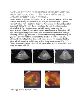
- 1. CASE 409: ECTOPIC FASCIOLIASIS, LÊ ĐÌNH VĨNH PHÚC, PHẠM CHÍ TOÀN, VÕ NGUYỄN THÀNH NHÂN, MEDIC MEDICAL CENTER, HCMC, VIETNAM Female patient, 31 year-old, accountant, in Gia Lai province. Onset 2 months with scattered bodyitching, no skin lesions, no fever, no abdominal pain. A private clinic in Gia Lai, with blood tests, diagnosed Toxocara sp infection, and gave her albendazole 800mg/day x 21 days. No itching, she went down to an another private clinic in Quy Nhon province, and with another blood tests, she was diagnosed infected Cysticercosis, treated with albendazole 800mg/day x 10 days. Then appearing right abdominal pain, ultrasound suspected liver damage caused by Fasciola spp. She went to Institute of Parasitology and Entomology in Quy Nhon province and then came to Medic Hoa Hoa in Ho Chi Minh city. Ultrasound detected right liver lesion with mixed echo, d = 5 cm, clear border, within a few hypoechoic nodules, and no liver tissue edema around. (Fig 1, 2). Further ultrasound detected colon wall thickness at liver region, hypoechoic, not lumen narrowing. (Fig 3). Endoscopyshowed transverse colitis.
- 2. Blood tests: WBC 14,500 cells/mm3 (Neutrophil 61.9%, Eosinophil 15.8%), hsCRP 14.53 mg/L. HBsAg (-), antiHCV (-), AFP (-), CEA (-), Fasciola sp IgG (+), stoolexam (-). Biopsy tissue in colon lesion was done and microscopic report was eosinophil mucosa colitis. MSCT CE presented liver lesion d = 4x6cm and transverse colon lesion with wall thickness d = 20mm (Fig 5, 6).
- 3. We diagnosed: liver abscess and pseudotumor colitis by Fasciola spp (Ectopic Fascioliasis), treated with Triclabendazol 10mg/kg/day x 2 days. Re-examination 4 weeks later, WBC 8,800 cells/mm3 (Eosinophil 2.5%), hsCRP 1.3 mg/L. Liver lesion in ultrasound and MSCT, wall thickness d = 8mm in MSCT (Fig 8, 9).
- 4. We represented an ectopic Fascioliasis case with hepatic and transverse colon lesions and an undifferential serodiagnosis. Endoscopic biopsyresult helped ruling out a colon tumor. But based on ultrasound findings of liver and colon lesions which were confirmed by MSCT we could chosed a concordantdiagnosis for this case.