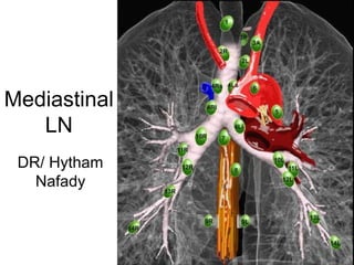
2. 1. Highest mediastinal LNs
Upper border: lower border or cricoid.
Lower border: upper border of manubrium
and clavicles.
Medial border: medial edge of CCA.
Lateral boder: first rib.
3.
4.
5.
6. 2R. Right upper paratracheal LNs
• Upper border: upper border of manubrium.
• Lower border: intersection between lower
border of left innominate vein and trachea.
• Medial border: midline of the trachea.
7.
8.
9. 2L. Left upper paratracheal LNs
• Upper border: superior border of the
manuibrium.
• Lower border: superior border of aortic
arch.
• Medial border: midline of the trachea.
10.
11.
12. 3. Prevascular & prevertebral LNs
• Prevascular LNs (3A) are located anterior
to the vessels.
• Prevertebral (retrotracheal) LNs (3B) are
located anterior to the spine.
• Prevertebral LNs are not accessible
through mediastnioscopy but only
available through Endoscopic U/S.
22. 6. para-aortic LNs
• Anterior & lateral to the ascending thoracic
aorta & aortic arch.
23.
24.
25. 7. Subcarinal LNs
• Upper border: carina of the trachea.
• Lower border: bronchus intermedius on
the right and left lower lobe bronchus on
the left.
29. 9. Pulmonary ligament LNs
• Pulmonary ligaments are inferior
extension of mediastinal pleural reflections
that surround the hila.
• Pulmonary ligaments LNs are located
posterior and inferior to the inferior
pulmonary veins.