1. 17
Poliomyelitis
249
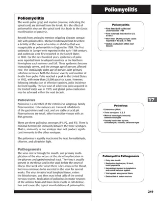
Poliomyelitis
The words polio (grey) and myelon (marrow, indicating the
spinal cord) are derived from the Greek. It is the effect of
poliomyelitis virus on the spinal cord that leads to the classic
manifestation of paralysis.
Records from antiquity mention crippling diseases compat-
ible with poliomyelitis. Michael Underwood first described
a debility of the lower extremities in children that was
recognizable as poliomyelitis in England in 1789. The first
outbreaks in Europe were reported in the early 19th century,
and outbreaks were first reported in the United States
in 1843. For the next hundred years, epidemics of polio
were reported from developed countries in the Northern
Hemisphere each summer and fall. These epidemics became
increasingly severe, and the average age of persons affected
rose. The increasingly older age of persons with primary
infection increased both the disease severity and number of
deaths from polio. Polio reached a peak in the United States
in 1952, with more than 21,000 paralytic cases. However,
following introduction of effective vaccines, polio incidence
declined rapidly. The last case of wild-virus polio acquired in
the United States was in 1979, and global polio eradication
may be achieved within the next decade.
Poliovirus
Poliovirus is a member of the enterovirus subgroup, family
Picornaviridae. Enteroviruses are transient inhabitants
of the gastrointestinal tract, and are stable at acid pH.
Picornaviruses are small, ether-insensitive viruses with an
RNA genome.
There are three poliovirus serotypes (P1, P2, and P3). There is
minimal heterotypic immunity between the three serotypes.
That is, immunity to one serotype does not produce signifi-
cant immunity to the other serotypes.
The poliovirus is rapidly inactivated by heat, formaldehyde,
chlorine, and ultraviolet light.
Pathogenesis
The virus enters through the mouth, and primary multi-
plication of the virus occurs at the site of implantation in
the pharynx and gastrointestinal tract. The virus is usually
present in the throat and in the stool before the onset of
illness. One week after onset there is less virus in the throat,
but virus continues to be excreted in the stool for several
weeks. The virus invades local lymphoid tissue, enters
the bloodstream, and then may infect cells of the central
nervous system. Replication of poliovirus in motor neurons
of the anterior horn and brain stem results in cell destruc-
tion and causes the typical manifestations of poliomyelitis.
2. Poliomyelitis
250
17
Clinical Features
The incubation period for poliomyelitis is commonly 6 to 20
days with a range of 3 to 35 days.
The response to poliovirus infection is highly variable and
has been categorized on the basis of the severity of clinical
presentation.
Up to 95% of all polio infections are inapparent or asymp-
tomatic. Estimates of the ratio of inapparent to paralytic
illness vary from 50:1 to 1,000:1 (usually 200:1). Infected
persons without symptoms shed virus in the stool and are
able to transmit the virus to others.
Approximately 4%–8% of polio infections consist of a minor,
nonspecific illness without clinical or laboratory evidence of
central nervous system invasion. This clinical presentation
is known as abortive poliomyelitis, and is characterized by
complete recovery in less than a week. Three syndromes
observed with this form of poliovirus infection are upper
respiratory tract infection (sore throat and fever), gastrointes-
tinal disturbances (nausea, vomiting, abdominal pain, consti-
pation or, rarely, diarrhea), and influenza-like illness. These
syndromes are indistinguishable from other viral illnesses.
Nonparalytic aseptic meningitis (symptoms of stiffness of the
neck, back, and/or legs), usually following several days after
a prodrome similar to that of minor illness, occurs in 1%–2%
of polio infections. Increased or abnormal sensations can
also occur. Typically these symptoms will last from 2 to 10
days, followed by complete recovery.
Fewer than 1% of all polio infections result in flaccid
paralysis. Paralytic symptoms generally begin 1 to 10 days
after prodromal symptoms and progress for 2 to 3 days.
Generally, no further paralysis occurs after the temperature
returns to normal. The prodrome may be biphasic, especially
in children, with initial minor symptoms separated by a
1- to 7-day period from more major symptoms. Additional
prodromal signs and symptoms can include a loss of
superficial reflexes, initially increased deep tendon reflexes
and severe muscle aches and spasms in the limbs or back.
The illness progresses to flaccid paralysis with diminished
deep tendon reflexes, reaches a plateau without change for
days to weeks, and is usually asymmetrical. Strength then
begins to return. Patients do not experience sensory losses or
changes in cognition.
Many persons with paralytic poliomyelitis recover completely
and, in most, muscle function returns to some degree.
Weakness or paralysis still present 12 months after onset is
usually permanent.
Paralytic polio is classified into three types, depending on
3. Poliomyelitis
251
17
the level of involvement. Spinal polio is most common, and
during 1969–1979, accounted for 79% of paralytic cases.
It is characterized by asymmetric paralysis that most often
involves the legs. Bulbar polio leads to weakness of muscles
innervated by cranial nerves and accounted for 2% of cases
during this period. Bulbospinal polio, a combination of
bulbar and spinal paralysis, accounted for 19% of cases.
The death-to-case ratio for paralytic polio is generally 2%–5%
among children and up to 15%–30% for adults (depending
on age). It increases to 25%–75% with bulbar involvement.
Laboratory Diagnosis
Viral Isolation
Poliovirus may be recovered from the stool or pharynx of a
person with poliomyelitis. Isolation of virus from the cere-
brospinal fluid (CSF) is diagnostic, but is rarely accomplished.
If poliovirus is isolated from a person with acute flaccid
paralysis, it must be tested further, using oligonucleotide
mapping (fingerprinting) or genomic sequencing, to deter-
mine if the virus is “wild type” (that is, the virus that causes
polio disease) or vaccine type (virus that could derive from a
vaccine strain).
Serology
Neutralizing antibodies appear early and may be at high
levels by the time the patient is hospitalized; therefore, a
fourfold rise in antibody titer may not be demonstrated.
Cerebrospinal Fluid
In poliovirus infection, the CSF usually contains an increased
number of white blood cells (10–200 cells/mm3, primarily
lymphocytes) and a mildly elevated protein (40–50 mg/100 mL).
Epidemiology
Occurrence
At one time poliovirus infection occurred throughout the
world. Transmission of wild poliovirus was interrupted
in the United States in 1979, or possibly earlier. A polio
eradication program conducted by the Pan American Health
Organization led to elimination of polio in the Western
Hemisphere in 1991. The Global Polio Eradication Program
has dramatically reduced poliovirus transmission throughout
the world. In 2009, only 1,579 confirmed cases of polio were
reported globally and polio was endemic in four countries.
4. Poliomyelitis
252
17
Reservoir
Humans are the only known reservoir of poliovirus, which
is transmitted most frequently by persons with inapparent
infections. There is no asymptomatic carrier state except in
immune deficient persons.
Transmission
Person-to-person spread of poliovirus via the fecal-oral route
is the most important route of transmission, although the
oral-oral route may account for some cases.
Temporal Pattern
Poliovirus infection typically peaks in the summer months in
temperate climates. There is no seasonal pattern in tropical
climates.
Communicability
Poliovirus is highly infectious, with seroconversion rates
among susceptible household contacts of children nearly
100%, and greater than 90% among susceptible household
contacts of adults. Persons infected with poliovirus are most
infectious from 7 to 10 days before and after the onset of
symptoms, but poliovirus may be present in the stool from
3 to 6 weeks.
Secular Trends in the United States
Before the 18th century, polioviruses probably circulated
widely. Initial infections with at least one type probably
occurred in early infancy, when transplacentally acquired
maternal antibodies were high. Exposure throughout life
probably provided continual boosting of immunity, and
paralytic infections were probably rare. (This view has been
recently challenged based on data from lameness studies
in developing countries).
In the immediate prevaccine era, improved sanitation
allowed less frequent exposure and increased the age of
primary infection. Boosting of immunity from natural
exposure became more infrequent and the number of
susceptible persons accumulated, ultimately resulting in
the occurrence of epidemics, with 13,000 to 20,000 para-
lytic cases reported annually.
In the early vaccine era, the incidence dramatically
decreased after the introduction of inactivated polio vaccine
(IPV) in 1955. The decline continued following oral polio
vaccine (OPV) introduction in 1961. In 1960, a total of 2,525
paralytic cases were reported, compared with 61 in 1965.
The last cases of paralytic poliomyelitis caused by endemic
transmission of wild virus in the United States were in
5. Poliomyelitis
253
17
1979, when an outbreak occurred among the Amish in
several Midwest states. The virus was imported from the
Netherlands.
From 1980 through 1999, a total of 152 confirmed cases
of paralytic poliomyelitis were reported, an average of 8
cases per year. Six cases were acquired outside the United
States and imported. The last imported case was reported
in 1993. Two cases were classified as indeterminant (no
poliovirus isolated from samples obtained from the patients,
and patients had no history of recent vaccination or direct
contact with a vaccine recipient). The remaining 144 (95%)
cases were vaccine-associated paralytic polio (VAPP) caused
by live oral polio vaccine.
In order to eliminate VAPP from the United States, ACIP
recommended in 2000 that IPV be used exclusively in the
United States. The last case of VAPP acquired in the United
States was reported in 1999. In 2005, an unvaccinated U.S.
resident was infected with polio vaccine virus in Costa Rica
and subsequently developed VAPP. A second case of VAPP
from vaccine-derived poliovirus was reported in 2009. Also
in 2005, several asymptomatic infections with a vaccine-
derived poliovirus were detected in unvaccinated children
in Minnesota. The source of the vaccine virus has not been
determined, but it appeared to have been circulating among
humans for at least 2 years based on genetic changes in the
virus. No VAPP has been reported from this virus.
Poliovirus Vaccines
Inactivated poliovirus vaccine (IPV) was licensed in 1955 and
was used extensively from that time until the early 1960s.
In 1961, type 1 and 2 monovalent oral poliovirus vaccine
(MOPV) was licensed, and in 1962, type 3 MOPV was licensed.
In 1963, trivalent OPV was licensed and largely replaced
IPV use. Trivalent OPV was the vaccine of choice in the
United States and most other countries of the world after its
introduction in 1963. An enhanced-potency IPV was licensed
in November 1987 and first became available in 1988. Use of
OPV was discontinued in the United States in 2000.
Characteristics
Inactivated poliovirus vaccine
Two enhanced forms of inactivated poliovirus vaccine are
currently licensed in the U.S., but only one vaccine (IPOL,
sanofi pasteur) is actually distributed. This vaccine contains
all three serotypes of polio vaccine virus. The viruses are
grown in a type of monkey kidney tissue culture (Vero
cell line) and inactivated with formaldehyde. The vaccine
contains 2-phenoxyethanol as a preservative, and trace
amounts of neomycin, streptomycin, and polymyxin B. It
6. Poliomyelitis
254
17
is supplied in a single-dose prefilled syringe and should be
administered by either subcutaneous or intramuscular injec-
tion.
Oral poliovirus vaccine
Trivalent OPV contains live attenuated strains of all three
serotypes of poliovirus in a 10:1:3 ratio. The vaccine viruses
are grown in monkey kidney tissue culture (Vero cell line).
The vaccine is supplied as a single 0.5-mL dose in a plastic
dispenser. The vaccine contains trace amounts of neomycin
and streptomycin. OPV does not contain a preservative.
Live attenuated polioviruses replicate in the intestinal
mucosa and lymphoid cells and in lymph nodes that drain
the intestine. Vaccine viruses are excreted in the stool of the
vaccinated person for up to 6 weeks after a dose. Maximum
viral shedding occurs in the first 1–2 weeks after vaccination,
particularly after the first dose.
Vaccine viruses may spread from the recipient to contacts.
Persons coming in contact with fecal material of a vaccinated
person may be exposed and infected with vaccine virus.
Immunogenicity and Vaccine Efficacy
Inactivated poliovirus vaccine
IPV is highly effective in producing immunity to poliovirus
and protection from paralytic poliomyelitis. Ninety percent
or more of vaccine recipients develop protective antibody
to all three poliovirus types after two doses, and at least
99% are immune following three doses. Protection against
paralytic disease correlates with the presence of antibody.
IPV appears to produce less local gastrointestinal immunity
than does OPV, so persons who receive IPV are more readily
infected with wild poliovirus than OPV recipients.
The duration of immunity with IPV is not known with
certainty, although it probably provides protection for many
years after a complete series.
Oral poliovirus vaccine
OPV is highly effective in producing immunity to poliovirus.
A single dose of OPV produces immunity to all three vaccine
viruses in approximately 50% of recipients. Three doses
produce immunity to all three poliovirus types in more than
95% of recipients. As with other live-virus vaccines, immunity
from oral poliovirus vaccine is probably lifelong. OPV
produces excellent intestinal immunity, which helps prevent
infection with wild virus.
7. Poliomyelitis
255
17
Serologic studies have shown that seroconversion following
three doses of either IPV or OPV is nearly 100% to all three
vaccine viruses. However, seroconversion rates after three
doses of a combination of IPV and OPV are lower, particu-
larly to type 3 vaccine virus (as low as 85% in one study).
A fourth dose (most studies used OPV as the fourth dose)
usually produces seroconversion rates similar to three doses
of either IPV or OPV.
Vaccination Schedule and Use
Trivalent OPV was the vaccine of choice in the United States
(and most other countries of the world) since it was licensed
in 1963. The nearly exclusive use of OPV led to elimination
of wild-type poliovirus from the United States in less than
20 years. However, one case of VAPP occurred for every 2
to 3 million doses of OPV administered, which resulted in
8 to 10 cases of VAPP each year in the United States (see
Adverse Reactions section for more details on VAPP). From
1980 through 1999, VAPP accounted for 95% of all cases of
paralytic poliomyelitis reported in the United States.
In 1996, ACIP recommended an increase in use of IPV
through a sequential schedule of IPV followed by OPV. This
recommendation was intended to reduce the occurrence of
vaccine-associated paralytic polio. The sequential schedule
was expected to eliminate VAPP among vaccine recipients
by producing humoral immunity to polio vaccine viruses
with inactivated polio vaccine prior to exposure to live
vaccine virus. Since OPV was still used for the third and
fourth doses of the polio vaccination schedule, a risk of VAPP
would continue to exist among contacts of vaccinees, who
were exposed to live vaccine virus in the stool of vaccine
recipients.
The sequential IPV–OPV polio vaccination schedule was
widely accepted by both providers and parents. Fewer
cases of VAPP were reported in 1998 and 1999, suggesting
an impact of the increased use of IPV. However, only
the complete discontinuation of use of OPV would lead
to complete elimination of VAPP. To further the goal of
complete elimination of paralytic polio in the United States,
ACIP recommended in July 1999 that inactivated polio
vaccine be used exclusively in the United States beginning
in 2000. OPV is no longer routinely available in the United
States. Exclusive use of IPV eliminated the shedding of live
vaccine virus, and eliminated any indigenous VAPP.
A primary series of IPV consists of three doses. In infancy,
these primary doses are integrated with the administration
of other routinely administered vaccines. The first dose may
be given as early as 6 weeks of age but is usually given at
2 months of age, with a second dose at 4 months of age.
The third dose should be given at 6–18 months of age. The
8. Poliomyelitis
256
17
recommended interval between the primary series doses is
2 months. However, if accelerated protection is needed, the
minimum interval between each of the first 3 doses of IPV is
4 weeks.
The final dose in the IPV series should be administered at 4
years of age or older. A dose of IPV on or after age 4 years is
recommended regardless of the number of previous doses.
The minimum interval from the next-to-last to final dose is 6
months.
When DTaP-IPV/Hib (Pentacel) is used to provide 4 doses at
ages 2, 4, 6, and 15-18 months, an additional booster dose
of age-appropriate IPV-containing vaccine (IPV or DTaP-IPV
[Kinrix]) should be administered at age 4-6 years. This will
result in a 5-dose IPV vaccine series, which is considered
acceptable by ACIP. DTaP-IPV/Hib is not indicated for the
booster dose at 4-6 years of age. ACIP recommends that the
minimum interval from dose 4 to dose 5 should be at least 6
months to provide an optimum booster response.
Shorter intervals between doses and beginning the series
at a younger age may lead to lower seroconversion rates.
Consequently, ACIP recommends the use of the minimum
age (6 weeks) and minimum intervals between doses in the
first 6 months of life only if the vaccine recipient is at risk for
imminent exposure to circulating poliovirus (e.g., during an
outbreak or because of travel to a polio-endemic region).
Only IPV is available for routine polio vaccination of children
in the United States. A polio vaccination schedule begun with
OPV should be completed with IPV. If a child receives both
types of vaccine, four doses of any combination of IPV or
OPV by 4–6 years of age is considered a complete poliovirus
vaccination series. A minimum interval of 4 weeks should
separate all doses of the series.
There are three combination vaccines that contain inacti-
vated polio vaccine. Pediarix is produced by GlaxoSmithKline
and contains DTaP, hepatitis B and IPV vaccines. Pediarix
is licensed for the first 3 doses of the DTaP series among
children 6 weeks through 6 years of age. Kinrix is also
produced by GSK and contains DTaP and IPV. Kinrix is
licensed only for the fifth dose of DTaP and fourth dose
of IPV among children 4 through 6 years of age. Pentacel
is produced by sanofi pasteur and contains DTaP, Hib and
IPV. It is licensed for the first four doses of the component
vaccines among children 6 weeks through 4 years of age.
Pentacel is not licensed for children 5 years or older.
Additional information about these combination vaccines is
in the Pertussis chapter of this book.
9. Poliomyelitis
257
17
Polio Vaccination of Adults
Routine vaccination of adults (18 years of age and older) who
reside in the United States is not necessary or recommended
because most adults are already immune and have a very
small risk of exposure to wild poliovirus in the United States.
Some adults, however, are at increased risk of infection with
poliovirus. These include travelers to areas where poliomy-
elitis is endemic or epidemic (currently limited to South Asia,
the eastern Mediterranean, and Africa), laboratory workers
handling specimens that may contain polioviruses.
Recommendations for poliovirus vaccination of adults in
the above categories depend upon the previous vaccination
history and the time available before protection is required.
For unvaccinated adults (including adults without a
written record of prior polio vaccination) at increased risk
of exposure to poliomyelitis, primary immunization with
IPV is recommended. The recommended schedule is two
doses separated by 1 to 2 months, and a third dose given
6 to 12 months after the second dose. The minimum
interval between the second and the third doses is 6
months.
In some circumstances time will not allow completion of this
schedule. If 8 weeks or more are available before protection
is needed, three doses of IPV should be given at least 4
weeks apart. If 4 to 8 weeks are available before protection
is needed, two doses of IPV should be given at least 4 weeks
apart. If less than 4 weeks are available before protection
is needed, a single dose of IPV is recommended. In all
instances, the remaining doses of vaccine should be given
later, at the recommended intervals, if the person remains at
increased risk.
Adults who have previously completed a primary series of
3 or more doses and who are at increased risk of exposure
to poliomyelitis should be given one dose of IPV. The need
for further supplementary doses has not been established.
Only one supplemental dose of polio vaccine is recom-
mended for adults who have received a complete series
(i.e., it is not necessary to administer additional doses for
subsequent travel to a polio endemic country).
Adults who have previously received less than a full
primary course of OPV or IPV and who are at increased
risk of exposure to poliomyelitis should be given the
remaining doses of IPV, regardless of the interval since
the last dose and type of vaccine previously received. It is
not necessary to restart the series of either vaccine if the
schedule has been interrupted.
10. Poliomyelitis
258
17
Contraindications And Precautions To
Vaccination
Severe allergic reaction (anaphylaxis) to a vaccine compo-
nent, or following a prior dose of vaccine, is a contraindica-
tion to further doses of that vaccine. Since IPV contains trace
amounts of streptomycin, neomycin, and polymyxin B, there
is a possibility of allergic reactions in persons sensitive to
these antibiotics. Persons with allergies that are not anaphy-
lactic, such as skin contact sensitivity, may be vaccinated.
Moderate or severe acute illness is a precaution for IPV.
Breastfeeding does not interfere with successful immuniza-
tion against poliomyelitis with IPV. IPV may be administered
to a child with diarrhea. Minor upper respiratory illnesses
with or without fever, mild to moderate local reactions to a
prior dose of vaccine, current antimicrobial therapy, and the
convalescent phase of an acute illness are not contraindica-
tions for vaccination with IPV.
Contraindications to combination vaccines that contain
IPV are the same as the contraindications to the individual
components (e.g., DTaP, hepatitis B).
Adverse Reactions Following
Vaccination
Minor local reactions (pain, redness) may occur following IPV.
No serious adverse reactions to IPV have been documented.
Because IPV contains trace amounts of streptomycin,
polymyxin B, and neomycin, allergic reactions may occur in
persons sensitive to these antibiotics.
Vaccine-Associated Paralytic Poliomyelitis
Vaccine-associated paralytic polio is a rare adverse reaction
following live oral poliovirus vaccine. Inactivated poliovirus
vaccine does not contain live virus, so it cannot cause VAPP.
The mechanism of VAPP is believed to be a mutation, or
reversion, of the vaccine virus to a more neurotropic form.
These mutated viruses are called revertants. Reversion is
believed to occur in almost all vaccine recipients, but it
only rarely results in paralytic disease. The paralysis that
results is identical to that caused by wild virus, and may be
permanent.
VAPP is more likely to occur in persons 18 years of age and
older than in children, and is much more likely to occur in
immunodeficient children than in those who are immuno-
competent. Compared with immunocompetent children,
the risk of VAPP is almost 7,000 times higher for persons
with certain types of immunodeficiencies, particularly
B-lymphocyte disorders (e.g., agammaglobulinemia and
hypogammaglobulinemia), which reduce the synthesis of
11. Poliomyelitis
259
17
immune globulins. There is no procedure available for iden-
tifying persons at risk of paralytic disease, except excluding
older persons and screening for immunodeficiency.
From 1980 through 1998, 152 cases of paralytic polio were
reported in the United States; 144 (95%) of these cases were
VAPP, and the remaining eight were in persons who acquired
documented or presumed wild-virus polio outside the United
States. Of the 144 VAPP cases, 59 (41%) occurred in healthy
vaccine recipients (average age 3 months). Forty-four (31%)
occurred in healthy contacts of vaccine recipients (average
age 26 years), and 7 (5%) were community acquired (i.e.,
vaccine virus was recovered but there was no known contact
with a vaccine recipient). Thirty-four (24%) of VAPP cases
occurred in persons with immunologic abnormalities (27 in
vaccine recipients and 7 in contacts of vaccine recipients).
None of the vaccine recipients were known to be immuno-
logically abnormal prior to vaccination.
The risk of VAPP is not equal for all OPV doses in the
vaccination series. The risk of VAPP is 7 to 21 times higher
for the first dose than for any other dose in the OPV
series. From 1980 through 1994, 303 million doses of OPV
were distributed and 125 cases of VAPP were reported,
for an overall risk of VAPP of one case per 2.4 million
doses. Forty-nine paralytic cases were reported among
immunocompetent recipients of OPV during this period.
The overall risk to these recipients was one VAPP case per
6.2 million OPV doses. However, 40 (82%) of these 49 cases
occurred following receipt of the first dose, making the risk
of VAPP one case per 1.4 million first doses. The risk for
all other doses was one per 27.2 million doses. The reason
for this difference by dose is not known with certainty, but
it is probably because the vaccine virus is able to replicate
longer in a completely nonimmune infant. This prolonged
replication increases the chance of the emergence of a
revertant virus that may cause paralysis. The situation is
similar for contacts. A nonimmune child may shed virus
longer, increasing the chance of exposure of a contact.
The last case of VAPP acquired in the United States was
reported in 1999. As noted previously, a U.S. resident with
VAPP was reported in 2005, but the vaccine virus infection
was acquired in Costa Rica.
Vaccine Storage and Handling
IPV may be shipped without refrigeration provided it is
delivered within 4 days. It should be maintained at 35°–46°F
(2°–8°C). The vaccine should be clear and colorless. Any
vaccine showing particulate matter, turbidity, or change in
color should be discarded.
12. Poliomyelitis
260
17
Outbreak Investigation and Control
Collect preliminary clinical and epidemiologic information
(including vaccine history and contact with OPV vaccines) on
any suspected case of paralytic polio. Notify CDC, (404-639-
8255) after appropriate local and state health authorities
have been notified. Intensify field investigation to verify
information and collect appropriate specimens for viral isola-
tion and serology.
A single case of paralytic poliomyelitis demands immediate
attention. If the evidence indicates vaccine-associated
disease, no outbreak control program is needed. If, however,
evidence indicates wild virus (for example, two cases in a
community), all unvaccinated persons in the epidemic area
who are 6 weeks of age and older and whose vaccine histo-
ries are uncertain should be vaccinated.
Polio Eradication
Following the widespread use of poliovirus vaccine in the
mid-1950s, the incidence of poliomyelitis declined rapidly
in many industrialized countries. In the United States, the
number of cases of paralytic poliomyelitis reported annually
declined from more than 20,000 cases in 1952 to fewer than
100 cases in the mid-1960s. The last documented indigenous
transmission of wild poliovirus in the United States was in 1979.
In 1985, the member countries of the Pan American Health
Organization adopted the goal of eliminating poliomyelitis
from the Western Hemisphere by 1990. The strategy to
achieve this goal included increasing vaccination coverage;
enhancing surveillance for suspected cases (i.e., surveillance
for acute flaccid paralysis); and using supplemental immu-
nization strategies such as national immunization days,
house-to-house vaccination, and containment activities.
Since 1991, when the last wild-virus–associated indigenous
case was reported from Peru, no additional cases of poliomy-
elitis have been confirmed despite intensive surveillance. In
September 1994, an international commission certified the
Western Hemisphere to be free of indigenous wild poliovirus.
The commission based its judgment on detailed reports from
national certification commissions that had been convened
in every country in the region.
In 1988, the World Health Assembly (the governing body of
the World Health Organization) adopted the goal of global
eradication of poliovirus by the year 2000. Although this goal
was not achieved, substantial progress has been made. One
type of poliovirus appears to have already been eradicated.
In 1988, an estimated 350,000 cases of paralytic polio
occurred, and the disease was endemic in more than 125
countries. By 2006, fewer than 2,000 cases were reported
globally—a reduction of more than 99% from 1988—and
polio remained endemic in only four countries. In addition,
13. Poliomyelitis
261
17
one type of poliovirus appears to have already been
eradicated. The last isolation of type 2 virus was in India in
October 1999.
The polio eradication initiative is led by a coalition
of international organizations that includes WHO, the
United Nations Children’s Fund (UNICEF), CDC, and Rotary
International. Other bilateral and multilateral organizations
also support the initiative. Rotary International has contrib-
uted more than $600 million to support the eradication
initiative. Current information on the status of the global
polio eradication initiative is available on the World Health
Organization website at www.polioeradication.org/.
Postpolio Syndrome
After an interval of 30–40 years, 25%–40% of persons who
contracted paralytic poliomyelitis in childhood experience
new muscle pain and exacerbation of existing weakness,
or develop new weakness or paralysis. This disease entity
is referred to as postpolio syndrome. Factors that increase
the risk of postpolio syndrome include increasing length of
time since acute poliovirus infection, presence of permanent
residual impairment after recovery from the acute illness,
and female sex. The pathogenesis of postpolio syndrome
is thought to involve the failure of oversized motor units
created during the recovery process of paralytic poliomy-
elitis. Postpolio syndrome is not an infectious process, and
persons experiencing the syndrome do not shed poliovirus.
For more information, or for support for persons with post-
polio syndrome and their families, contact:
Post-Polio Health International
4207 Lindell Boulevard #110
St. Louis, MO 63108-2915
314-534-0475
www.post-polio.org
Selected References
CDC. Imported vaccine-associated paralytic poliomyelitis—
United States, 2005. MMWR 2006;55:97–9.
CDC. Tracking Progress Toward Global Polio Eradication —
Worldwide, 2009–2010. MMWR 2011;60(No. 14):441-5.
CDC. Poliomyelitis prevention in the United States:
updated recommendations of the Advisory Committee
on Immunization Practices. (ACIP). MMWR 2000;49 (No.
RR-5):1–22.
CDC. Apparent global interruption of wild poliovirus type 2
transmission. MMWR 2001;50:222–4.