

Recommandé
Contenu connexe
Tendances
Tendances (20)
En vedette
En vedette (20)
Similaire à Paeds
Similaire à Paeds (20)
Plus de Pratik Kumar
Paeds
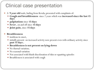
- 1. Clinical case presentation A 9 year old male, hailing from Kerala, presented with complaints of: Cough and breathlessness since 2 years which was increased since the last 15 days palpitations since 15 days Fever , on and off since 15 days Joint pain, since 15 days Breathlessness insidious in onset, initially present on increased activity now present even with ordinary activity since past 15 days . Breathlesness is not present on lying down No diurnal variation. No seasonal variation Not associated with bluish discoloration of skin or squatting episodes Breathlesness is associated with cough
- 2. Cough associated with scanty mucoid expectoration since the past 15 dyas, non blood stained. Not associated with rise in temperature Not associated with wheeze No diurnal variation , not increased on exposure to cold(seasonal variation) Cough is associated with chest pain, which is present only during coughing, pricking in type, present diffusely, non radiating. Palpitations Present even at rest. Not associated with chest pain Associated with dizziness but there was no loss of consciousness.
- 3. Fever High grade Present on and off since 15 days Not associated with rigors Pain and swelling of the left knee joint since 15 days Associated with limitation of movement, the child could not walk due to the pain Not history of trauma after a week the pain in the knee joint subsided, and the child had pain in the left ankle joint and right elbow joint. No history of morning stiffness. pain subsided immediately after the initiation of treatment
- 4. Negative history no history of reduced urine output no history of pedal oedema No history of abdominal pain No history of recurrent sore throat. No history of rash or bleeding manifestations (petechial hemorrhages, purpura) No history of recurrent respiratory tract infections history of weight loss present but could not be quantified
- 5. PAST HISTORY History of similar complaints in the past. 3 years back he presented with fever associated with fleeting type of joint pain , and was admitted in the hospital and treated for the same. He was advised to take injections every 3 weeks until the age of twenty five(suggestive of acute rheumatic fever) but the injections were discontinued one year back due to reasons unknown. No history of asthma. No history of contact with tuberculosis
- 6. BIRTH HISTORY No complications in the antenatal period Institutional delivery at term, normal vaginal delivery, baby cried immediately. Birth weight- not known. No complications during the delivery No post natal complications. NO ICU admissions after birth. No problems during infancy.
- 7. DEVELOPMENTAL HISTORY all milestones were achieved on time. IMMUNISATION HISTORY Immunisation up to date OPV, BCG, DPT and boosters , Measles vaccine given.
- 8. DIET HISTORY MEAL FOOD ITEM CALORIES PROTEIN Morning 1 cup milk with sugar 127 4.1 2 biscuits Breakfast 2 dosa , half cup dal 200 6 Lunch 1 cup rice 376 10 1 cup sambar 1 cup of vegetables Evening snack 1 cup milk with sugar 87 3.3 dinner 2 roti 287 24 Half cup dal 1 fish Calories requirement = 1620 kcal Protein requiremnt=33.2 g/day Calories obtained= 1047 Obtained= 35 g Defecit= 573 No defecit
- 9. SOCIAL HISTORY He is in 3rd standard. He performs averagely at school. He has been held back a year due to missing school due to poor health and his performance.
- 10. FAMILY HISTORY Non consanguineous marriage. No history of similar complaints in the family Socioeconomic status- Upper lower class as per modified Kuppuswamy scale. Total family members- 4. The child has two siblings, both healthy and active, going to school. 9 yrs 7 yrs 5 yrs
- 11. Summary A 9 year old boy, with previous history suggestive of rheumatic fever, advised monthly injections since the last 3 years which were discontinued the past 1 year, presented with cough, breathlessness, palpitations, along with fever and fleeting type of joint pain and swelling of knee, ankle and elbow joint since past 15 days. Probable diagnosis Recurrence of rheumatic fever with rheumatic carditis.
- 13. GENERAL PHYSICAL EXAMINATION Patient is conscious, cooperative, well oriented with time, place and person 1. Pallor present 2. No Icterus, Clubbing, Cyanosis, Lymphadenopathy or Edema
- 14. Signs of Infective endocarditis: Absent(except pallor) Head to toe examination: Sunken eyes Thyroid- normal. Spine- normal. No skeletal deformities.
- 15. Anthropometry: 1. Height: 126cm Inference: Normal (Between 10th and 25th centiles) 2. Weight: 18.5kg Inference: Below the 3rd centile. BMI=12.85Kg/m2 Impression:UNDERWEIGHT 3.Arm span:125cm. 4.US:LS=1:1
- 16. Vitals: 1. BP:90/60mmHg Right arm supine position. 2. Pulse: 120bpm, Increased rate, regular rhythm, normal volume & character. No radioradial or radiofemoral delay.All peripheral pulses felt. 3. Respiratory rate: 30 cycles/minute. Abdominothoracic. 4. Temperature: 37.2°C 5. JVP: Not raised.
- 18. Inspection: Precordium appears normal. Apical impulse: diffuse, Left 6th ICS 1cm lateral to MCL. Visible pulsation seen in Left 2nd ,3rd,4th ICS. No visible epigastric pulsations. No scars, sinuses or dilated veins. Palpation: Apex beat is in Left 6th ICS 1cm lateral to MCL, Hyperdynamic in character, Systolic thrill present. Parasternal heave present. Palpable P2. No epigastric pulsations. No carotid thrill. No palpable pericardial rub/tracheal tug.
- 19. Percussion: Right heart border corresponds to sternum, Left heart border corresponds to the apex. Auscultation: Mitral area: S1normal, S2 muffled. A high pitched, pansystolic murmur of grade IV intensity,soft blowing in character,heard with the diaphragm of stethoscope, in left lateral position of the patient and at the height of expiration.The murmur is radiated to the left axilla and the back.
- 20. Pulmonary area: S1 heard, loud P2,ejection systolic murmur of grade III intensity heard. Aortic area: S1 S2 heard. Tricuspid area:S1 S2 heard. Pansystolic murmur of gradeIII intensity. No carotid bruit.
- 21. Respiratory system Inspection: Upper respiratory tract: normal Lower respiratory tract: Trachea central Movements B/L symmetrical No signs of volume loss. No scars, sinuses or dilated veins
- 22. Palpation: Trachea central Movements B/L symmetrical Vocal fremitus: B/L equal Percussion: Normal resonant note B/L Auscultation: Normal vesicular breath sounds heard in all areas. No added sounds.
- 23. Gastrointestinal system Oral cavity:Normal Per Abdomen: Soft, nontender Liver is palpable 2cm below the RCM. Liver span 8cm. Spleen not palpable. No shifting dullness. Traube’s space tympanic on percussion. Normal bowel sounds heard
- 24. 1. Nervous system examination: Higher mental functions: No abnormalities detected No cranial nerve abnormalities Motor system: No abnormalities detected; Bilateral flexor plantar Sensory system: No abnormalities detected Stance and Gait: No abnormalities detected Co-ordination: No abnormalities detected Signs of meningeal irritation: Absent Skull and spine: No abnormalities detected
- 25. Summary A 9 yr old child with dyspnoea,on& off fever,cough, chest pain,palpitation since the past10days. O/E: Found to have tachycardia, pallor, apex is shifted outwards & is hyperdynamic, systolic thrill at the apex, parasternal heave, palpable P2, pansystolic murmur (grade IV)in mitral area radiated to the left axilla and the back. loud P2
- 26. Diagnosis ACUTE RHEUMATIC CARDITIS WITH MITRAL REGURGITATION WITH FEATURES OF PULMONARY HYPERTENSION. PATIENT IS IN SINUS RHYTHM AT PRESESNT NOT IN CCF OR INFECTIVE ENDOCRDITIS.
- 27. Investigation
- 28. Complete blood counts: 1. Total count: increased 2. Differential count: polymorphonuclear leucocytosis 3. Hb: anemia 4. Peripheral smear 5. ESR: Raised: acute rheumatic fever Decreased: CCF, mild carditis, chorea 6. Acute phase reactants: ESR: increased CRP: increased (beta-globulin in a/c rheumatic fever) 7. Blood culture Liver Function Tests Renal Function Tests Urinalysis ABG
- 29. Evidence of streptococcal infection 1. ASO titer 2. Other antibodies: Antihyaluronidase (AH) Anti-streptokinase (ASK) Antistreptozyme (ASTZ) Anti-DNAse B 3. Positive throat culture 4. Rapid streptococcal antigen detection test Evidence of carditis 1. CXR 2. ECG 3. ECHO
- 30. Investigations Results Inference Hemoglobin 9.5 g% Moderate anemia Total Count 15,800 cells/cumm Elevated Differential Count N70, L17, M1.3 Normal ESR 13mm/L/hr ??? Normal Platelets 3.79lakhs Normal Serum urea 22mg/dl Normal Serum Creatinine 1.5mg/dl Raised Na+ 131mEq/L Slightly low K+ 4.4mEq/L Normal Cl- 93.3mEq/L Slightly low HCO3- 17.5mEq/L Low Ca2+ 8.6mg/dl Normal Phosphate 4.2mg/dl Normal
- 31. Investigation Result Inference Total bilirubin 0.6g/dl Normal Direct bilirubin 0.17g/dl Normal Serum globulin 3.7g/dl Slightly elevated SGOT/AST 29IU/L Normal SPGT/ALT 30IU/L Normal ALP 215IU.L Normal Urinanalysis Albumin: 2+ Albuminuria 2-4 pus cells Normal 8-10 RBCs Elevated pH 7.45 Normal pCO2 28.2 Low pO2 187 High SpO2 99.7% Normal HCO3- 19.3 Low
- 32. Peripheral smear: Blood culture: Negative CXR: Cardiomegaly present No signs of pulmonary congestion ECHO: Rheumatic heart disease Mildly dilated LA/LV Severe mitral regurgitation Mild pericardial effusion No pulmonary arterial hypertension No vegetation
- 33. Impression Acute rheumatic carditis with severe MR, cardiomegaly & mild pericardial effusion Anemia Respiratory alkalosis - compensated (low pCO2, low HCO3- and normal pH)
- 35. Management of Acute Episode Bed Rest For carditis and arthritis Prednisolone 2 mg/kg/day for 2 weeks Taper over next 2-4 weeks Start Aspirin 50-75 mg/kg/day simultaneously to complete total 12 weeks Antistreptococcal therapy 200,000 units/ kg/ day for 10 days
- 36. Infective Endocarditis Based on culture and sensitivity Empirical Therapy : Add aminoglycoside
- 37. Secondary Prophylaxis Up to 40 years of age or Lifelong Benzathine Penicillin 0.6 MU single dose every 15 days
- 38. Management Of Malnutrition And Anemia Health Education – non compliance!! Increase Calorie intake Increase frequency Vitamins Oral Iron – 3-6 mg/kg/day. Continue for 4-6 months after correction Dietary counseling
- 39. Current Medications Inj. Crystalline Penicillin 1 MU IV 6 hourly Inj. Furosemide 20 mg IV BD Inj. Ranitidine 20 mg IV 8 hourly Tab. Paracetamol 500 mg (1/2) SOS Inj. Gentamicin 60 mg IV OD (3 mg/kg/day) Inj. Digoxin Tab. Prednisolone 10 mg 6 hourly Neb. Asthalin 1 respule 4 hourly
- 40. Atrial Fibrillation Rate Control – Digoxin Rhythm Control – Amiodarone Anticoagulants
- 41. THANK YOU!