Class iii malocclusion /certified fixed orthodontic courses by Indian dental academy
•
38 j'aime•9,756 vues

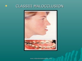
Class III malocclusion is characterized by the mandible being positioned forward in relation to the maxilla and cranial base. It can be caused by mandibular prognathism, maxillary retrognathism, or a combination. Treatment depends on whether the malocclusion has a dentoalveolar or skeletal component, and the patient's growth stage. For skeletal class III issues, early intervention like facemask therapy or chin cup therapy can encourage more favorable growth. Later treatment may involve orthodontics alone or combined with orthognathic surgery.

Recommandé
Contenu connexe
Tendances
Tendances (20)
En vedette
En vedette (20)
Similaire à Class iii malocclusion /certified fixed orthodontic courses by Indian dental academy
Similaire à Class iii malocclusion /certified fixed orthodontic courses by Indian dental academy (20)
Plus de Indian dental academy
Plus de Indian dental academy (20)
Dernier
Dernier (20)
Class iii malocclusion /certified fixed orthodontic courses by Indian dental academy
- 2. INDIAN DENTAL ACADEMY Leader in continuing dental education www.indiandentalacademy.com www.indiandentalacademy.com
- 3. CONTENTS INTRODUCTION ETIOLOGY OF CLASSIII MALOCCLUSION COMPONENTS DIAGNOSIS OF CLASSIII MALOCCLUSION PSEUDO CLASSIII MALOCCUSION TREATMENT TIME GROWTH PREDICTION TREATMENT STRATERGY FACEMASK THERAPY CHINCUP THERAPY COMBINED ORTHODONTIC AND SURGICAL CONCLUSION REFERENCES www.indiandentalacademy.com
- 4. Introduction Class III malocclusion can be defined as skeletofacial deformity characterized by a forward mandibular position with respect to the cranial base and for maxilla. The facial dysplasia can be classified into mandibular prognathism, maxillary retrognathism or combination of both depending variation of the anteroposterior jaw relation. www.indiandentalacademy.com
- 5. Vertically they can be classified as, long average and short face. To obtain an accurate diagnosis of class III malocclusions, a through evaluation of the clinical data is necessary. 1) Age, Sex, and family history of patients. 2) Molar relationship; careful assessment. 3) Craniofacial morphologic characteristics: i.e. maxilla and mandible relation to cranial base, intermaxillary relationship, mandibular plane angle, gonial angle and vertical dimension. www.indiandentalacademy.com
- 6. 4) Position of maxillary and mandibular incisor. 5) Soft tissue appearance : frontal and profile views can identify the skeletal class III problem. Functional Soft :- Some ant crossbite and skeletal class III patients shows functional shift, due to premature contact between maxillary and mandibular incisors. www.indiandentalacademy.com
- 7. Angle published his classification of malocclusion in 1899 based on the dental arch relation using study casts. According to Angle Class III malocclusion occurred when the lower teeth occluded measial to their normal relationship by the width of one premolar or even more in extreme cases. By cephalomatric radiograph, it is possible to make out underlying skeletal pattern of class III malocclusion. Tweed divided class III malocclusion into two categories Pseudo class III malocclusion with normally shaped mandible and under developed maxilla and skeletal class III malocclusion with large mandibles. www.indiandentalacademy.com
- 8. Moyers further classified class III malocclusion according to cause of the problem. Osseous, muscular or dental in origin. The frequency of Class III malocclusion varies among different ethnic groups. The incidence of Caucasians ranges between 1% and 4%. In Swedish children about 4.2%. In African American the frequency of Class III between 5 - 8%. www.indiandentalacademy.com
- 9. In Asian society the frequency of Class III malocclusion is higher because of a large percentage of patients with maxillary deficiency The incidence is 4-13% among Japanese and413% among Japanese and 4-14% of Chinese. www.indiandentalacademy.com
- 10. Etiology of Class III malocclusion: The main etiology of Class III malocclusion is heridity. McGuigan described the most well known example of inheritance of the Hapsburg family having the distinct characteristic of prognathic lower jaw. In 1970 Litton et al studied the families of 51 individuals with Class III anomalies and conncluded that the dental Class III were related to genetic inheritance in offspring and sibling. www.indiandentalacademy.com
- 11. In additions Rakosi and Sehilli suggested a role for environmental influences such as habits and mouth breathing in the etiology of Class III malocclusion. They hypothesized that excessive mandibular growth could arise as a result of abnormal mandibular posture because constant distraction of the mandibular condyle from the fossa may be a growth stimulus. www.indiandentalacademy.com
- 12. Components of Class III malocclusion : Individuals with Class III malocclusion may have combination of skeletal and dentoalveolar components. Various components are essential to know the under lying cause of the discrepancy. The position of the maxilla, mandible, maxillary alveolus, mandibular alveolus and vertical development of all these components give three possible values i.e. plus, zero and minus. www.indiandentalacademy.com
- 13. Guyer et all conducted a cephalotometric study to identify the various types of skeletal Class III pattern between 13-15 year old children. They found 57% of patients with either a normal or prognathic mandible. Masaki reported that maxillary skeletal retrusion occurred more in Assians. The Assian patients with Class III malocclusion typically had a more retrussive facial profile and a longer lower anterior facial height. A backward rotation of the mandible with relatively smaller maxilla. www.indiandentalacademy.com
- 14. Differential diagnosis of class III In evaluating the class IIII relationship during primary a mixed dentition period, it is important to consider whether the problem is dento alveolar or skeletal in origin. In the diagnosis of class III malocclusion patients may present with class III symptom such as multiple teeth in anterior cross bite. Minimal over jet or lingually inclined lower incisor. Anterior cross bites may be caused by the improper inclination of the maxillary and mandibular incisors, occlusal interferences or skeletal discrepancy of the maxilla or the mandible. www.indiandentalacademy.com
- 15. Class III malocclusion caused by a dento alveolar malrelationship. In the dento alveolar class III malocclusion there is no apparent skeletal discrepancy. The ANB angle is normal. The problem is primarily caused by lingual tipping of maxillary incisors and labial tipping of mandibular incisors. Skeletal Class III malocclusion with mandibular protrusion, maxillary retrusion or a combination of both: www.indiandentalacademy.com
- 16. The ANB angle in patients with skeletal Class III malocclusion is generally negative & decreased SNA angle and increased SNB angle. If there is any variations in cranial base flexure and anteroposterior displacement of nasion alters the ANB angle. So alternate measurement include nasion perpendicular to the point A, wits appraisal and effective maxillary and mandibular length. Vertically, patients with a long mandibular base usually have a large gonial angle. The incisal inclination in skeletal Class III, upper incisors are tipped labialy and lower incisors are tipped lingually. www.indiandentalacademy.com
- 17. Pseudo Class III malocclusion : Kwavang and Lin conducted a cephalometric study comparing the characteristics of patients with Class I, pseudo ClassIII and skeletal Class III malocclusion. Most of the ceephalometric measurements suggested that pseudo Class III malocclusion is an intermediate form between class I & III malocclusion. The only exception was the gonial angle, which was more obtuse in skeletal Class III sample. measurement of gonial angle in pseudo Class III was found to be similar to Class I sample. This is main key point in pseudo and Class III malocclusion. www.indiandentalacademy.com
- 18. Class III skeletal growth pattern Cranial base :- Battagel found the linear and angular measurements of the cranial base were decreased in patients with Class III malocclusion. Cranial base angle is acute and exhibited a more anteriorly positioned articulare compared with Class I malocclusion. Middle cranial fossa is in Class III patients has posterior and superior alignmet. This alignment positions the nasomaxillary complex in more retrusive relation and contributes to a forward rotation of the mandible. www.indiandentalacademy.com
- 19. Maxilla: Patients with a Class III malocclusion commonly exhibited decreased horizontal maxillary growth when compared with the patients with a Class I malocclusion. Mandible :- The individual with a Class III malocclusion exhibits an increased length of the mandible, where as mandibular articulation more anteriorly positioned, resulting in a more prominent lower jaw. The gonial angle is obtuse in Class III malocclusion than in class I malocclusion www.indiandentalacademy.com
- 20. The malocclusion prominences along with the decreased length of the maxillary complex may accentuate the typical straight to concave profile in these cases. Patients with Class III malocclusion display dento alveolar compensation in the form of proclination of maxillary incisors accompanied with retroclination of the mandibular incisors. www.indiandentalacademy.com
- 21. Treatment timing of Class III malocclusion Class III malocclusion is established early in life and is not a self correcting disharmony. Cephalometric and morphometric gives treatment of Class III malocclusion. It is carried out more efficiently during early mixed dentition than late mixed dentition. At post pubertal observation (Cs5 and Cs6) when active growth of the skeleton is completed. Class III subjects treated with rapid maxillary expander and facial mask well before the growth (CS1) present and there will be peak mandibular growth at cs3 stage. www.indiandentalacademy.com
- 22. Rationale for early treatment of Class III malocclusion 1) To prevent irreversible soft tissue or on bony changes. Often associated with anterior crossbite leads to abnormal wear of lower incisor. Dental decompensation of mandibular incisors leads to tinning of alveolar place and gingival recession. 2) To improve skeletal discrepancies. Early orthopedic treatment using facemask or chin cup therapy improve skeletal relations which minimize excessive dental decompensation i.e. over closure of mandible and retroclination of mandibular incisor. www.indiandentalacademy.com
- 23. 3) Early treatment eliminate the functional shift, CR.Co discrepancies and prevents severe orthognathic surgeries . 4) Early treatment provides pleasing profile thus provides psycosocial development of a child. www.indiandentalacademy.com
- 24. Class III Growth Predictions In 1970 Dietrich reported that Class III skeletal discrepancies with age. Children with a negative ANB angle were examined in three stages. Stage I - Primary - 23% Stage II - Mixed - 30% Stage III - Permanent dentition - 34% www.indiandentalacademy.com
- 25. Maxillary anteroposterior deficiency problem 26% - 44% -37%. These all results indicates that the abnormal skeletal characteristical can become move pronounced with time. Growth prediction can be used to differentiate Class III tendency and identify specific skeletal morphologic pattern. Certain cerphalometric measurements such as cranial flexura. Porion location and ramus position have been used predict normal or abnormal growth. Mito and Cowarkers suggested that accuracy of prediction is around 70-80% is by use of cervical vertebral bone age to predicted mandibular growth-potential. www.indiandentalacademy.com
- 26. Growth treatment Response vector (GTRV) Analysis: GTRV analysis is performed in early permanent dentition. This gives clinicians to decide whether the malocclusion can be camouflaged by orthodontic or by surgical intervention once the growth is completed. The GTRV ratio was calculated by using formula GIRV =Horizontal growth changes of maxilla Horizontal growth changes of mandible GRTV ratio normal indivisual 0.77 mm at age 8-16 year. www.indiandentalacademy.com
- 27. In case of Class III patient have GTRV Ratio 0.330.88 maxillary deficiency and can be successfully treated by camouflaged with orthodontic Rx. Class III patient with excessive mandibular growth with GTRV<0.38 then it indicated orthrognathic surgery. www.indiandentalacademy.com
- 28. Growth in patients with Class III malocclusions The craniofacial skeletal pattern of children with Class III malocclusion is evident in the early deciduous dentition. A sample of 69 Class III subjects was compared with 60 subjects exhibiting normal occlusion. They showed both maxillary retrusion and mandibular protrusion with additional other skeletal characteristics are short anterior cranial base length, larger mandibular ramus height and corpus length. www.indiandentalacademy.com
- 29. The mean annual growth increment for the maxilla was 0.8mm in Class III subjects and1.1 mm in normal subjects during early mixed dentition and late mixed dentition 1.1 mm in Class III and 1.4 mm normal subjects. The mean annual growth of mandible is 4.5mm vs 2.6 mm in early mixed dentition and 4.4 vs 2.8 mm in late mixed dentition. Class III skeletal imbalance shows either edge to edge incisor relationship or an anterior crossbite in deciduous dentition. The skeletal components of the class III malocclusion tend to worsen along with subsequent growth. www.indiandentalacademy.com
- 30. The selection of treatment stratergies: When a patient is diagnosed as a Class III malocclusion in the permanent dentition and if there is a strong skeletal component to the Class III malocclusion then treatment options are lesss. Such treatment usually includes comprehensive orthodontic therapy, either combined with extraction or orthognathic surgery. www.indiandentalacademy.com
- 31. The orthognathic surgical procedure is designed to address the imbalance of the skeletal component (eg: mandibular set back in patient with mandibular prognataism and lefort I advancement in maxillary skeletal retrusion.) In patients who are expected to have excessive skeletal growth in the future, the surgical procedure is usually deferred until the end of active growth period. In the diagnosis and treatment planning of patients who present with a Class III malocclusion in the late deciduous or in the mixed dentition, several treatment options are available. www.indiandentalacademy.com
- 32. The Orthopedic facial Mask Though the facial mask was developed over 100 yrs ago it was reintroduced by Delair in 1960 for the treatment of cleft patients again it is modified by Petit. The most young Class III patients were candidates for facial mask treatment. Thus this treatment protocol can be applied to most developing Class III patients regardless of the specific etiology. www.indiandentalacademy.com
- 35. Treatment timing for Orthopedic facial Mask Therapy: Recent studies showed first treatment of Class III malocclusion with facial mask in early mixed dentition results in more favorable for craniofacial changes than in late mixed dentition. This is mainly due to changes in maxillary suture which leads to forward displacement of maxilla in early mixed dentition. All these observation suggest that the early mixed dentition phase of dental development is most appropriate period to perform treatment of Class III malocclusion with the orthopedic facial mask. www.indiandentalacademy.com
- 36. Chincup therapy Skeletal Class III malocclusion with relatively normal maxilla and moderately protrusive mandible can be treated with the use of a chin cup. Early treatment with chincup provides better grown inhibition or redirection and post positioning of the mandible. www.indiandentalacademy.com
- 37. Effects on mandibular growth: The orthopedic effects of a chin cap on mandible includes 1) redirection of mandibular growth vertically. backward rotation of mandible. Remodeling of mandible with closure of the gonial angle. 2) 3) www.indiandentalacademy.com
- 38. Effects on Maxillary growth: Some studied have indicated that a chincap appliance has no effect on antero posterior growth of the maxilla. But early correction of an anterior crossbite with chincap prevents retardation of A-P maxillary growth. Force magnitude and duration:Chin caps are divided into two types 1) occipital chincap that is used for patients with mandibular protrusion. www.indiandentalacademy.com
- 39. 2) vertical pad chincap used in patient with steep mandibular plane angle and excessive anterior facial height. Orthopedic force is about 300-500 gm / side. Patient is in instructed to wear 14 hr/day. The orthopedic force is usually dilevered either through the condyle or below the condyle. www.indiandentalacademy.com
- 40. Treatment timing and duration: Patient with mandibular excess usually recognized in the primary dentition because most of children will have retrusive mandible. To reduce the mandibular protrusion is more successful when treatment is started in primary or early mixed dentition. The treatment time varies from 1 year to as long as 4 year depending on the severity of the original malocclusion. www.indiandentalacademy.com
- 41. Stability of treatment: The stability of chincap treatment is still unclear. Some studies showed mandible has tendency to retain to original growth pattern after chincap is discontinued. When treatment is started at an early age mandible is displaced farward and downward direction before the growth is completed. So they concluded that chincap therapy should be extended over the growth period for best skeletal results. www.indiandentalacademy.com
- 42. Effects on TMJ: There is some adverse effect of chincap therapy on the TMJ. Some studies showed temporary soreness of the TMJ during retention period. They have some degree of difficult in opening mouth after the end of active treatment. www.indiandentalacademy.com
- 43. Treatment of Class III malocclusion Reverse twin Blocks:- www.indiandentalacademy.com
- 46. Two piece corrector for classIII skeletal and dental malocclusion www.indiandentalacademy.com
- 49. Functional orthopedic magnetic appliance (FOMAIII) www.indiandentalacademy.com
- 50. EARLY ORTHOPEDIC CLASSIII TREATMENT WITH MODIFIED TANDEM APPLIANCE www.indiandentalacademy.com
- 51. Mini maxillary protractor for classIII CORRECTION www.indiandentalacademy.com
- 52. Non surgical treatment of an adult skeletal classIII patient. www.indiandentalacademy.com
- 54. Tooth borne orthopedic maxillary protraction in class III patients www.indiandentalacademy.com
- 55. Combined orthodontics and orthognathic surgery. Early surgery is possible in maxillary defecient but surgical intervention in young child may further adversely affect the growth of maxilla. Patients with true mandibular prognatism may continue to grow for several years beyond the puberty so that two lateral cepholograms are taken at least 1 year alpart demonstrate no significant growth occurring over that period. www.indiandentalacademy.com
- 56. The current surgical methods for correcting skeletal Clalss III problems include names osteotomy to set back a prognathic mandible. Mandibular inferior border osteotomy to reduce chin height or prominence. Lefort I osteotomy to advance a deficient maxilla. www.indiandentalacademy.com
- 57. Treatment to camouflage the Class III skeletal discrepancy. Skeletal discrepancies can not be resolved during mixed dentition by growth modification may require comprehensive appliance therapy or surgical correction. Some patients treated in early childhood may recur malocclusion during adolescence. Treatment in adolescence is indicated to alleviate the potential psychosocial problems and reduce the need for surgery. Malocclusion with mild mandibular prognathism and moderate overbite can be corrected by dento alveolar movements. www.indiandentalacademy.com
- 58. Class III elastics with or without extraction of teeth are used to camouflaged the skeletal discrepancy, resulting in acceptable facial profile. As early as 1907 Edward Angle suggested that the only way to correct severe Class III malocclusion in adult was to combine surgery and orthodontic treatment. Before 1970s must thought that Class III malocclusion were primarily caused by excessive anteroposterior growth of the mandible and most were corrected by mandibular setbacks procedures. www.indiandentalacademy.com
- 59. However later studies indicated 20-25% of mandibular protrusion,but also around 75% of maxilla deficient cases also leads to Class III malocclusion so that clinician should analyse where the fault,whether in maxilla or mandible or combination. Maxillary growth may be completed at age 15 or 14 years were as mandibular growth may continue until early 20 ears. After this surgical procedure can be carried out. www.indiandentalacademy.com
- 60. Pre surgical orthodontics:for mandibular prognathism Eliminate anterior and posterior dental compensation with guideline from orthodontic visual treatment objectives. Establish appriate anteroposterior and vertical incisor position. Achieve compatible arch forms and inter canine widths, which are essential to make dental midlines compatible at surgery. Correct tooth size discrepancy www.indiandentalacademy.com
- 61. Orthodontic mechanics: Mandibular arch: Class III mechanics, molar tie backs are not used when leveling and teeth are allowed to level forward. The orthodontic VTO should be referred to confirm the extent of incisor decompensation required. On the completion of leveling, ClassII elestics may be used to advance the mandibular buccal segment and further to procline the mandibular incisors. www.indiandentalacademy.com
- 62. When decompensating the mandibular incisors, clinician should keep in mind, patient with mandibular antero posterior excess often have a very thin bony symphysis and a small area of attached gingiva in the incisor region. www.indiandentalacademy.com
- 63. Surgical treatment: Bilateral sagittal split osteoteomcy is the procedure of choice, although transoral vertical ramus osteotomy may be indicated in large setback procedures. Correct positioning of condyle is important. The surgeon should carefully free the medial pterygoid an a stylo mandibular ligament from the medial side of the ramus. Otherwise proximal segment will be pushed back by distal segment with return of mascle function, the patient will tend to position mandible forward again. www.indiandentalacademy.com
- 64. The incidence of neurosensary morbidity with transoral Vertical ramus osteotomy is less associated with bilateral sagittal split ramus osteo-tomy. A genioplasty is often indicated to place the chin in most esthetic antero posterior, vertical and midsagittal position. www.indiandentalacademy.com
- 65. Post surgical orthodontic: If f tendency to relapse is noticed, light (2.5-3.5 oz) Class III elastics should be placed immediately. A rectangular arch wire should be placed in maxilla to prevent the molar extrusion. The clinician should design the retention plan according to original malocclusion and its possible relapse tendency. www.indiandentalacademy.com
- 67. Maxillary Anteroposterior deficiency: Maxillary anteroposterior deficiency often misdiagnosed as mandibular anteroposterior excess. So therefore clinician must carefully distinguish between the two deformities. Pre surgical orthodontics: Same as done previous mandibular prognathism. i.e. 1) Eliminate compoensation 2) Establish ideal incisor position 3) establish arch compatibility 4)level and align the arches. www.indiandentalacademy.com
- 68. Cases of maxillary deficiency often involve crowding in the maxilla and retraction of incisor is indicated. This will require extraction of teeth. 1.If maximum retraction is necessary or significant crowding patient present then extn of 1st premolar is indicated 2.If little retractionis necessary and crowding is slight then second premolar is indicated. 3. Advancement of mandibular incisors from an upright or lingualy tipped position may be limited by lack of attached gingiva or thiin alveolar bone and symphasis mandibular second premolar is necessary to provide require space to manage crowding. www.indiandentalacademy.com
- 69. The most common retraction of Class III case maxillary first premolar and mandibular second premolar (Reverse in Class II) Orthodontic Mechanics:Pre surgically, the maxillary incisors should be placed in good angulation in the central trough of bone. To achieve best esthetic results. Surgical treatment:-The maxilla is advanced by means of Lefort I Ostevtomy. So surgeon can correct discrepancies in the vertical transverse and occlusal planes. www.indiandentalacademy.com
- 70. Undesirable soft tissue changes may occur, including widening of, tipping up of the nose and increasing nasolabial angle. So that patient should be informed about expected soft tissue changes. Post surgical orthodonties:Similar to treat of mandibular set back surgical splint is given only in multipiece Lefort I maxillary osteotomy is performed or bijaw surgeries. Once the splint is removed immediately the orthodontist should place orthodontic palatal bar and continuous arch wire to maintain achieved results. www.indiandentalacademy.com
- 72. Conclusion To improve skeletal discrepancy and provide a more favorable environment for future growth. Early orthopedic treatment using face mask or chin cup therapy improve skeletal relations which in turn minimizes excessive dental decompensation. Early treatment provides more pleasing facial profile, thus improves psyco-social development of child. It eliminates orthognathic surgery maximizing growth potential of maxilla may minimize the extent of surgical procedures in cases of severe Class III malocclusion. www.indiandentalacademy.com
- 73. References Contemporary orthodontics; william R. profit Early orthodontic treatment, J daniel subtenly Orthodontics current priciplesand techniques, T.M Graberand vanarsdal Biomechanics and esthetic statergies In clinical orthodontics, Ravindra nanda Text of orthodontics, samier bishara Graber petrovic rakosi Seminar in orthodontics 2005 www.indiandentalacademy.com
- 74. THANK YOU www.indiandentalacademy.com Leader in continuing dental education www.indiandentalacademy.com