SlideShare une entreprise Scribd logo
1  sur  307
TEMPOROMANDIBULARTEMPOROMANDIBULAR
JOINTJOINT
INDIAN DENTAL ACADEMY
Leader in continuing dental education
www.indiandentalacademy.com
www.indiandentalacademy.comwww.indiandentalacademy.com
CONTENTSCONTENTS
 IntroductionIntroduction
 Classification of the jointClassification of the joint
 Development of the jointDevelopment of the joint
 Special features of the jointSpecial features of the joint
www.indiandentalacademy.comwww.indiandentalacademy.com
 Anatomy of the jointAnatomy of the joint
 Bony componentBony component
 Articular discArticular disc
 Articular cartilageArticular cartilage
 Histology of articular surfacesHistology of articular surfaces
 Joint capsuleJoint capsule
 Synovial membraneSynovial membrane
 Mechanism of lubricationMechanism of lubrication
 Muscles involved in the jaw movementsMuscles involved in the jaw movements
 LigamentsLigaments
www.indiandentalacademy.comwww.indiandentalacademy.com
 Innervation of the jointInnervation of the joint
 Blood supply to the jointBlood supply to the joint
 Biomechanics of the temporomandibular jointBiomechanics of the temporomandibular joint
 Movements of the jointMovements of the joint
 Age changes in the temporomandibular jointAge changes in the temporomandibular joint
 Clinical examinationClinical examination
 Functions of temporomandibular jointFunctions of temporomandibular joint
 Functional and clinical considerationsFunctional and clinical considerations
 Prosthodontic considerationsProsthodontic considerations
 ConclusionConclusion
 ReferencesReferences
www.indiandentalacademy.comwww.indiandentalacademy.com
INTRODUCTIONINTRODUCTION
 Most human bones are joined with oneMost human bones are joined with one
another. These connections of bones to eachanother. These connections of bones to each
other are termed articulation’s or joints.other are termed articulation’s or joints.
www.indiandentalacademy.comwww.indiandentalacademy.com
 The articulation of the lower jaw with theThe articulation of the lower jaw with the
cranium and upper facial skeleton involvescranium and upper facial skeleton involves
two separate joints and the teeth when intwo separate joints and the teeth when in
occlusion.occlusion.
 The mandibular articulation, is a bilateralThe mandibular articulation, is a bilateral
diarthrosis between the condyles of thediarthrosis between the condyles of the
mandible and the articular eminences of themandible and the articular eminences of the
temporal bone, anteriorly and madibular fossaetemporal bone, anteriorly and madibular fossae
posteriorly.posteriorly.
www.indiandentalacademy.comwww.indiandentalacademy.com
 Temporomandibular joint (TMJ) is aTemporomandibular joint (TMJ) is a
complex cranio mandibular articulationcomplex cranio mandibular articulation
formed between the condyle of theformed between the condyle of the
mandible and the glenoid fossamandible and the glenoid fossa
www.indiandentalacademy.comwww.indiandentalacademy.com
 The bones involved areThe bones involved are
thethe MANDIBLEMANDIBLE and theand the
OS TEMPORALEOS TEMPORALE andand
the joint is thereforethe joint is therefore
designated as thedesignated as the
TemporomandibularTemporomandibular
joint. – Other wisejoint. – Other wise
called as ginglymo –called as ginglymo –
arthroidal jointarthroidal joint
www.indiandentalacademy.comwww.indiandentalacademy.com
 It provides for hinging movement on one sideIt provides for hinging movement on one side
and hence called a ginglymoid joint. At theand hence called a ginglymoid joint. At the
other side it also provides for glidingother side it also provides for gliding
movements, which classifies it as an arthroidalmovements, which classifies it as an arthroidal
joint. Thus TMJ is considered ajoint. Thus TMJ is considered a
ginglymoarthroidal joint.ginglymoarthroidal joint.
www.indiandentalacademy.comwww.indiandentalacademy.com
CLASSIFICATION OF THE JOINTSCLASSIFICATION OF THE JOINTS
Joints are classified into three different kinds:Joints are classified into three different kinds:
 FIBROUS JOINTFIBROUS JOINT
 CARTILAGENOUS JOINTCARTILAGENOUS JOINT
 SYNOVIAL JOINTSYNOVIAL JOINT
www.indiandentalacademy.comwww.indiandentalacademy.com
FIBROUS JOINTSFIBROUS JOINTS
 In this type, two bonesIn this type, two bones
are connected by fibrousare connected by fibrous
tissue. Three types aretissue. Three types are
described: The first is thedescribed: The first is the
suture, a joint thatsuture, a joint that
permits little or nopermits little or no
movement.movement.
www.indiandentalacademy.comwww.indiandentalacademy.com
 Its function is to permit growth because itsIts function is to permit growth because its
articulating surfaces are covered by anarticulating surfaces are covered by an
osteogenetic layer responsible for new boneosteogenetic layer responsible for new bone
formation to maintain the suture as the skullformation to maintain the suture as the skull
bones which are separated by the expandingbones which are separated by the expanding
brain.brain.
www.indiandentalacademy.comwww.indiandentalacademy.com
 The second type of fibrous joint is theThe second type of fibrous joint is the
gomphosis, the socketed attachment of tooth togomphosis, the socketed attachment of tooth to
bone by the fibrous periodontal ligament. Herebone by the fibrous periodontal ligament. Here
functional movement is restricted to intrusionfunctional movement is restricted to intrusion
and recovery in response to biting forces.and recovery in response to biting forces.
www.indiandentalacademy.comwww.indiandentalacademy.com
 The third type of fibrous type is theThe third type of fibrous type is the
syndesmosis: examples of which are the jointssyndesmosis: examples of which are the joints
between the fibula and the tibia and betweenbetween the fibula and the tibia and between
the radius and the ulna.the radius and the ulna.
www.indiandentalacademy.comwww.indiandentalacademy.com
 Here the two bony components are someHere the two bony components are some
distance apart but are joined by andistance apart but are joined by an
interosseous ligament that permits limitedinterosseous ligament that permits limited
movement.movement.
www.indiandentalacademy.comwww.indiandentalacademy.com
CARTILAGENOUS JOINTSCARTILAGENOUS JOINTS
 In a primaryIn a primary
cartilaginous joint,cartilaginous joint,
bone and cartilagebone and cartilage
are in directare in direct
apposition.apposition.
www.indiandentalacademy.comwww.indiandentalacademy.com
 An example is the costochondral junction. In aAn example is the costochondral junction. In a
secondary cartilaginous joint, the tissues of thesecondary cartilaginous joint, the tissues of the
articulation occur in the sequence of bone-articulation occur in the sequence of bone-
cartilage-fibrous tissue-cartilage-bone. Ancartilage-fibrous tissue-cartilage-bone. An
example is the pubic symphysis.example is the pubic symphysis.
www.indiandentalacademy.comwww.indiandentalacademy.com
SYNOVIAL JOINTSSYNOVIAL JOINTS
 In a synovial joint, twoIn a synovial joint, two
bones are united andbones are united and
surrounded by a capsule,surrounded by a capsule,
thereby creating a jointthereby creating a joint
cavity. This cavity iscavity. This cavity is
filled with synovial fluidfilled with synovial fluid
formed by a synovialformed by a synovial
membrane that lines themembrane that lines the
nonarticular surfaces.nonarticular surfaces.
www.indiandentalacademy.comwww.indiandentalacademy.com
 The cavity is dividedThe cavity is divided
by an articular disk.by an articular disk.
Various ligaments areVarious ligaments are
associated with theassociated with the
joints. These couldjoints. These could
further be classifiedfurther be classified
into uni-axial, biaxialinto uni-axial, biaxial
and multi-axial.and multi-axial.
 Example :Example :
TemporomandibularTemporomandibular
jointjoint
www.indiandentalacademy.comwww.indiandentalacademy.com
DEVELOPMENT OFDEVELOPMENT OF
TEMPOROMANDIBULAR JOINTTEMPOROMANDIBULAR JOINT
www.indiandentalacademy.comwww.indiandentalacademy.com
EARLY DEVELOPMENT OF TMJEARLY DEVELOPMENT OF TMJ
 In the first 8 weeks of intrauterineIn the first 8 weeks of intrauterine
development, the mandible consists of adevelopment, the mandible consists of a
cylindrical rod of cartilage, which iscylindrical rod of cartilage, which is
surrounded by mesenchymal tissue.surrounded by mesenchymal tissue.
 This cartilaginous rod provides the skeletalThis cartilaginous rod provides the skeletal
support for the lower jaw.support for the lower jaw.
www.indiandentalacademy.comwww.indiandentalacademy.com
 It extends from the midline of the fusedIt extends from the midline of the fused
mandibular processes, posteriorly, into themandibular processes, posteriorly, into the
future middle ear cavity.future middle ear cavity.
 At the posterior end, Meckel’s cartilage isAt the posterior end, Meckel’s cartilage is
fused to the malleus cartilage.fused to the malleus cartilage.
www.indiandentalacademy.comwww.indiandentalacademy.com
 This round piece of cartilage articulates withThis round piece of cartilage articulates with
another small cartilage termed the incus. Foranother small cartilage termed the incus. For
the first 16 to 18 weeks of prenatal life, thethe first 16 to 18 weeks of prenatal life, the
articulation fo the malleus and the incusarticulation fo the malleus and the incus
functions as the primary temporomandibularfunctions as the primary temporomandibular
joint.joint.
 As far as is known, this is a hinge joint with noAs far as is known, this is a hinge joint with no
lateral excursion. During this time, these twolateral excursion. During this time, these two
cartilages become enclosed in the otic capsule.cartilages become enclosed in the otic capsule.
www.indiandentalacademy.comwww.indiandentalacademy.com
 Later they undergo endochondral boneLater they undergo endochondral bone
formation to become middle ear bones.formation to become middle ear bones.
 From the sixteenth to the eighteenth prenatalFrom the sixteenth to the eighteenth prenatal
week, the secondary joint, consisting of theweek, the secondary joint, consisting of the
head of the condyle, which articulates in thehead of the condyle, which articulates in the
glenoid fossa, functions through out life byglenoid fossa, functions through out life by
means of a sliding and rotating condylar head.means of a sliding and rotating condylar head.
www.indiandentalacademy.comwww.indiandentalacademy.com
CONDYLAR CARTILAGECONDYLAR CARTILAGE
DEVELOPMENTDEVELOPMENT
 Between the eigth and the twelfth week of theBetween the eigth and the twelfth week of the
fetal life, the cartilagenous condyles developfetal life, the cartilagenous condyles develop
anteriorly to the malleus and incus articulation.anteriorly to the malleus and incus articulation.
The early cone – shaped cartilagenous condyleThe early cone – shaped cartilagenous condyle
soon is altered by endochondral bonesoon is altered by endochondral bone
formation and fuses with the posterior part offormation and fuses with the posterior part of
the bony body of the mandible.the bony body of the mandible.
www.indiandentalacademy.comwww.indiandentalacademy.com
 At this same time, the lateral pterygoid muscleAt this same time, the lateral pterygoid muscle
attaches to the medial surface of the condyle.attaches to the medial surface of the condyle.
 Meckel’s cartilage, the malleus and the incus are seenMeckel’s cartilage, the malleus and the incus are seen
medial to the developing mandible by now.medial to the developing mandible by now.
 The secondary joint thus lies lateral to theThe secondary joint thus lies lateral to the
cartilagenous rod and is anterior to the primary joint.cartilagenous rod and is anterior to the primary joint.
The condyle is seen as a small condensation of theThe condyle is seen as a small condensation of the
cartilage cells with a mesenchymal cap.cartilage cells with a mesenchymal cap.
www.indiandentalacademy.comwww.indiandentalacademy.com
 By the twelfth prenatal week, the condyleBy the twelfth prenatal week, the condyle
consists of a large mass of hyaline cartilageconsists of a large mass of hyaline cartilage
covered by a thin fibrous cap . In this, bonycovered by a thin fibrous cap . In this, bony
ramus is outlined by connective tissue, and aramus is outlined by connective tissue, and a
thin core of bone is seen. Meckel’s cartilage isthin core of bone is seen. Meckel’s cartilage is
positioned medially to the ramus.positioned medially to the ramus.
www.indiandentalacademy.comwww.indiandentalacademy.com
ARTICULAR DISC FORMATIONARTICULAR DISC FORMATION
 The first appearance of a temporomandibularThe first appearance of a temporomandibular
joint cavity is seen in the 12- week old fetus.joint cavity is seen in the 12- week old fetus.
The first of the two compartments to form isThe first of the two compartments to form is
the inferior or mandibular compartment.the inferior or mandibular compartment.
 A split first appears in the synovialA split first appears in the synovial
mesenchyme overlying the condylar head andmesenchyme overlying the condylar head and
extends into a small cleft.extends into a small cleft.
www.indiandentalacademy.comwww.indiandentalacademy.com
 The precise mechanism of cavitation stillThe precise mechanism of cavitation still
remains unknown, although the split outlinesremains unknown, although the split outlines
the head of the condyle.the head of the condyle.
 The process probably is due to theThe process probably is due to the
programmed cell death along the path of theprogrammed cell death along the path of the
movement of the condyle and adjacentmovement of the condyle and adjacent
connective tissue.connective tissue.
www.indiandentalacademy.comwww.indiandentalacademy.com
 Within another week or so, the superior orWithin another week or so, the superior or
temporal compartment is formed by the sametemporal compartment is formed by the same
process. The presence of both superior andprocess. The presence of both superior and
inferior cavities thus creates the articular disc.inferior cavities thus creates the articular disc.
 The primodium consists of dense, highlyThe primodium consists of dense, highly
cellular mesenchyme pads.cellular mesenchyme pads.
www.indiandentalacademy.comwww.indiandentalacademy.com
 Each blends circumferentially into aEach blends circumferentially into a
developing capsule at its borders.developing capsule at its borders.
 With continued growth of the condyle andWith continued growth of the condyle and
fossa, the articular disc becomes fibrous infossa, the articular disc becomes fibrous in
character and assumes a typical shape, i.e. acharacter and assumes a typical shape, i.e. a
very thin central zone and a much thickervery thin central zone and a much thicker
peripheral area.peripheral area.
www.indiandentalacademy.comwww.indiandentalacademy.com
FATE OF MECKEL’SFATE OF MECKEL’S
CARTILAGECARTILAGE
 When the anterior aspect of Meckel’s cartilageWhen the anterior aspect of Meckel’s cartilage
fuses in the medial wall of the mandible in thefuses in the medial wall of the mandible in the
tenth prenatal week, the cartilage undergoestenth prenatal week, the cartilage undergoes
endochondral bone formation along the medialendochondral bone formation along the medial
aspect of the mandible.aspect of the mandible.
 As the mandible enlarges, the remnants ofAs the mandible enlarges, the remnants of
Meckel’s cartilage becomes relatively smaller.Meckel’s cartilage becomes relatively smaller.
www.indiandentalacademy.comwww.indiandentalacademy.com
 By the sixteenth prenatal week, the malleusBy the sixteenth prenatal week, the malleus
and the incus have begun transformation intoand the incus have begun transformation into
middle ear bones by endochondral bonemiddle ear bones by endochondral bone
formation.formation.
 As Meckel’s cartilage degenerates in the areaAs Meckel’s cartilage degenerates in the area
anterior to the ear, two ligaments,anterior to the ear, two ligaments, the anteriorthe anterior
malleus and sphenomandibular ,malleus and sphenomandibular , form withinform within
its pathway.its pathway.
www.indiandentalacademy.comwww.indiandentalacademy.com
 When the secondary temporomandibular jointWhen the secondary temporomandibular joint
becomes functional at 18 to 20 prenatal weeks,becomes functional at 18 to 20 prenatal weeks,
Meckel’s cartilage loses its function andMeckel’s cartilage loses its function and
disappears.disappears.
www.indiandentalacademy.comwww.indiandentalacademy.com
DIFFERENTIATION OF THE TMJDIFFERENTIATION OF THE TMJ
 Once the component parts of the joint haveOnce the component parts of the joint have
been established, at the fourteenth prenatalbeen established, at the fourteenth prenatal
week, no major alterations occur, except inweek, no major alterations occur, except in
differentiation of the joint tissues and thedifferentiation of the joint tissues and the
increase in size of the joint.increase in size of the joint.
 The growth of the condyle up to 14 prenatalThe growth of the condyle up to 14 prenatal
weeks consists of both interstitial andweeks consists of both interstitial and
appositional growth of the condylar cartilage.appositional growth of the condylar cartilage.
www.indiandentalacademy.comwww.indiandentalacademy.com
 From this time on, and continuing intoFrom this time on, and continuing into
maturity in the middle twenties, thematurity in the middle twenties, the
mandibular condlye increases in size bymandibular condlye increases in size by
endochondral like bone formation.endochondral like bone formation.
 This process of bone formation helps providedThis process of bone formation helps provided
for the elongation of the ramus of thefor the elongation of the ramus of the
mandible.mandible.
www.indiandentalacademy.comwww.indiandentalacademy.com
 The gradual formation of the temporal fossaThe gradual formation of the temporal fossa
starts in the twelfth prenatal week, when thestarts in the twelfth prenatal week, when the
synovial cavity outlines the condylar head.synovial cavity outlines the condylar head.
 Then a heavy spicule of the temporal boneThen a heavy spicule of the temporal bone
develops superior to the forming articular disc.develops superior to the forming articular disc.
With continued intramembranous boneWith continued intramembranous bone
formation, the small segments soon coalesce toformation, the small segments soon coalesce to
form the glenoid fossa.form the glenoid fossa.
www.indiandentalacademy.comwww.indiandentalacademy.com
 The temporal bone as well as the condyleThe temporal bone as well as the condyle
increases in size by 16 weeeks prenatally.increases in size by 16 weeeks prenatally.
 There is further bone formation in the temporalThere is further bone formation in the temporal
region, so that at 22 weeks prenatally, theregion, so that at 22 weeks prenatally, the
glenoid fossa has developed a medial wall asglenoid fossa has developed a medial wall as
well as extension of bone laterally.well as extension of bone laterally.
 This joint becomes functional at 18 to 20This joint becomes functional at 18 to 20
weeks prenatally. At this time also, theweeks prenatally. At this time also, the
articular eminence develops.articular eminence develops.
www.indiandentalacademy.comwww.indiandentalacademy.com
 The articular disc further takes on itsThe articular disc further takes on its
characteristic shape during this period,characteristic shape during this period,
appearing very thin centrally and thicker at itsappearing very thin centrally and thicker at its
peripheral region where it blends into theperipheral region where it blends into the
articular capsule.articular capsule.
 The capsule is well differentiated at 26 weeksThe capsule is well differentiated at 26 weeks
or 6 ½ months prenatally.or 6 ½ months prenatally.
www.indiandentalacademy.comwww.indiandentalacademy.com
 The disc now consists of a dense network ofThe disc now consists of a dense network of
collagenous fibers. The fossa is formed bycollagenous fibers. The fossa is formed by
typical endochondral bone.typical endochondral bone.
 Thus, cartilage forms in the joint as well as inThus, cartilage forms in the joint as well as in
the condylar head which articulates with it.the condylar head which articulates with it.
 the true surface lining of both the upper andthe true surface lining of both the upper and
the lower compartment is fibrous tissue orthe lower compartment is fibrous tissue or
perichondrium. It is seen as the dark bandperichondrium. It is seen as the dark band
covering the fossa and condyle.covering the fossa and condyle.
www.indiandentalacademy.comwww.indiandentalacademy.com
LATE PRENATALLATE PRENATAL
DEVELOPMENTDEVELOPMENT
 There are few major changes in theThere are few major changes in the
temporomandibular joint after function beginstemporomandibular joint after function begins
in the eighteenth to the twentieth prenatalin the eighteenth to the twentieth prenatal
week.week.
 The condylar bone increases in size andThe condylar bone increases in size and
density, and the mandible undergoes changesdensity, and the mandible undergoes changes
in shape and size associated within shape and size associated with
differentiation and function of the muscles ofdifferentiation and function of the muscles of
mastication.mastication.
www.indiandentalacademy.comwww.indiandentalacademy.com
 One noteworthy feature, occuring in the last 3One noteworthy feature, occuring in the last 3
prenatal months, is the appearance of clefts inprenatal months, is the appearance of clefts in
the condylar head.the condylar head.
 These connective tissue ingrowths originateThese connective tissue ingrowths originate
from the fibrous perichondrium covering thefrom the fibrous perichondrium covering the
cartilagenous condylar head.cartilagenous condylar head.
www.indiandentalacademy.comwww.indiandentalacademy.com
 They carry blood vessels into the rapidly growingThey carry blood vessels into the rapidly growing
cartilage. This is unusual;other cartilages in thecartilage. This is unusual;other cartilages in the
human body are considered to be avascular. Some ofhuman body are considered to be avascular. Some of
the vascular ingrowths extend to the front of thethe vascular ingrowths extend to the front of the
endochondral bone, forming the major end in theendochondral bone, forming the major end in the
hyaline cartilage of the condyle. This feature ishyaline cartilage of the condyle. This feature is
probably rekated to the rapid increase in size andprobably rekated to the rapid increase in size and
function of the temporomandibular joint. Another latefunction of the temporomandibular joint. Another late
prenatal change is the general thinning of the cartlageprenatal change is the general thinning of the cartlage
on the condylar head.on the condylar head.
www.indiandentalacademy.comwww.indiandentalacademy.com
 During this period, endochondral boneDuring this period, endochondral bone
replaces the cartilage more rapidly than thereplaces the cartilage more rapidly than the
formation of new cartilage more rapidly thanformation of new cartilage more rapidly than
the formation of new cartilage occurs on thethe formation of new cartilage occurs on the
condylar surface.condylar surface.
 A narrow band of cartilage does persist,A narrow band of cartilage does persist,
however, on the head of the condyle untilhowever, on the head of the condyle until
approximately the twenty – fifth postnatal yearapproximately the twenty – fifth postnatal year
of life.of life.
www.indiandentalacademy.comwww.indiandentalacademy.com
SECONDARY GROWTHSECONDARY GROWTH
CARTILAGESCARTILAGES
 It is noteworthy that the bony mandibleIt is noteworthy that the bony mandible
develops by both endochondral formation ofdevelops by both endochondral formation of
the condyle and intra-membranousthe condyle and intra-membranous
development of the mandibular body.development of the mandibular body.
 There also appears several other cartilageThere also appears several other cartilage
growth centres, known as secondary growthgrowth centres, known as secondary growth
cartilages, in the mandible.cartilages, in the mandible.
www.indiandentalacademy.comwww.indiandentalacademy.com
 These includeThese include
1.1. The coronoid cartilagesThe coronoid cartilages
2.2. The tooth follicle cartilages andThe tooth follicle cartilages and
3.3. The symphseal cartilagesThe symphseal cartilages
www.indiandentalacademy.comwww.indiandentalacademy.com
 The carilage in the coronoid process is a smallThe carilage in the coronoid process is a small
island that appears between the fourteenth andisland that appears between the fourteenth and
sixteenth week and disappears by the twentiethsixteenth week and disappears by the twentieth
week.week.
 Tiny sites of cartilage appear near and aroundTiny sites of cartilage appear near and around
early forming tooth buds, but these cartilageearly forming tooth buds, but these cartilage
sites soon disappear. Only the symphysealsites soon disappear. Only the symphyseal
cartilage persists until birth or longer.cartilage persists until birth or longer.
www.indiandentalacademy.comwww.indiandentalacademy.com
 Two bars of cartilage appear in theTwo bars of cartilage appear in the
symphyseal region of the mandible by thesymphyseal region of the mandible by the
twelfth prenatal week.twelfth prenatal week.
 The two cartilages are separated by theThe two cartilages are separated by the
perichondrium in the midline. On cessation ofperichondrium in the midline. On cessation of
the growth of these cartilages, the suturethe growth of these cartilages, the suture
persists at the midline.persists at the midline.
www.indiandentalacademy.comwww.indiandentalacademy.com
POST NATAL GROWTH ANDPOST NATAL GROWTH AND
MATURATION OF TMJMATURATION OF TMJ
 Further growth of the TMJ continues into theFurther growth of the TMJ continues into the
second decade of postnatal life. The temporalsecond decade of postnatal life. The temporal
fossa deepens as bone forms laterally on thefossa deepens as bone forms laterally on the
skull and the articular tubercle enlarges byskull and the articular tubercle enlarges by
further bone formation.further bone formation.
 The articular surface of the fossa is covered byThe articular surface of the fossa is covered by
fibrous tissue and a synovial membrane.fibrous tissue and a synovial membrane.
www.indiandentalacademy.comwww.indiandentalacademy.com
 The dense fibrous nature of the disc andThe dense fibrous nature of the disc and
capsule is apparent.capsule is apparent.
 The condyle continues to exhibit endochondralThe condyle continues to exhibit endochondral
– like bone growth, however.– like bone growth, however.
 The perichondrial covering of the condyleThe perichondrial covering of the condyle
consists of two layers.consists of two layers.
www.indiandentalacademy.comwww.indiandentalacademy.com
 The portion lying next to the cartilage isThe portion lying next to the cartilage is
highly cellular, and the outer layer is morehighly cellular, and the outer layer is more
fibrous.fibrous.
 The cartilaginous condyle consists of a numberThe cartilaginous condyle consists of a number
of indistinct layers.of indistinct layers.
 The outer articular surface is a highly cellularThe outer articular surface is a highly cellular
zone cosisting of undifferentiated spindle –zone cosisting of undifferentiated spindle –
shaped cells and fine collagen fibers and isshaped cells and fine collagen fibers and is
termed thetermed the reserve or resting zonereserve or resting zone containingcontaining
prechondroblasts.prechondroblasts.
www.indiandentalacademy.comwww.indiandentalacademy.com
 The next layer is theThe next layer is the multiplication zonemultiplication zone
where dividing chondrocytes appear.where dividing chondrocytes appear.
 TheThe maturation zonematuration zone is where theis where the
chondrocytes begin to enlarge.chondrocytes begin to enlarge.
 The next layer is theThe next layer is the hypertrophy zonehypertrophy zone wherewhere
the large cells accumulate glycogen and fatthe large cells accumulate glycogen and fat
droplets. As these cells die, the matrix arounddroplets. As these cells die, the matrix around
them mineralizes, which forms thethem mineralizes, which forms the calcifiedcalcified
matrix zone.matrix zone.
www.indiandentalacademy.comwww.indiandentalacademy.com
 TheThe resorption zoneresorption zone is next as osteoclastsis next as osteoclasts
appear.appear.
 Bone formationBone formation then occures on the calcifiedthen occures on the calcified
cartilage trabeculae. As the bony trabeculaecartilage trabeculae. As the bony trabeculae
fuse together and increase in size, vascularfuse together and increase in size, vascular
sprouts are seen invading the resorbed areas.sprouts are seen invading the resorbed areas.
www.indiandentalacademy.comwww.indiandentalacademy.com
 The cycle continues with cartilageThe cycle continues with cartilage
proliferation and bone formation which areproliferation and bone formation which are
necessary for the ramus to grow.necessary for the ramus to grow.
 During the second decade of life, there is aDuring the second decade of life, there is a
reduction in the width of the proliferationreduction in the width of the proliferation
layer, which indicates a slowing – growth ratelayer, which indicates a slowing – growth rate
of the condylar head as well as of the ramus.of the condylar head as well as of the ramus.
www.indiandentalacademy.comwww.indiandentalacademy.com
 With time, there is a complete “sealing off” ofWith time, there is a complete “sealing off” of
the cartilagenous region by bone. By the latterthe cartilagenous region by bone. By the latter
part of the second decade, the cartilaginouspart of the second decade, the cartilaginous
zone becomes mineralized and exhibitszone becomes mineralized and exhibits
osteoclasts on its inner zone.osteoclasts on its inner zone.
 At this time, the condyle has nearly reachedAt this time, the condyle has nearly reached
maturity. The blood vessels are now limited tomaturity. The blood vessels are now limited to
the nutrient canals of the adjacent bone.the nutrient canals of the adjacent bone.
www.indiandentalacademy.comwww.indiandentalacademy.com
 The cartilage of the condylar head varies inThe cartilage of the condylar head varies in
several ways from that of the head of a largeseveral ways from that of the head of a large
bone.bone.
 First, the condyles do not develop secondaryFirst, the condyles do not develop secondary
ossification centers, as do long bones, whichossification centers, as do long bones, which
results in their developing cartilage in tworesults in their developing cartilage in two
directions and forming an epiphyseal line.directions and forming an epiphyseal line.
www.indiandentalacademy.comwww.indiandentalacademy.com
 Each condyle forms a single front of cartilage andEach condyle forms a single front of cartilage and
bone.bone.
 Second, the chondrocytes in the condyles do not formSecond, the chondrocytes in the condyles do not form
in rows, as do those in long bone growth..in rows, as do those in long bone growth..
 Third, the condyles form a thick fibrousThird, the condyles form a thick fibrous
perichondrium covering the cartilage and lining theperichondrium covering the cartilage and lining the
joint cavity.joint cavity.
 Fourth, vascular tracts enter the forming cartilageFourth, vascular tracts enter the forming cartilage
which does not occur in other cartilages in our boides.which does not occur in other cartilages in our boides.
www.indiandentalacademy.comwww.indiandentalacademy.com
 Once the TMJ attains adult size, the articularOnce the TMJ attains adult size, the articular
eminence becomes prominent and cartilageeminence becomes prominent and cartilage
thins over the condylar head.thins over the condylar head.
 During the second decade of post natal life, theDuring the second decade of post natal life, the
reduction in the width of the proliferating layerreduction in the width of the proliferating layer
causes the slowing growth rate of the condylarcauses the slowing growth rate of the condylar
head as well as of the ramus.head as well as of the ramus.
www.indiandentalacademy.comwww.indiandentalacademy.com
CHRONOLOGICALCHRONOLOGICAL
DEVELOPMENT OF THE TMJDEVELOPMENT OF THE TMJ
 A. BirthA. Birth
1. The joint displays the structural components1. The joint displays the structural components
of the adult joint, but it lies parallel with theof the adult joint, but it lies parallel with the
occlusal plane instead of several mm superiorocclusal plane instead of several mm superior
to it as in the adult.to it as in the adult.
www.indiandentalacademy.comwww.indiandentalacademy.com
 2. The articular eminence is inferiorly2. The articular eminence is inferiorly
positioned, the fossa is relatively flat and thepositioned, the fossa is relatively flat and the
joint functions as a pure hinge system with nojoint functions as a pure hinge system with no
translation.translation.
www.indiandentalacademy.comwww.indiandentalacademy.com
 3. Throughout fetal life, the fibrous articular3. Throughout fetal life, the fibrous articular
surfaces and interposing disc are vascularizedsurfaces and interposing disc are vascularized
and innervated. This will disappear as functionand innervated. This will disappear as function
induces compression of the disc between theinduces compression of the disc between the
condyle and temporal bone.condyle and temporal bone.
www.indiandentalacademy.comwww.indiandentalacademy.com
 B. ChildhoodB. Childhood
1. With the occlusion of the primary dentition1. With the occlusion of the primary dentition
at age three, pressure is placed on the jointat age three, pressure is placed on the joint
which results in the change to nonvascularitywhich results in the change to nonvascularity
of the articular surfaces. Before age three, aof the articular surfaces. Before age three, a
fall or other trauma to the joint will oftenfall or other trauma to the joint will often
result in a hemarthrosis and possible fibrousresult in a hemarthrosis and possible fibrous
ankylosis.ankylosis.
www.indiandentalacademy.comwww.indiandentalacademy.com
SPECIAL FEATURES OF CRANIO -SPECIAL FEATURES OF CRANIO -
MANDIBULAR ARTICULATIONMANDIBULAR ARTICULATION
 A. Bilateral:A. Bilateral: also known as bipedalism. Thealso known as bipedalism. The
craniomandibular articulation is composed ofcraniomandibular articulation is composed of
two joints, a right and a lefttwo joints, a right and a left
temporomandibular joint, which is atemporomandibular joint, which is a
functioning unit. What affects one joint mustfunctioning unit. What affects one joint must
affect the other.affect the other.
www.indiandentalacademy.comwww.indiandentalacademy.com
 B. Each of the joints functions as aB. Each of the joints functions as a compoundcompound
jointjoint. For the human mandible, this is a. For the human mandible, this is a
sliding-hinge joint which would imply threesliding-hinge joint which would imply three
bones. Instead, the articular disc functions asbones. Instead, the articular disc functions as
the third bone, supplying true articularthe third bone, supplying true articular
surfaces superior and inferior.surfaces superior and inferior.
www.indiandentalacademy.comwww.indiandentalacademy.com
 The TMJ is a double joint composed of anThe TMJ is a double joint composed of an
inferiorly positioned hinge joint and ainferiorly positioned hinge joint and a
superiorly positioned sliding joint. The disc-superiorly positioned sliding joint. The disc-
condyle complex is simple hinge joint and thecondyle complex is simple hinge joint and the
superior joint is designed for slidingsuperior joint is designed for sliding
movement in any direction, the range beingmovement in any direction, the range being
limited by structural restraints.limited by structural restraints.
www.indiandentalacademy.comwww.indiandentalacademy.com
 C. The articular surfaces of theC. The articular surfaces of the
temporomandibular joints are different fromtemporomandibular joints are different from
other synovial joints in that they are notother synovial joints in that they are not
composed ofcomposed of hyaline cartilage.hyaline cartilage. These surfacesThese surfaces
are composed of nonvascularized andare composed of nonvascularized and
noninnervated fibrous connective tissue.noninnervated fibrous connective tissue.
www.indiandentalacademy.comwww.indiandentalacademy.com
 The difference becomes important in theThe difference becomes important in the
matter of the regenerative capabilities of thematter of the regenerative capabilities of the
joint. In general, the temporomandibular jointsjoint. In general, the temporomandibular joints
have somewhat greater potential for repair.have somewhat greater potential for repair.
www.indiandentalacademy.comwww.indiandentalacademy.com
 D. Joint position is determined byD. Joint position is determined by musclemuscle
action until the moment of intercuspationaction until the moment of intercuspation,,
when an irresistible force suddenly determineswhen an irresistible force suddenly determines
that position (the occlusion). This is the onlythat position (the occlusion). This is the only
joint in the body whose position is determinedjoint in the body whose position is determined
by other hard structures.by other hard structures.
www.indiandentalacademy.comwww.indiandentalacademy.com
 There will frequently be muscle problemsThere will frequently be muscle problems
associated with this joint, since the jointassociated with this joint, since the joint
position is not determined by resting muscleposition is not determined by resting muscle
tonus or postural position. When occlusaltonus or postural position. When occlusal
position and muscular position are not inposition and muscular position are not in
harmony, dysfunctional problems will occur.harmony, dysfunctional problems will occur.
www.indiandentalacademy.comwww.indiandentalacademy.com
ANATOMY OFANATOMY OF
TEMPOROMANDIBULAR JOINTTEMPOROMANDIBULAR JOINT
www.indiandentalacademy.comwww.indiandentalacademy.com
BONES OF THE JOINTBONES OF THE JOINT
 The bones of the temporomandibularThe bones of the temporomandibular
articulation are thearticulation are the glenoid fossaglenoid fossa (on the(on the
undersurface of the squmous part of theundersurface of the squmous part of the
temporal bone) andtemporal bone) and the condylethe condyle (supported by(supported by
the condylar process of the mandible).the condylar process of the mandible).
www.indiandentalacademy.comwww.indiandentalacademy.com
 The squamotympanic and petrotympanicThe squamotympanic and petrotympanic
fissures limit the glenoid fossa posteriorly.fissures limit the glenoid fossa posteriorly.
 Medially it is limited by the spine of sphenoid,Medially it is limited by the spine of sphenoid,
and laterally by the root of zygomatic process ofand laterally by the root of zygomatic process of
the temporal bone.the temporal bone.
www.indiandentalacademy.comwww.indiandentalacademy.com
 Anteriorly it is bounded by a ridge of boneAnteriorly it is bounded by a ridge of bone
described as the articular eminence, alsodescribed as the articular eminence, also
involved in the articulation. The middle part isinvolved in the articulation. The middle part is
a fairly thin plate of bone whose upper surfacea fairly thin plate of bone whose upper surface
forms the middle cranial fossa.forms the middle cranial fossa.
www.indiandentalacademy.comwww.indiandentalacademy.com
Temporal bone / Glenoid fossaTemporal bone / Glenoid fossa
 The mandibular condyle articulates at the base of theThe mandibular condyle articulates at the base of the
cranium, with the squamous portion of the temporalcranium, with the squamous portion of the temporal
bone. The specific locations is on the posterior slopebone. The specific locations is on the posterior slope
of articular eminence.of articular eminence.
 This portion of the temporal bone is made up of aThis portion of the temporal bone is made up of a
concave mandibular fossa, in which condyle isconcave mandibular fossa, in which condyle is
situated and which has also been called the articularsituated and which has also been called the articular
or glenoid fossa.or glenoid fossa.
www.indiandentalacademy.comwww.indiandentalacademy.com
 Anterior to the fossa is a convex bonyAnterior to the fossa is a convex bony
prominence called the articular eminence.prominence called the articular eminence.
 Posterior to mandibular fossa is thePosterior to mandibular fossa is the
squamotympanic fissure.squamotympanic fissure.
 Articular eminence is defined as a stronglyArticular eminence is defined as a strongly
convex bony elevation on the root of theconvex bony elevation on the root of the
zygomatic process.zygomatic process.
www.indiandentalacademy.comwww.indiandentalacademy.com
Mandibular condyleMandibular condyle
 The mandible possesses two articularThe mandible possesses two articular
surfaces a condyle located on the superiorsurfaces a condyle located on the superior
extremity of each of the bilateral condylarextremity of each of the bilateral condylar
processes.processes.
 The condyle is the portion of the mandibleThe condyle is the portion of the mandible
that articulates with the cranium, around whichthat articulates with the cranium, around which
movement occurs.movement occurs.
www.indiandentalacademy.comwww.indiandentalacademy.com
 From the anterior view it has a medial and aFrom the anterior view it has a medial and a
lateral projection called poles.lateral projection called poles.
 The medial pole is generally moreThe medial pole is generally more
prominent than the lateral.prominent than the lateral.
 From above if a line drawn through theFrom above if a line drawn through the
centres of the poles of the condyle will usuallycentres of the poles of the condyle will usually
extend medially and posteriorly toward theextend medially and posteriorly toward the
anterior border of the foramen magnum.anterior border of the foramen magnum.
www.indiandentalacademy.comwww.indiandentalacademy.com
 Each condyle articulates with a meniscusEach condyle articulates with a meniscus
(disk) which is interposed between it and(disk) which is interposed between it and
temporal bone.temporal bone.
 The articulating surface of the condyle is quiteThe articulating surface of the condyle is quite
convex anteroposteriorly and only slightlyconvex anteroposteriorly and only slightly
convex mediolaterally.convex mediolaterally.
www.indiandentalacademy.comwww.indiandentalacademy.com
 The articular surface of the condyle is covered by aThe articular surface of the condyle is covered by a
thick layer of fibrocartilage.thick layer of fibrocartilage.
 The mediolateral length of the condyle is 15 to 20The mediolateral length of the condyle is 15 to 20
mm and the anteroposterior width is between 8 tomm and the anteroposterior width is between 8 to
10 mm.10 mm.
www.indiandentalacademy.comwww.indiandentalacademy.com
Different shapes of mandibularDifferent shapes of mandibular
condylescondyles
www.indiandentalacademy.comwww.indiandentalacademy.com
1 Mandibular condyle
2 Articular disk
3 Superior joint cavity
4 Articular eminence
5 External earwww.indiandentalacademy.comwww.indiandentalacademy.com
ARTICULAR DISKARTICULAR DISK
 A fibrous disk divides the joint cavity into twoA fibrous disk divides the joint cavity into two
compartments and is a structure with ancompartments and is a structure with an
important functional role: it provides, in effect,important functional role: it provides, in effect,
an largely passive movable articular surfacean largely passive movable articular surface
accommodating the translatory movementaccommodating the translatory movement
made by the head of the condyle. It consists ofmade by the head of the condyle. It consists of
dense fibrous tissue and its shape conforms todense fibrous tissue and its shape conforms to
that of the opposed articular surfaces.that of the opposed articular surfaces.
www.indiandentalacademy.comwww.indiandentalacademy.com
 Its lower surface is concave and matches theIts lower surface is concave and matches the
convex contour of the condyle. Its upperconvex contour of the condyle. Its upper
surface is a concavo-convex.surface is a concavo-convex. AnteriorlyAnteriorly thethe
disk divides into two lamellae-disk divides into two lamellae-
www.indiandentalacademy.comwww.indiandentalacademy.com
 upper one running forward to fuse with theupper one running forward to fuse with the
capsule and periosteum on the anterior slopecapsule and periosteum on the anterior slope
of the eminence, and a lower one runningof the eminence, and a lower one running
down to attach to the front of the neck of thedown to attach to the front of the neck of the
condyle. In between is the foot, merging eithercondyle. In between is the foot, merging either
with the capsule or with the upper fiberswith the capsule or with the upper fibers
constituting the superior head of the lateralconstituting the superior head of the lateral
pterygoid muscle.pterygoid muscle.
www.indiandentalacademy.comwww.indiandentalacademy.com
 PosteriorlyPosteriorly the disk again divides into twothe disk again divides into two
lamellae- an upper one, consisting of fibrouslamellae- an upper one, consisting of fibrous
and elastic tissues, that fuses with the capsuleand elastic tissues, that fuses with the capsule
and inserts into the squamotypanic fissure, andand inserts into the squamotypanic fissure, and
a lower one consisting of collagen only.a lower one consisting of collagen only.
www.indiandentalacademy.comwww.indiandentalacademy.com
 The disc is avascular in the central region.The disc is avascular in the central region.
During function it makes only relatively shortDuring function it makes only relatively short
movements in a passive manner to fit best withmovements in a passive manner to fit best with
the changing relationships of the condylarthe changing relationships of the condylar
head and the glenoid fossa and articularhead and the glenoid fossa and articular
eminence.eminence.
www.indiandentalacademy.comwww.indiandentalacademy.com
Disk positionDisk position
 Normal position -12Normal position -12
o’clock positiono’clock position
www.indiandentalacademy.comwww.indiandentalacademy.com
CARTILAGE ASSOCIATED WITHCARTILAGE ASSOCIATED WITH
THE JOINTTHE JOINT
 The surface coverings of the joint consist ofThe surface coverings of the joint consist of
fibro cartilage. A secondary growth cartilagefibro cartilage. A secondary growth cartilage
associated with the developing TMJ formsassociated with the developing TMJ forms
within the blastema the condylar cartilage.within the blastema the condylar cartilage.
www.indiandentalacademy.comwww.indiandentalacademy.com
 It consists essentially of a proliferating layer ofIt consists essentially of a proliferating layer of
replicating cells that function as progenitorreplicating cells that function as progenitor
cells for the growth cartilage. These cellscells for the growth cartilage. These cells
become chondroblasts and elaborate an extrabecome chondroblasts and elaborate an extra
cellular matrix consisting of proteoglycans andcellular matrix consisting of proteoglycans and
type II collagen to form the extra cellulartype II collagen to form the extra cellular
matrix of cartilage.matrix of cartilage.
www.indiandentalacademy.comwww.indiandentalacademy.com
 At the same time there is an increase in size ofAt the same time there is an increase in size of
the chondroblasts (hypertrophy). Followingthe chondroblasts (hypertrophy). Following
this cartilage production endochondralthis cartilage production endochondral
ossification occurs involving mineralization ofossification occurs involving mineralization of
the cartilage, vascular invasion, loss ofthe cartilage, vascular invasion, loss of
chondrocytes and differentiation of osteoblastschondrocytes and differentiation of osteoblasts
to produce bone on the mineralizedto produce bone on the mineralized
cartilaginous framework.cartilaginous framework.
www.indiandentalacademy.comwww.indiandentalacademy.com
 In summary:In summary: Although fibrocartilage isAlthough fibrocartilage is
associated with the temporomandibularassociated with the temporomandibular
articulation, it does not form part of thearticulation, it does not form part of the
articulation and has no formal functional rolearticulation and has no formal functional role
to play in the everyday movements occurringto play in the everyday movements occurring
between the two bones of the joint.between the two bones of the joint.
www.indiandentalacademy.comwww.indiandentalacademy.com
Histology of the articular surfacesHistology of the articular surfaces
The articular surfaces of the mandibularThe articular surfaces of the mandibular
condyle and fossa are composed of fourcondyle and fossa are composed of four
distinct layers (or) zones.distinct layers (or) zones.
 Articular zoneArticular zone
 Proliferative zoneProliferative zone
 Fibrocartilaginous zoneFibrocartilaginous zone
 Calcified cartilage zoneCalcified cartilage zone
www.indiandentalacademy.comwww.indiandentalacademy.com
 Articular zone :Articular zone : the most superficial layer isthe most superficial layer is
called the articular zone. It is found adjacent tocalled the articular zone. It is found adjacent to
the joint cavity and forms the outermostthe joint cavity and forms the outermost
functional surface. This articular layer is madefunctional surface. This articular layer is made
up of dense fibrous connective tissue.up of dense fibrous connective tissue.
www.indiandentalacademy.comwww.indiandentalacademy.com
 The second zone is theThe second zone is the proliferative zone.proliferative zone. ThisThis
is mainly cellular. This area hasis mainly cellular. This area has
undifferentiated mesenchymal tissue. Thisundifferentiated mesenchymal tissue. This
tissue is responsible for the proliferation oftissue is responsible for the proliferation of
articular cartilage in response to the functionalarticular cartilage in response to the functional
demands placed on it.demands placed on it.
www.indiandentalacademy.comwww.indiandentalacademy.com
 TheThe fibrocartilaginous zonefibrocartilaginous zone is the third zone.is the third zone.
In this zone collagen fibrils are arranged inIn this zone collagen fibrils are arranged in
bundles in a crossing pattern. They offerbundles in a crossing pattern. They offer
resistance against compressive and lateralresistance against compressive and lateral
forces.forces.
www.indiandentalacademy.comwww.indiandentalacademy.com
 TheThe calcified cartilage zonecalcified cartilage zone is the fourth andis the fourth and
deepest zone. This zone is made up ofdeepest zone. This zone is made up of
chondrocytes and chondroblasts distributedchondrocytes and chondroblasts distributed
throughout the articular cartilage.throughout the articular cartilage.
www.indiandentalacademy.comwww.indiandentalacademy.com
CAPSULE, SYNOVIALCAPSULE, SYNOVIAL
MEMBRANE AND DISK OF THEMEMBRANE AND DISK OF THE
JOINTJOINT
 The TMJ is divided into two compartments byThe TMJ is divided into two compartments by
a disk and surrounded by a capsule lined witha disk and surrounded by a capsule lined with
synovial membrane.synovial membrane.
www.indiandentalacademy.comwww.indiandentalacademy.com
CAPSULECAPSULE
 The capsule, as it surrounds the joint, includes theThe capsule, as it surrounds the joint, includes the
articualar eminence and consists of densearticualar eminence and consists of dense
collagenous tissue, with its upper half (above thecollagenous tissue, with its upper half (above the
articular disc) forming a loose envelope that isarticular disc) forming a loose envelope that is
attached to the squamotympanic fissure behind, theattached to the squamotympanic fissure behind, the
articular eminence in front, and the margins of thearticular eminence in front, and the margins of the
glenoid fossa elsewhere.glenoid fossa elsewhere.
www.indiandentalacademy.comwww.indiandentalacademy.com
 It is attached at its medial and lateral marginsIt is attached at its medial and lateral margins
to the articular disk. Below the disk it attachesto the articular disk. Below the disk it attaches
tightly to the neck of the condyle.tightly to the neck of the condyle.
www.indiandentalacademy.comwww.indiandentalacademy.com
SYNOVIAL MEMBRANESYNOVIAL MEMBRANE
 The capsule is lined on its inner surface by aThe capsule is lined on its inner surface by a
synovial membrane, with folds or villi of thesynovial membrane, with folds or villi of the
membrane protruding into the joint cavity,membrane protruding into the joint cavity,
especially in its fornices and its upper posteriorespecially in its fornices and its upper posterior
aspect.aspect.
www.indiandentalacademy.comwww.indiandentalacademy.com
 These folds increase in number with age andThese folds increase in number with age and
are also more prominent in joints affected by aare also more prominent in joints affected by a
pathologic process. Synovial membrane doespathologic process. Synovial membrane does
not cover the articular surfaces of the joint ornot cover the articular surfaces of the joint or
the disk, except for its bilaminar posteriorthe disk, except for its bilaminar posterior
region.region.
www.indiandentalacademy.comwww.indiandentalacademy.com
 Histologically, it consists of two layers, a cellularHistologically, it consists of two layers, a cellular
intima resting on a vascular subintima, which inintima resting on a vascular subintima, which in
turn blends with the fibrous tissue of the capsule.turn blends with the fibrous tissue of the capsule.
The subinitma is a loose connective tissueThe subinitma is a loose connective tissue
containing vascular elements together withcontaining vascular elements together with
scattered fibroblasts, macrophages, mast cells andscattered fibroblasts, macrophages, mast cells and
fat cells.fat cells.
www.indiandentalacademy.comwww.indiandentalacademy.com
 The intima has one to four layers of synovialThe intima has one to four layers of synovial
cells embedded in an amorphous fiber-freecells embedded in an amorphous fiber-free
intercellular matrix. The cells are notintercellular matrix. The cells are not
connected by junctional complexes and do notconnected by junctional complexes and do not
rest on basement membrane.rest on basement membrane.
www.indiandentalacademy.comwww.indiandentalacademy.com
 The synovial membrane is responsible for theThe synovial membrane is responsible for the
production of the synovial fluid, which isproduction of the synovial fluid, which is
characterized by the physical properties ofcharacterized by the physical properties of
viscosity, elasticity and plasticity.viscosity, elasticity and plasticity.
www.indiandentalacademy.comwww.indiandentalacademy.com
 The synovial fluid contains a small populationThe synovial fluid contains a small population
of varying cell types such as monocytes,of varying cell types such as monocytes,
lymphocytes, free synovial cells andlymphocytes, free synovial cells and
occasionally polymorphonuclear leucocytes.occasionally polymorphonuclear leucocytes.
The chemical composition is a dialysate ofThe chemical composition is a dialysate of
plasma with some added protein and mucin.plasma with some added protein and mucin.
www.indiandentalacademy.comwww.indiandentalacademy.com
Functions of synovial fluidFunctions of synovial fluid
 A liquid enivornment for the joint surfacesA liquid enivornment for the joint surfaces
 Lubrication to increase efficiency and reduceLubrication to increase efficiency and reduce
erosion.erosion.
www.indiandentalacademy.comwww.indiandentalacademy.com
1 and 5 = Bone
2 = Cartilage
3 = Membrane
4 = Fluid
www.indiandentalacademy.comwww.indiandentalacademy.com
Mechanism of lubricationMechanism of lubrication
 The first is called boundary lubrication, whichThe first is called boundary lubrication, which
occurs when the joint is moved and theoccurs when the joint is moved and the
synovial fluid is forced from one area of thesynovial fluid is forced from one area of the
cavity into another. Boundary lubricationcavity into another. Boundary lubrication
prevents friction in the moving joint.prevents friction in the moving joint.
www.indiandentalacademy.comwww.indiandentalacademy.com
 The second lubricating mechanism is calledThe second lubricating mechanism is called
weeping lubrication. This refers to the abilityweeping lubrication. This refers to the ability
of the articualr surfaces to absorb a smallof the articualr surfaces to absorb a small
amount of synovial fluid. During function of aamount of synovial fluid. During function of a
joint forces are created between the articularjoint forces are created between the articular
surfaces.surfaces.
www.indiandentalacademy.comwww.indiandentalacademy.com
 These forces drive a small amount of synovialThese forces drive a small amount of synovial
fluid in and out of the articular tissues.fluid in and out of the articular tissues.
 Under compressive forces a small amount ofUnder compressive forces a small amount of
synovial fluid is released.synovial fluid is released.
www.indiandentalacademy.comwww.indiandentalacademy.com
 This synovial fluid acts as a lubricant betweenThis synovial fluid acts as a lubricant between
articular tissues to prevent sticking.articular tissues to prevent sticking.
 Only a small amount of friction is eliminatedOnly a small amount of friction is eliminated
as a result of weeping lubrication.as a result of weeping lubrication.
www.indiandentalacademy.comwww.indiandentalacademy.com
 Its function is to provideIts function is to provide
1.1. A liquid environment for the joint surfacesA liquid environment for the joint surfaces
2.2. Lubrication to increase efficiency and reduceLubrication to increase efficiency and reduce
erosion.erosion.
3.3. Nutrition fro the disk and articular surfaces ofNutrition fro the disk and articular surfaces of
the joint.the joint.
4.4. Phagocytic properties by removal ofPhagocytic properties by removal of
extraneous material shed into the joint cavity.extraneous material shed into the joint cavity.
www.indiandentalacademy.comwww.indiandentalacademy.com
MUSCLES INVOLVED IN THEMUSCLES INVOLVED IN THE
MOVEMENTMOVEMENT
 Muscles involved are the muscles ofMuscles involved are the muscles of
mastication namely the masseter, temporalis,mastication namely the masseter, temporalis,
lateral pterygoid and the medial pterygoidlateral pterygoid and the medial pterygoid
 Along with the digastric, geniohyoid and theAlong with the digastric, geniohyoid and the
genioglossus.genioglossus.
www.indiandentalacademy.comwww.indiandentalacademy.com
 Muscles are contractile structures speciallyMuscles are contractile structures specially
adapted to produce movement.adapted to produce movement.
Three major categories.Three major categories.
 –– Cardiac muscle.Cardiac muscle.
 –– Smooth muscle (visceral,involuntary )Smooth muscle (visceral,involuntary )
 –– Skeletal muscle (striated, voluntary).Skeletal muscle (striated, voluntary).
 The masticatory muscles belong to the thirdThe masticatory muscles belong to the third
categorycategory
www.indiandentalacademy.comwww.indiandentalacademy.com
Masticatory Muscles are otherwise called suprahyoidMasticatory Muscles are otherwise called suprahyoid
group of musclesgroup of muscles
 MasseterMasseter
 TemporalTemporal
 Lateral PterygoidLateral Pterygoid
 Medial PterygoidMedial Pterygoid
 -Less direct associations – Infrahyoid muscles-Less direct associations – Infrahyoid muscles
 MylohyoidMylohyoid
 Anterior belly of digastricAnterior belly of digastric
 Tensor veli palatini and tensor veli tympaniTensor veli palatini and tensor veli tympani
www.indiandentalacademy.comwww.indiandentalacademy.com
EmbryologyEmbryology
 Derived from the mesoderm of the firstDerived from the mesoderm of the first
brachial arch.brachial arch.
 Formed around ninth week in utero.Formed around ninth week in utero.
 The mesenchymal condensations of theThe mesenchymal condensations of the
mandibular arch forms the primitive cellsmandibular arch forms the primitive cells
called myoblasts. These configure to formcalled myoblasts. These configure to form
multinucleated myofibrils.multinucleated myofibrils.
 These start forming much before the skeletalThese start forming much before the skeletal
components to which they insert.components to which they insert.
www.indiandentalacademy.comwww.indiandentalacademy.com
Anatomy of MasseterAnatomy of Masseter
 Quadrilateral muscle which covers the lateralQuadrilateral muscle which covers the lateral
surface of the ramus of the mandible. Its fiberssurface of the ramus of the mandible. Its fibers
are arranged in three layers.are arranged in three layers.
www.indiandentalacademy.comwww.indiandentalacademy.com
OriginOrigin
 Superficial layer : from the anterior 2/3rd of the lowerSuperficial layer : from the anterior 2/3rd of the lower
border of the zygomatic arch and from the zygomaticborder of the zygomatic arch and from the zygomatic
process of the maxillaprocess of the maxilla
 Middle layer : from anterior 2/3rd of deep surfaceMiddle layer : from anterior 2/3rd of deep surface
and posterior 1/3rd of the lower border of theand posterior 1/3rd of the lower border of the
zygomatic archzygomatic arch
 Deep layer : from the deep surface of the zygomaticDeep layer : from the deep surface of the zygomatic
arch.arch.
www.indiandentalacademy.comwww.indiandentalacademy.com
InsertionInsertion
 Superficial fibers pass downwards andSuperficial fibers pass downwards and
backwards at an angle of 45 degrees. They arebackwards at an angle of 45 degrees. They are
inserted in the lower part of the lateral surfaceinserted in the lower part of the lateral surface
of the ramus of the mandibleof the ramus of the mandible
 Middle and deep fibers pass verticallyMiddle and deep fibers pass vertically
downwards. Middle fiber is inserted in thedownwards. Middle fiber is inserted in the
middle part of the ramus and into the coronoidmiddle part of the ramus and into the coronoid
process.process.
www.indiandentalacademy.comwww.indiandentalacademy.com
RelationsRelations
 Superficial : Skin, platysma, risorious,Superficial : Skin, platysma, risorious,
zygomaticus major, parotid gland, parotidzygomaticus major, parotid gland, parotid
duct, facial nerve, transverse facial vessels.duct, facial nerve, transverse facial vessels.
 Medial : Temporalis and mandibular ramus.Medial : Temporalis and mandibular ramus.
Fat separates it anteriorly from buccinator andFat separates it anteriorly from buccinator and
the buccal nervethe buccal nerve
 Posterior : margin overlap by parotid glandPosterior : margin overlap by parotid gland
 Anterior : margin projects over buccinator andAnterior : margin projects over buccinator and
is crossed below by the facial veinis crossed below by the facial vein
www.indiandentalacademy.comwww.indiandentalacademy.com
 Nerve Supply– Masseteric nerveNerve Supply– Masseteric nerve
 Actions : elevates the mandible to close theActions : elevates the mandible to close the
mouth and clenches teeth.mouth and clenches teeth.
www.indiandentalacademy.comwww.indiandentalacademy.com
TemporalisTemporalis
 This muscle is fan shaped and fills theThis muscle is fan shaped and fills the
temporal fossatemporal fossa
www.indiandentalacademy.comwww.indiandentalacademy.com
OriginOrigin
 Temporal fossa excluding the zygomaticTemporal fossa excluding the zygomatic
bonebone
 ––Temporal fasciaTemporal fascia
www.indiandentalacademy.comwww.indiandentalacademy.com
InsertionInsertion
 It converges and pass through the gap deep toIt converges and pass through the gap deep to
the zygomatic archthe zygomatic arch
 The margins and deep surface of coronoidThe margins and deep surface of coronoid
process andprocess and
 Anterior border of the ramus of the mandibleAnterior border of the ramus of the mandible
www.indiandentalacademy.comwww.indiandentalacademy.com
RelationsRelations
 Superficial are skin, auricularis anterior and superior,Superficial are skin, auricularis anterior and superior,
temporal fascia,superficial temporal vessels, auriculotemporal fascia,superficial temporal vessels, auriculo
temporal nerve,temporal branches of facialtemporal nerve,temporal branches of facial
nerve,zygomatico temporal nerve,zygomatic arch andnerve,zygomatico temporal nerve,zygomatic arch and
massetermasseter
 Medial : temporal fossa,lateral Pterygoid,superficialMedial : temporal fossa,lateral Pterygoid,superficial
head of medial Pterygoid, a small part of buccinator,head of medial Pterygoid, a small part of buccinator,
maxillary artery, deep temporal nerves,buccal nervemaxillary artery, deep temporal nerves,buccal nerve
and vesselsand vessels
 Posterior : Masseteric vessels and nerve,fat separatesPosterior : Masseteric vessels and nerve,fat separates
its anterior border from zygomatic boneits anterior border from zygomatic bone
www.indiandentalacademy.comwww.indiandentalacademy.com
 Nerve SupplyNerve Supply
–– Deep temporal branches from the anteriorDeep temporal branches from the anterior
division of the mandibular nervedivision of the mandibular nerve
www.indiandentalacademy.comwww.indiandentalacademy.com
ActionAction
 Elevates the mandibleElevates the mandible
 Posterior fibers retract the protruded mandiblePosterior fibers retract the protruded mandible
www.indiandentalacademy.comwww.indiandentalacademy.com
Lateral PterygoidLateral Pterygoid
 -This muscle has upper and lower heads-This muscle has upper and lower heads
 Origin:Origin:
 -Both heads arise from the sphenoid bone-Both heads arise from the sphenoid bone
 -Upper head is small. It arises from the infra-Upper head is small. It arises from the infra
temporal surface and crest of the greater wingtemporal surface and crest of the greater wing
of the sphenoid bone.of the sphenoid bone.
 -Lower head is large. It arises from the lateral-Lower head is large. It arises from the lateral
Pterygoid plate(2).Pterygoid plate(2).
www.indiandentalacademy.comwww.indiandentalacademy.com
InsertionInsertion
 Fibers run backwards and laterally andFibers run backwards and laterally and
converge to be inserted into Pterygoid foveaconverge to be inserted into Pterygoid fovea
on the anterior surface of the neck condyle ofon the anterior surface of the neck condyle of
the mandiblethe mandible
 Anterior margin of the articular disc andAnterior margin of the articular disc and
capsule of the TMJcapsule of the TMJ
www.indiandentalacademy.comwww.indiandentalacademy.com
RelationsRelations
 Superficial : Masseter, ramus of the mandible, tendonSuperficial : Masseter, ramus of the mandible, tendon
of Temporalis, maxillary arteryof Temporalis, maxillary artery
 Deep : Mandibular nerve, middle meningial artery,Deep : Mandibular nerve, middle meningial artery,
sphenomandibular ligament, deep head of medialsphenomandibular ligament, deep head of medial
PterygoidPterygoid
 Structures emerging at the upper border : DeepStructures emerging at the upper border : Deep
temporal nerves and Masseteric nervetemporal nerves and Masseteric nerve
 Structures emerging at the lower border : inferiorStructures emerging at the lower border : inferior
alveolar nerve and the middle meningial artery.alveolar nerve and the middle meningial artery.
 Structures passing through the gap between the twoStructures passing through the gap between the two
heads are maxillary artery,buccal branch ofheads are maxillary artery,buccal branch of
mandibular nervemandibular nerve
www.indiandentalacademy.comwww.indiandentalacademy.com
ActionsActions
 Depresses the mandible to open the mouthDepresses the mandible to open the mouth
with supra hyoid muscleswith supra hyoid muscles
 Lateral and medial Pterygoid muscles of bothLateral and medial Pterygoid muscles of both
sides acting together protrude the mandiblesides acting together protrude the mandible
 Medial and lateral Pterygoid muscles of theMedial and lateral Pterygoid muscles of the
two sides contract alternately to produce sidetwo sides contract alternately to produce side
to side movement of the mandible as into side movement of the mandible as in
chewingchewing
www.indiandentalacademy.comwww.indiandentalacademy.com
 Nerve Supply : branch from the anteriorNerve Supply : branch from the anterior
division of the mandibular nervedivision of the mandibular nerve
www.indiandentalacademy.comwww.indiandentalacademy.com
Medial PterygoidMedial Pterygoid
 Quadrilateral muscle with two headsQuadrilateral muscle with two heads
 OriginOrigin
 -Superficial head (small) : from the tuberosity-Superficial head (small) : from the tuberosity
of the maxilla and the adjoining bone.of the maxilla and the adjoining bone.
 -Deep head from the medial surface of the-Deep head from the medial surface of the
lateral Pterygoid plate and adjoining part oflateral Pterygoid plate and adjoining part of
the palatine bone.the palatine bone.
www.indiandentalacademy.comwww.indiandentalacademy.com
InsertionInsertion
 The fibers run backwards and downwards andThe fibers run backwards and downwards and
laterally to be inserted into the roughened arealaterally to be inserted into the roughened area
on the medial surface of angle and the ramuson the medial surface of angle and the ramus
of the mandible i.e. below and behind theof the mandible i.e. below and behind the
mandibular foramen and the mylohyhoidmandibular foramen and the mylohyhoid
groovegroove
www.indiandentalacademy.comwww.indiandentalacademy.com
RelationRelation
 Superficial : Lateral Pterygoid plate, lingualSuperficial : Lateral Pterygoid plate, lingual
nerve, inferior alveolar nervenerve, inferior alveolar nerve
 Deep : relations are tensor palati, superiorDeep : relations are tensor palati, superior
constrictor of pharynx, styloglossus and styloconstrictor of pharynx, styloglossus and stylo
phayngiousphayngious
www.indiandentalacademy.comwww.indiandentalacademy.com
 Nerve Supply : Nerve to medial Pterygoid :Nerve Supply : Nerve to medial Pterygoid :
which is a branch of the main trunk of thewhich is a branch of the main trunk of the
mandibular nervemandibular nerve
www.indiandentalacademy.comwww.indiandentalacademy.com
ActionsActions
 ElevationElevation
 ProtrusionProtrusion
 Side to side movementSide to side movement
www.indiandentalacademy.comwww.indiandentalacademy.com
INFRAMANDIBULAR MUSCLESINFRAMANDIBULAR MUSCLES
 DigastricDigastric
 GeniohyoidGeniohyoid
 MylohyoidMylohyoid
 StylohyoidStylohyoid
www.indiandentalacademy.comwww.indiandentalacademy.com
 Their function is either to elevate hyoid boneTheir function is either to elevate hyoid bone
or depress the mandibleor depress the mandible
 If mandible is fixed the inframandibularIf mandible is fixed the inframandibular
muscles elevate the hyoid bone and larynxmuscles elevate the hyoid bone and larynx
 If these muscles contracts, the hyoid isIf these muscles contracts, the hyoid is
stabilized, the mandible is depressed andstabilized, the mandible is depressed and
retracts the lower jaw.retracts the lower jaw.
www.indiandentalacademy.comwww.indiandentalacademy.com
LIGAMENTS ASSOCIATED WITHLIGAMENTS ASSOCIATED WITH
THE JOINTTHE JOINT
Four ligaments have been identified:Four ligaments have been identified:
1.1. Capsular ligamentCapsular ligament
2.2. Temporomandibular ligamentTemporomandibular ligament
3.3. Sphenomandibular ligamentSphenomandibular ligament
4.4. Stylomandibular ligament.Stylomandibular ligament.
www.indiandentalacademy.comwww.indiandentalacademy.com
Capsular ligamentCapsular ligament
 The entire TMJ is surrounded andThe entire TMJ is surrounded and
encompassed by the capsular ligament. Theencompassed by the capsular ligament. The
fibers of the capsular ligament are attachedfibers of the capsular ligament are attached
superiorly to the temporal bone along thesuperiorly to the temporal bone along the
borders of the articular surfaces of theborders of the articular surfaces of the
mandibular fossa and articular eminence.mandibular fossa and articular eminence.
www.indiandentalacademy.comwww.indiandentalacademy.com
 Inferiorly, the fibers of the capsularInferiorly, the fibers of the capsular
ligament attach to the neck of the condyle.ligament attach to the neck of the condyle.
 Capsular ligament acts to resist any medial,Capsular ligament acts to resist any medial,
lateral or inferior forces that tend to separate orlateral or inferior forces that tend to separate or
dislocate the articular surfaces.dislocate the articular surfaces.
 A significant function of the capsularA significant function of the capsular
ligament is that as it encompass the joint, thusligament is that as it encompass the joint, thus
retaining the synovial fluid.retaining the synovial fluid.
www.indiandentalacademy.comwww.indiandentalacademy.com
 TheThe temporomandibular ligamenttemporomandibular ligament is a fanis a fan
shaped reinforcement of the lateral wall of theshaped reinforcement of the lateral wall of the
capsule running obliquely backward andcapsule running obliquely backward and
downward from the lateral aspect o thedownward from the lateral aspect o the
articular eminence to the posterior aspect ofarticular eminence to the posterior aspect of
the condylar neck.the condylar neck.
www.indiandentalacademy.comwww.indiandentalacademy.com
 Its functions in a similar way to collateralIts functions in a similar way to collateral
ligaments of other joints because of theligaments of other joints because of the
bilateral nature of the articulation. Thus bybilateral nature of the articulation. Thus by
preventing lateral dislocation it also preventspreventing lateral dislocation it also prevents
medial dislocation of the opposite joint.medial dislocation of the opposite joint.
www.indiandentalacademy.comwww.indiandentalacademy.com
 The ligament consists of two parts – anThe ligament consists of two parts – an
oblique portion arising from the outer surfaceoblique portion arising from the outer surface
of the articular eminence and extendingof the articular eminence and extending
backward and downward to insert into thebackward and downward to insert into the
outer surface of the condylar neck and in innerouter surface of the condylar neck and in inner
horizontal portion with the same origin buthorizontal portion with the same origin but
inserting into the lateral pole of the condyleinserting into the lateral pole of the condyle
and the lateral margin of the disk.and the lateral margin of the disk.
www.indiandentalacademy.comwww.indiandentalacademy.com
 The position of the ligament is such that itsThe position of the ligament is such that its
oblique portion limits the amount of inferioroblique portion limits the amount of inferior
displacement that can occur whereas itsdisplacement that can occur whereas its
horizontal portion prevents or at least limitshorizontal portion prevents or at least limits
the posterior displacement of the condyle andthe posterior displacement of the condyle and
disk.disk.
www.indiandentalacademy.comwww.indiandentalacademy.com
 TheThe sphenomandibular ligamentsphenomandibular ligament runningrunning
from the linguala and shielding the opening offrom the linguala and shielding the opening of
the inferior alveolar canal to the spine of thethe inferior alveolar canal to the spine of the
sphenoid. It represents the residualsphenoid. It represents the residual
perichondrium of the Meckel’s cartilage.perichondrium of the Meckel’s cartilage.
www.indiandentalacademy.comwww.indiandentalacademy.com
 TheThe stylomandibular ligamentstylomandibular ligament running fromrunning from
the spine of the sphenoid to the angle of thethe spine of the sphenoid to the angle of the
mandible. It represents the free border of themandible. It represents the free border of the
deep cervical fascia.deep cervical fascia.
www.indiandentalacademy.comwww.indiandentalacademy.com
 Trigeminal nerveTrigeminal nerve provide the afferentprovide the afferent
innervation to this joint.innervation to this joint.
 Branches of the mandibular division of theBranches of the mandibular division of the
fifth cranial nerve- the auriculotemporal, deepfifth cranial nerve- the auriculotemporal, deep
temporal and masseteric supply the joint.temporal and masseteric supply the joint.
INNERVATION OF THE JOINTINNERVATION OF THE JOINT
www.indiandentalacademy.comwww.indiandentalacademy.com
BLOOD SUPPLY TO THE JOINTBLOOD SUPPLY TO THE JOINT
 The vascular supply the joint comes fromThe vascular supply the joint comes from
branches of the superficial temporal, deepbranches of the superficial temporal, deep
auricular, anterior tympanic and ascendingauricular, anterior tympanic and ascending
pharyngeal arteries, all branches of thepharyngeal arteries, all branches of the
external carotid.external carotid.
www.indiandentalacademy.comwww.indiandentalacademy.com
BIOMECHANICS OF THE JOINTBIOMECHANICS OF THE JOINT
 The muscles act on the TMJ to achieveThe muscles act on the TMJ to achieve
opening and closure of the jaw, protrusion andopening and closure of the jaw, protrusion and
retrusion, and alternate lateral movements andretrusion, and alternate lateral movements and
to provide stability.to provide stability.
www.indiandentalacademy.comwww.indiandentalacademy.com
Based on the anatomic configuration of theBased on the anatomic configuration of the
muscles, and remembering that mostmuscles, and remembering that most
movements of the joint involve both rotatorymovements of the joint involve both rotatory
and translatory movement, we can now groupand translatory movement, we can now group
muscle function as follows:-muscle function as follows:-
www.indiandentalacademy.comwww.indiandentalacademy.com
 Protrusion – Lateral and medial pterygoid muscles.Protrusion – Lateral and medial pterygoid muscles.
 Retraction – Temporalis (posterior fibers), assisted byRetraction – Temporalis (posterior fibers), assisted by
middle and deep parts of masseter, digastric andmiddle and deep parts of masseter, digastric and
geniohyoid muscles.geniohyoid muscles.
 Elevation – Temporalis, masseter, medial pterygoidElevation – Temporalis, masseter, medial pterygoid
of both sides.of both sides.
 Depression – Lateral pterygoidDepression – Lateral pterygoid
 Lateral movements – Medial and lateral pterygoids ofLateral movements – Medial and lateral pterygoids of
each side acting alternatively.each side acting alternatively.
www.indiandentalacademy.comwww.indiandentalacademy.com
MOVEMENTS OF THE JOINTMOVEMENTS OF THE JOINT
 The mandible can be depressed or elevated,The mandible can be depressed or elevated,
protruded or retracted since both joints alwaysprotruded or retracted since both joints always
act together but may differ in actualact together but may differ in actual
movement. Some rotation may occur. Thesemovement. Some rotation may occur. These
actions involve gliding, spin, roll andactions involve gliding, spin, roll and
angulation.angulation.
www.indiandentalacademy.comwww.indiandentalacademy.com
 In the position of rest upper and lower teethIn the position of rest upper and lower teeth
are slightly apart; in closure they are apposed,are slightly apart; in closure they are apposed,
thethe occlusal position.occlusal position.
www.indiandentalacademy.comwww.indiandentalacademy.com
 When theWhen the mouth opensmouth opens the mandible condylesthe mandible condyles
rotate on a common horizontal axis and alsorotate on a common horizontal axis and also
glide forwards and downwards on the inferiorglide forwards and downwards on the inferior
surfaces of their articular discs, which slide insurfaces of their articular discs, which slide in
the same direction on the temporal bones duethe same direction on the temporal bones due
to their attachments to the mandibular hyeadsto their attachments to the mandibular hyeads
and to contraction of the lateral pterygoidsand to contraction of the lateral pterygoids
drawing the heads and to contraction of thedrawing the heads and to contraction of the
lateral pterygoids drawing the heads and discslateral pterygoids drawing the heads and discs
as the mouth opens fully.as the mouth opens fully.
www.indiandentalacademy.comwww.indiandentalacademy.com
 In closureIn closure the movements are reversed: eachthe movements are reversed: each
head glides back and hinges on its disc, stillhead glides back and hinges on its disc, still
held by the lateral pterygoid, which relaxes toheld by the lateral pterygoid, which relaxes to
allow the disc to glide back and up into theallow the disc to glide back and up into the
mandibular fossa.mandibular fossa.
www.indiandentalacademy.comwww.indiandentalacademy.com
 InIn protrusionprotrusion the teeth are parallel to thethe teeth are parallel to the
occlusal plane but variably separated, theocclusal plane but variably separated, the
lower carried forwards by both laterallower carried forwards by both lateral
pterygoid muscles.pterygoid muscles.
 InIn retractionretraction,, the mandible is returned to thethe mandible is returned to the
position of rest.position of rest.
www.indiandentalacademy.comwww.indiandentalacademy.com
 In rotatory movements or mastication, oneIn rotatory movements or mastication, one
head with its disc glides forwards, rotatinghead with its disc glides forwards, rotating
around a vertical axis immediately behind thearound a vertical axis immediately behind the
opposite head, then glides backwards rotatingopposite head, then glides backwards rotating
in the opposite direction, as the opposite headin the opposite direction, as the opposite head
comes forward in turn. This alteration swingscomes forward in turn. This alteration swings
the mandible from side to side.the mandible from side to side.
www.indiandentalacademy.comwww.indiandentalacademy.com
FORCESFORCES
 While chewing sudden waves of impact is sent fromWhile chewing sudden waves of impact is sent from
articular surface to body of bony component.articular surface to body of bony component.
 These waves are dissipated by gradual increase inThese waves are dissipated by gradual increase in
resistance provided by the elegant design ofresistance provided by the elegant design of
underlying architecture.underlying architecture.
 These thrusts are first met by layer of maxillaryThese thrusts are first met by layer of maxillary
flexibility in lubricating film and fibrous articularflexibility in lubricating film and fibrous articular
covering.covering.
 Then passed on to calcified cartilage – subchondralThen passed on to calcified cartilage – subchondral
bone – bony trabeculae-in the end to the hard cortexbone – bony trabeculae-in the end to the hard cortex
of body of bones.of body of bones.
www.indiandentalacademy.comwww.indiandentalacademy.com
POSTURESPOSTURES
 Rest postureRest posture
 Hinge postureHinge posture
 Centric postureCentric posture
 Occlusal postureOcclusal posture
www.indiandentalacademy.comwww.indiandentalacademy.com
RESTING POSTURERESTING POSTURE
 Position of mandible when all the mandibularPosition of mandible when all the mandibular
musculature are at restmusculature are at rest
 To achieve it, the subject must stand / sit inTo achieve it, the subject must stand / sit in
upright posture with head held so that the gazeupright posture with head held so that the gaze
is toward the horizonis toward the horizon
 If head is flexed, slightly forward soft tissuesIf head is flexed, slightly forward soft tissues
between chin and neck push the mandiblebetween chin and neck push the mandible
forward from resting posture.forward from resting posture.
www.indiandentalacademy.comwww.indiandentalacademy.com
 Teeth are not in occlusal contactTeeth are not in occlusal contact
 Free way space / interocclusal clearance is 2-Free way space / interocclusal clearance is 2-
5mm, lips touch lightly5mm, lips touch lightly
 Therefore, posture is independent of presenceTherefore, posture is independent of presence
or absence of teethor absence of teeth
 Dependant on resting tonus of muscles ofDependant on resting tonus of muscles of
gravity.gravity.
 Resting tonus means residual tension ofResting tonus means residual tension of
muscle at rest.muscle at rest.
www.indiandentalacademy.comwww.indiandentalacademy.com
Clinical significanceClinical significance
 If bite is raised upper and lower teeth touch andIf bite is raised upper and lower teeth touch and
mandibular muscles are constantly stressed. So nomandibular muscles are constantly stressed. So no
resting position.resting position.
 This stimulate neural end organs in periodontiumThis stimulate neural end organs in periodontium
which gives signal to the muscles of 5which gives signal to the muscles of 5thth
cranial nervecranial nerve
spreading impulses across the brain stem.spreading impulses across the brain stem.
 So muscles go into spasm and causes trauma to teeth,So muscles go into spasm and causes trauma to teeth,
supporting structures and joint structures resulting insupporting structures and joint structures resulting in
pain and tissue destruction.pain and tissue destruction.
www.indiandentalacademy.comwww.indiandentalacademy.com
HINGE POSTUREHINGE POSTURE
 Posture in which condyles stop at their mostPosture in which condyles stop at their most
retrudable limits when teeth are free of occlusion.retrudable limits when teeth are free of occlusion.
 Here condyles press against disks and retrodiscalHere condyles press against disks and retrodiscal
pads are in articular fossa.pads are in articular fossa.
 Hinge movements rotate around a transverse axis thatHinge movements rotate around a transverse axis that
runs through the centre of condyle.runs through the centre of condyle.
 In this hinge swing of mandible the incisal point ofIn this hinge swing of mandible the incisal point of
lower central describes a short arc.lower central describes a short arc.
 Resting posture is anterior to hinge posture.Resting posture is anterior to hinge posture.
www.indiandentalacademy.comwww.indiandentalacademy.com
CENTRIC OCCLUSAL POSTURECENTRIC OCCLUSAL POSTURE
 Normal mandibular posture when the dentition isNormal mandibular posture when the dentition is
occluded with all teeth fully interdigitated.occluded with all teeth fully interdigitated.
 Condyles rotate slightly backward from restingCondyles rotate slightly backward from resting
posture.posture.
 Found in healthy young adult with normal occlusionFound in healthy young adult with normal occlusion
 If teeth are free of contact mandible can be retrudedIf teeth are free of contact mandible can be retruded
from this position to 0.5-1.0mm.from this position to 0.5-1.0mm.
 This is a sign of normal balanced oral apparatus.This is a sign of normal balanced oral apparatus.
www.indiandentalacademy.comwww.indiandentalacademy.com
Tmj / fellowships in orthodontics

Contenu connexe

Tendances

Temporo Mandibular Joint /certified fixed orthodontic courses by Indian denta...
Temporo Mandibular Joint /certified fixed orthodontic courses by Indian denta...Temporo Mandibular Joint /certified fixed orthodontic courses by Indian denta...
Temporo Mandibular Joint /certified fixed orthodontic courses by Indian denta...Indian dental academy
 
Temporomandibular joint /certified fixed orthodontic courses by Indian denta...
Temporomandibular joint  /certified fixed orthodontic courses by Indian denta...Temporomandibular joint  /certified fixed orthodontic courses by Indian denta...
Temporomandibular joint /certified fixed orthodontic courses by Indian denta...Indian dental academy
 
Temporomandibular joint 1
Temporomandibular joint 1Temporomandibular joint 1
Temporomandibular joint 1ANIL KUMAR
 
Internal disk derangement/ dental crown & bridge courses
Internal disk derangement/ dental crown & bridge coursesInternal disk derangement/ dental crown & bridge courses
Internal disk derangement/ dental crown & bridge coursesIndian dental academy
 
TMJ and Its Applied Aspects in Prosthodontics
TMJ and Its Applied Aspects in ProsthodonticsTMJ and Its Applied Aspects in Prosthodontics
TMJ and Its Applied Aspects in ProsthodonticsTanmay Popat
 
Tmj disorders 1 /certified fixed orthodontic courses by Indian dental academy
Tmj disorders 1 /certified fixed orthodontic courses by Indian   dental academy Tmj disorders 1 /certified fixed orthodontic courses by Indian   dental academy
Tmj disorders 1 /certified fixed orthodontic courses by Indian dental academy Indian dental academy
 
Temporomandibular Joint
Temporomandibular JointTemporomandibular Joint
Temporomandibular JointMaylord Demol
 
Examination of tmj &muscles of mastication (2)
Examination of tmj &muscles of mastication (2)Examination of tmj &muscles of mastication (2)
Examination of tmj &muscles of mastication (2)rachitajainr
 
Temporo mandibular joint /certified fixed orthodontic courses by Indian denta...
Temporo mandibular joint /certified fixed orthodontic courses by Indian denta...Temporo mandibular joint /certified fixed orthodontic courses by Indian denta...
Temporo mandibular joint /certified fixed orthodontic courses by Indian denta...Indian dental academy
 
Internal disc derangement/dental courses
Internal disc derangement/dental coursesInternal disc derangement/dental courses
Internal disc derangement/dental coursesIndian dental academy
 
Diagnosing TMJ /certified fixed orthodontic courses by Indian dental academy
Diagnosing TMJ  /certified fixed orthodontic courses by Indian dental academy Diagnosing TMJ  /certified fixed orthodontic courses by Indian dental academy
Diagnosing TMJ /certified fixed orthodontic courses by Indian dental academy Indian dental academy
 
MRI TMJ temporo mandibular jiont Dr Ahmed Esawy
MRI TMJ  temporo mandibular jiont Dr Ahmed EsawyMRI TMJ  temporo mandibular jiont Dr Ahmed Esawy
MRI TMJ temporo mandibular jiont Dr Ahmed EsawyAHMED ESAWY
 

Tendances (20)

Temporo Mandibular Joint /certified fixed orthodontic courses by Indian denta...
Temporo Mandibular Joint /certified fixed orthodontic courses by Indian denta...Temporo Mandibular Joint /certified fixed orthodontic courses by Indian denta...
Temporo Mandibular Joint /certified fixed orthodontic courses by Indian denta...
 
Temporomandibular joint /certified fixed orthodontic courses by Indian denta...
Temporomandibular joint  /certified fixed orthodontic courses by Indian denta...Temporomandibular joint  /certified fixed orthodontic courses by Indian denta...
Temporomandibular joint /certified fixed orthodontic courses by Indian denta...
 
TMJ
TMJTMJ
TMJ
 
Temporomandibular joint 1
Temporomandibular joint 1Temporomandibular joint 1
Temporomandibular joint 1
 
Tmj (4)
Tmj (4)Tmj (4)
Tmj (4)
 
Internal disk derangement/ dental crown & bridge courses
Internal disk derangement/ dental crown & bridge coursesInternal disk derangement/ dental crown & bridge courses
Internal disk derangement/ dental crown & bridge courses
 
TMJ and Its Applied Aspects in Prosthodontics
TMJ and Its Applied Aspects in ProsthodonticsTMJ and Its Applied Aspects in Prosthodontics
TMJ and Its Applied Aspects in Prosthodontics
 
TMJ
TMJ TMJ
TMJ
 
Tmj disorders 1 /certified fixed orthodontic courses by Indian dental academy
Tmj disorders 1 /certified fixed orthodontic courses by Indian   dental academy Tmj disorders 1 /certified fixed orthodontic courses by Indian   dental academy
Tmj disorders 1 /certified fixed orthodontic courses by Indian dental academy
 
TMJ
TMJTMJ
TMJ
 
temporomandibular joint
temporomandibular jointtemporomandibular joint
temporomandibular joint
 
Temporomandibular Joint
Temporomandibular JointTemporomandibular Joint
Temporomandibular Joint
 
Examination of tmj &muscles of mastication (2)
Examination of tmj &muscles of mastication (2)Examination of tmj &muscles of mastication (2)
Examination of tmj &muscles of mastication (2)
 
Temporo mandibular joint /certified fixed orthodontic courses by Indian denta...
Temporo mandibular joint /certified fixed orthodontic courses by Indian denta...Temporo mandibular joint /certified fixed orthodontic courses by Indian denta...
Temporo mandibular joint /certified fixed orthodontic courses by Indian denta...
 
Tmj & ankylosis ppt
Tmj & ankylosis pptTmj & ankylosis ppt
Tmj & ankylosis ppt
 
Internal disc derangement/dental courses
Internal disc derangement/dental coursesInternal disc derangement/dental courses
Internal disc derangement/dental courses
 
Diagnosing TMJ /certified fixed orthodontic courses by Indian dental academy
Diagnosing TMJ  /certified fixed orthodontic courses by Indian dental academy Diagnosing TMJ  /certified fixed orthodontic courses by Indian dental academy
Diagnosing TMJ /certified fixed orthodontic courses by Indian dental academy
 
anatomy of TMJ
anatomy of TMJanatomy of TMJ
anatomy of TMJ
 
Tmj (7)
Tmj (7)Tmj (7)
Tmj (7)
 
MRI TMJ temporo mandibular jiont Dr Ahmed Esawy
MRI TMJ  temporo mandibular jiont Dr Ahmed EsawyMRI TMJ  temporo mandibular jiont Dr Ahmed Esawy
MRI TMJ temporo mandibular jiont Dr Ahmed Esawy
 

Similaire à Tmj / fellowships in orthodontics

Tmj  / dental implant courses by Indian dental academy 
Tmj    / dental implant courses by Indian dental academy Tmj    / dental implant courses by Indian dental academy 
Tmj  / dental implant courses by Indian dental academy Indian dental academy
 
Tmj  /orthodontic courses by Indian dental academy 
Tmj   /orthodontic courses by Indian dental academy Tmj   /orthodontic courses by Indian dental academy 
Tmj  /orthodontic courses by Indian dental academy Indian dental academy
 
biological consideration for maxillary denture bearing areas / dental courses
 biological consideration for maxillary denture bearing areas / dental courses biological consideration for maxillary denture bearing areas / dental courses
biological consideration for maxillary denture bearing areas / dental coursesIndian dental academy
 
Temporomandibular joint/prosthodontic courses
Temporomandibular joint/prosthodontic coursesTemporomandibular joint/prosthodontic courses
Temporomandibular joint/prosthodontic coursesIndian dental academy
 
temporomandibular joint-development and anatomy
temporomandibular joint-development and anatomytemporomandibular joint-development and anatomy
temporomandibular joint-development and anatomyspsangeetaporiya
 
Copy of biology and biomechanics /certified fixed orthodontic courses by Indi...
Copy of biology and biomechanics /certified fixed orthodontic courses by Indi...Copy of biology and biomechanics /certified fixed orthodontic courses by Indi...
Copy of biology and biomechanics /certified fixed orthodontic courses by Indi...Indian dental academy
 
Final anatomic landmarks/ online orthodontic courses
Final anatomic landmarks/ online orthodontic coursesFinal anatomic landmarks/ online orthodontic courses
Final anatomic landmarks/ online orthodontic coursesIndian dental academy
 
Alveolar bone and its relavance in prosthodontics / dental courses
Alveolar bone and its relavance in prosthodontics / dental coursesAlveolar bone and its relavance in prosthodontics / dental courses
Alveolar bone and its relavance in prosthodontics / dental coursesIndian dental academy
 
Growth & development of cranium /fixed orthodontic courses
Growth  & development of cranium   /fixed orthodontic coursesGrowth  & development of cranium   /fixed orthodontic courses
Growth & development of cranium /fixed orthodontic coursesIndian dental academy
 
Osteology of max n mand/prosthodontic courses
Osteology of max n mand/prosthodontic coursesOsteology of max n mand/prosthodontic courses
Osteology of max n mand/prosthodontic coursesIndian dental academy
 
Jaw relation /certified fixed orthodontic courses by Indian dental academy
Jaw relation /certified fixed orthodontic courses by Indian dental academy Jaw relation /certified fixed orthodontic courses by Indian dental academy
Jaw relation /certified fixed orthodontic courses by Indian dental academy Indian dental academy
 
C r growth ash /certified fixed orthodontic courses by Indian dental academy
C r growth  ash /certified fixed orthodontic courses by Indian dental academy C r growth  ash /certified fixed orthodontic courses by Indian dental academy
C r growth ash /certified fixed orthodontic courses by Indian dental academy Indian dental academy
 
Biology of tooth movement 1.12.2004 /certified fixed orthodontic courses by...
Biology of tooth movement   1.12.2004 /certified fixed orthodontic courses by...Biology of tooth movement   1.12.2004 /certified fixed orthodontic courses by...
Biology of tooth movement 1.12.2004 /certified fixed orthodontic courses by...Indian dental academy
 

Similaire à Tmj / fellowships in orthodontics (20)

Tmj  / dental implant courses by Indian dental academy 
Tmj    / dental implant courses by Indian dental academy Tmj    / dental implant courses by Indian dental academy 
Tmj  / dental implant courses by Indian dental academy 
 
Tmj  /orthodontic courses by Indian dental academy 
Tmj   /orthodontic courses by Indian dental academy Tmj   /orthodontic courses by Indian dental academy 
Tmj  /orthodontic courses by Indian dental academy 
 
biological consideration for maxillary denture bearing areas / dental courses
 biological consideration for maxillary denture bearing areas / dental courses biological consideration for maxillary denture bearing areas / dental courses
biological consideration for maxillary denture bearing areas / dental courses
 
biological consideration for maxillary denture bearing areas / dental courses
 biological consideration for maxillary denture bearing areas / dental courses biological consideration for maxillary denture bearing areas / dental courses
biological consideration for maxillary denture bearing areas / dental courses
 
Alveolar process/dental courses
Alveolar process/dental coursesAlveolar process/dental courses
Alveolar process/dental courses
 
Temporomandibular joint/prosthodontic courses
Temporomandibular joint/prosthodontic coursesTemporomandibular joint/prosthodontic courses
Temporomandibular joint/prosthodontic courses
 
temporomandibular joint-development and anatomy
temporomandibular joint-development and anatomytemporomandibular joint-development and anatomy
temporomandibular joint-development and anatomy
 
Cranio facial musculature
Cranio facial musculatureCranio facial musculature
Cranio facial musculature
 
Craniofacial musculature
Craniofacial musculatureCraniofacial musculature
Craniofacial musculature
 
Craniofacial muscle
Craniofacial muscleCraniofacial muscle
Craniofacial muscle
 
Copy of biology and biomechanics /certified fixed orthodontic courses by Indi...
Copy of biology and biomechanics /certified fixed orthodontic courses by Indi...Copy of biology and biomechanics /certified fixed orthodontic courses by Indi...
Copy of biology and biomechanics /certified fixed orthodontic courses by Indi...
 
Final anatomic landmarks/ online orthodontic courses
Final anatomic landmarks/ online orthodontic coursesFinal anatomic landmarks/ online orthodontic courses
Final anatomic landmarks/ online orthodontic courses
 
Alveolar bone and its relavance in prosthodontics / dental courses
Alveolar bone and its relavance in prosthodontics / dental coursesAlveolar bone and its relavance in prosthodontics / dental courses
Alveolar bone and its relavance in prosthodontics / dental courses
 
Growth & development of cranium /fixed orthodontic courses
Growth  & development of cranium   /fixed orthodontic coursesGrowth  & development of cranium   /fixed orthodontic courses
Growth & development of cranium /fixed orthodontic courses
 
Osteology of max n mand/prosthodontic courses
Osteology of max n mand/prosthodontic coursesOsteology of max n mand/prosthodontic courses
Osteology of max n mand/prosthodontic courses
 
Jaw relation /certified fixed orthodontic courses by Indian dental academy
Jaw relation /certified fixed orthodontic courses by Indian dental academy Jaw relation /certified fixed orthodontic courses by Indian dental academy
Jaw relation /certified fixed orthodontic courses by Indian dental academy
 
Biology of tooth movement
Biology of tooth movement  Biology of tooth movement
Biology of tooth movement
 
C r growth ash /certified fixed orthodontic courses by Indian dental academy
C r growth  ash /certified fixed orthodontic courses by Indian dental academy C r growth  ash /certified fixed orthodontic courses by Indian dental academy
C r growth ash /certified fixed orthodontic courses by Indian dental academy
 
Biology of tooth movement 1.12.2004 /certified fixed orthodontic courses by...
Biology of tooth movement   1.12.2004 /certified fixed orthodontic courses by...Biology of tooth movement   1.12.2004 /certified fixed orthodontic courses by...
Biology of tooth movement 1.12.2004 /certified fixed orthodontic courses by...
 
Bone /cosmetic dentistry courses
Bone /cosmetic dentistry coursesBone /cosmetic dentistry courses
Bone /cosmetic dentistry courses
 

Plus de Indian dental academy

Indian Dentist - relocate to united kingdom
Indian Dentist - relocate to united kingdomIndian Dentist - relocate to united kingdom
Indian Dentist - relocate to united kingdomIndian dental academy
 
1ST, 2ND AND 3RD ORDER BENDS IN STANDARD EDGEWISE APPLIANCE SYSTEM /Fixed ort...
1ST, 2ND AND 3RD ORDER BENDS IN STANDARD EDGEWISE APPLIANCE SYSTEM /Fixed ort...1ST, 2ND AND 3RD ORDER BENDS IN STANDARD EDGEWISE APPLIANCE SYSTEM /Fixed ort...
1ST, 2ND AND 3RD ORDER BENDS IN STANDARD EDGEWISE APPLIANCE SYSTEM /Fixed ort...Indian dental academy
 
Invisalign -invisible aligners course in india
Invisalign -invisible aligners course in india Invisalign -invisible aligners course in india
Invisalign -invisible aligners course in india Indian dental academy
 
Invisible aligners for your orthodontics pratice
Invisible aligners for your orthodontics praticeInvisible aligners for your orthodontics pratice
Invisible aligners for your orthodontics praticeIndian dental academy
 
Development of muscles of mastication / dental implant courses
Development of muscles of mastication / dental implant coursesDevelopment of muscles of mastication / dental implant courses
Development of muscles of mastication / dental implant coursesIndian dental academy
 
Corticosteriods uses in dentistry/ oral surgery courses  
Corticosteriods uses in dentistry/ oral surgery courses  Corticosteriods uses in dentistry/ oral surgery courses  
Corticosteriods uses in dentistry/ oral surgery courses  Indian dental academy
 
Cytotoxicity of silicone materials used in maxillofacial prosthesis / dental ...
Cytotoxicity of silicone materials used in maxillofacial prosthesis / dental ...Cytotoxicity of silicone materials used in maxillofacial prosthesis / dental ...
Cytotoxicity of silicone materials used in maxillofacial prosthesis / dental ...Indian dental academy
 
Diagnosis and treatment planning in completely endntulous arches/dental courses
Diagnosis and treatment planning in completely endntulous arches/dental coursesDiagnosis and treatment planning in completely endntulous arches/dental courses
Diagnosis and treatment planning in completely endntulous arches/dental coursesIndian dental academy
 
Properties of Denture base materials /rotary endodontic courses
Properties of Denture base materials /rotary endodontic coursesProperties of Denture base materials /rotary endodontic courses
Properties of Denture base materials /rotary endodontic coursesIndian dental academy
 
Use of modified tooth forms in complete denture occlusion / dental implant...
Use of modified  tooth forms  in  complete denture occlusion / dental implant...Use of modified  tooth forms  in  complete denture occlusion / dental implant...
Use of modified tooth forms in complete denture occlusion / dental implant...Indian dental academy
 
Dental luting cements / oral surgery courses  
Dental   luting cements / oral surgery courses  Dental   luting cements / oral surgery courses  
Dental luting cements / oral surgery courses  Indian dental academy
 
Dental casting alloys/ oral surgery courses  
Dental casting alloys/ oral surgery courses  Dental casting alloys/ oral surgery courses  
Dental casting alloys/ oral surgery courses  Indian dental academy
 
Dental casting investment materials/endodontic courses
Dental casting investment materials/endodontic coursesDental casting investment materials/endodontic courses
Dental casting investment materials/endodontic coursesIndian dental academy
 
Dental casting waxes/ oral surgery courses  
Dental casting waxes/ oral surgery courses  Dental casting waxes/ oral surgery courses  
Dental casting waxes/ oral surgery courses  Indian dental academy
 
Dental ceramics/prosthodontic courses
Dental ceramics/prosthodontic coursesDental ceramics/prosthodontic courses
Dental ceramics/prosthodontic coursesIndian dental academy
 
Dental implant/ oral surgery courses  
Dental implant/ oral surgery courses  Dental implant/ oral surgery courses  
Dental implant/ oral surgery courses  Indian dental academy
 
Dental perspective/cosmetic dentistry courses
Dental perspective/cosmetic dentistry coursesDental perspective/cosmetic dentistry courses
Dental perspective/cosmetic dentistry coursesIndian dental academy
 
Dental tissues and their replacements/ oral surgery courses  
Dental tissues and their replacements/ oral surgery courses  Dental tissues and their replacements/ oral surgery courses  
Dental tissues and their replacements/ oral surgery courses  Indian dental academy
 

Plus de Indian dental academy (20)

Indian Dentist - relocate to united kingdom
Indian Dentist - relocate to united kingdomIndian Dentist - relocate to united kingdom
Indian Dentist - relocate to united kingdom
 
1ST, 2ND AND 3RD ORDER BENDS IN STANDARD EDGEWISE APPLIANCE SYSTEM /Fixed ort...
1ST, 2ND AND 3RD ORDER BENDS IN STANDARD EDGEWISE APPLIANCE SYSTEM /Fixed ort...1ST, 2ND AND 3RD ORDER BENDS IN STANDARD EDGEWISE APPLIANCE SYSTEM /Fixed ort...
1ST, 2ND AND 3RD ORDER BENDS IN STANDARD EDGEWISE APPLIANCE SYSTEM /Fixed ort...
 
Invisalign -invisible aligners course in india
Invisalign -invisible aligners course in india Invisalign -invisible aligners course in india
Invisalign -invisible aligners course in india
 
Invisible aligners for your orthodontics pratice
Invisible aligners for your orthodontics praticeInvisible aligners for your orthodontics pratice
Invisible aligners for your orthodontics pratice
 
online fixed orthodontics course
online fixed orthodontics courseonline fixed orthodontics course
online fixed orthodontics course
 
online orthodontics course
online orthodontics courseonline orthodontics course
online orthodontics course
 
Development of muscles of mastication / dental implant courses
Development of muscles of mastication / dental implant coursesDevelopment of muscles of mastication / dental implant courses
Development of muscles of mastication / dental implant courses
 
Corticosteriods uses in dentistry/ oral surgery courses  
Corticosteriods uses in dentistry/ oral surgery courses  Corticosteriods uses in dentistry/ oral surgery courses  
Corticosteriods uses in dentistry/ oral surgery courses  
 
Cytotoxicity of silicone materials used in maxillofacial prosthesis / dental ...
Cytotoxicity of silicone materials used in maxillofacial prosthesis / dental ...Cytotoxicity of silicone materials used in maxillofacial prosthesis / dental ...
Cytotoxicity of silicone materials used in maxillofacial prosthesis / dental ...
 
Diagnosis and treatment planning in completely endntulous arches/dental courses
Diagnosis and treatment planning in completely endntulous arches/dental coursesDiagnosis and treatment planning in completely endntulous arches/dental courses
Diagnosis and treatment planning in completely endntulous arches/dental courses
 
Properties of Denture base materials /rotary endodontic courses
Properties of Denture base materials /rotary endodontic coursesProperties of Denture base materials /rotary endodontic courses
Properties of Denture base materials /rotary endodontic courses
 
Use of modified tooth forms in complete denture occlusion / dental implant...
Use of modified  tooth forms  in  complete denture occlusion / dental implant...Use of modified  tooth forms  in  complete denture occlusion / dental implant...
Use of modified tooth forms in complete denture occlusion / dental implant...
 
Dental luting cements / oral surgery courses  
Dental   luting cements / oral surgery courses  Dental   luting cements / oral surgery courses  
Dental luting cements / oral surgery courses  
 
Dental casting alloys/ oral surgery courses  
Dental casting alloys/ oral surgery courses  Dental casting alloys/ oral surgery courses  
Dental casting alloys/ oral surgery courses  
 
Dental casting investment materials/endodontic courses
Dental casting investment materials/endodontic coursesDental casting investment materials/endodontic courses
Dental casting investment materials/endodontic courses
 
Dental casting waxes/ oral surgery courses  
Dental casting waxes/ oral surgery courses  Dental casting waxes/ oral surgery courses  
Dental casting waxes/ oral surgery courses  
 
Dental ceramics/prosthodontic courses
Dental ceramics/prosthodontic coursesDental ceramics/prosthodontic courses
Dental ceramics/prosthodontic courses
 
Dental implant/ oral surgery courses  
Dental implant/ oral surgery courses  Dental implant/ oral surgery courses  
Dental implant/ oral surgery courses  
 
Dental perspective/cosmetic dentistry courses
Dental perspective/cosmetic dentistry coursesDental perspective/cosmetic dentistry courses
Dental perspective/cosmetic dentistry courses
 
Dental tissues and their replacements/ oral surgery courses  
Dental tissues and their replacements/ oral surgery courses  Dental tissues and their replacements/ oral surgery courses  
Dental tissues and their replacements/ oral surgery courses  
 

Dernier

ECONOMIC CONTEXT - LONG FORM TV DRAMA - PPT
ECONOMIC CONTEXT - LONG FORM TV DRAMA - PPTECONOMIC CONTEXT - LONG FORM TV DRAMA - PPT
ECONOMIC CONTEXT - LONG FORM TV DRAMA - PPTiammrhaywood
 
ECONOMIC CONTEXT - PAPER 1 Q3: NEWSPAPERS.pptx
ECONOMIC CONTEXT - PAPER 1 Q3: NEWSPAPERS.pptxECONOMIC CONTEXT - PAPER 1 Q3: NEWSPAPERS.pptx
ECONOMIC CONTEXT - PAPER 1 Q3: NEWSPAPERS.pptxiammrhaywood
 
Full Stack Web Development Course for Beginners
Full Stack Web Development Course  for BeginnersFull Stack Web Development Course  for Beginners
Full Stack Web Development Course for BeginnersSabitha Banu
 
Q4 English4 Week3 PPT Melcnmg-based.pptx
Q4 English4 Week3 PPT Melcnmg-based.pptxQ4 English4 Week3 PPT Melcnmg-based.pptx
Q4 English4 Week3 PPT Melcnmg-based.pptxnelietumpap1
 
Judging the Relevance and worth of ideas part 2.pptx
Judging the Relevance  and worth of ideas part 2.pptxJudging the Relevance  and worth of ideas part 2.pptx
Judging the Relevance and worth of ideas part 2.pptxSherlyMaeNeri
 
ANG SEKTOR NG agrikultura.pptx QUARTER 4
ANG SEKTOR NG agrikultura.pptx QUARTER 4ANG SEKTOR NG agrikultura.pptx QUARTER 4
ANG SEKTOR NG agrikultura.pptx QUARTER 4MiaBumagat1
 
MULTIDISCIPLINRY NATURE OF THE ENVIRONMENTAL STUDIES.pptx
MULTIDISCIPLINRY NATURE OF THE ENVIRONMENTAL STUDIES.pptxMULTIDISCIPLINRY NATURE OF THE ENVIRONMENTAL STUDIES.pptx
MULTIDISCIPLINRY NATURE OF THE ENVIRONMENTAL STUDIES.pptxAnupkumar Sharma
 
Barangay Council for the Protection of Children (BCPC) Orientation.pptx
Barangay Council for the Protection of Children (BCPC) Orientation.pptxBarangay Council for the Protection of Children (BCPC) Orientation.pptx
Barangay Council for the Protection of Children (BCPC) Orientation.pptxCarlos105
 
Computed Fields and api Depends in the Odoo 17
Computed Fields and api Depends in the Odoo 17Computed Fields and api Depends in the Odoo 17
Computed Fields and api Depends in the Odoo 17Celine George
 
ISYU TUNGKOL SA SEKSWLADIDA (ISSUE ABOUT SEXUALITY
ISYU TUNGKOL SA SEKSWLADIDA (ISSUE ABOUT SEXUALITYISYU TUNGKOL SA SEKSWLADIDA (ISSUE ABOUT SEXUALITY
ISYU TUNGKOL SA SEKSWLADIDA (ISSUE ABOUT SEXUALITYKayeClaireEstoconing
 
call girls in Kamla Market (DELHI) 🔝 >༒9953330565🔝 genuine Escort Service 🔝✔️✔️
call girls in Kamla Market (DELHI) 🔝 >༒9953330565🔝 genuine Escort Service 🔝✔️✔️call girls in Kamla Market (DELHI) 🔝 >༒9953330565🔝 genuine Escort Service 🔝✔️✔️
call girls in Kamla Market (DELHI) 🔝 >༒9953330565🔝 genuine Escort Service 🔝✔️✔️9953056974 Low Rate Call Girls In Saket, Delhi NCR
 
USPS® Forced Meter Migration - How to Know if Your Postage Meter Will Soon be...
USPS® Forced Meter Migration - How to Know if Your Postage Meter Will Soon be...USPS® Forced Meter Migration - How to Know if Your Postage Meter Will Soon be...
USPS® Forced Meter Migration - How to Know if Your Postage Meter Will Soon be...Postal Advocate Inc.
 
What is Model Inheritance in Odoo 17 ERP
What is Model Inheritance in Odoo 17 ERPWhat is Model Inheritance in Odoo 17 ERP
What is Model Inheritance in Odoo 17 ERPCeline George
 
Science 7 Quarter 4 Module 2: Natural Resources.pptx
Science 7 Quarter 4 Module 2: Natural Resources.pptxScience 7 Quarter 4 Module 2: Natural Resources.pptx
Science 7 Quarter 4 Module 2: Natural Resources.pptxMaryGraceBautista27
 
Choosing the Right CBSE School A Comprehensive Guide for Parents
Choosing the Right CBSE School A Comprehensive Guide for ParentsChoosing the Right CBSE School A Comprehensive Guide for Parents
Choosing the Right CBSE School A Comprehensive Guide for Parentsnavabharathschool99
 
DATA STRUCTURE AND ALGORITHM for beginners
DATA STRUCTURE AND ALGORITHM for beginnersDATA STRUCTURE AND ALGORITHM for beginners
DATA STRUCTURE AND ALGORITHM for beginnersSabitha Banu
 

Dernier (20)

Raw materials used in Herbal Cosmetics.pptx
Raw materials used in Herbal Cosmetics.pptxRaw materials used in Herbal Cosmetics.pptx
Raw materials used in Herbal Cosmetics.pptx
 
YOUVE_GOT_EMAIL_PRELIMS_EL_DORADO_2024.pptx
YOUVE_GOT_EMAIL_PRELIMS_EL_DORADO_2024.pptxYOUVE_GOT_EMAIL_PRELIMS_EL_DORADO_2024.pptx
YOUVE_GOT_EMAIL_PRELIMS_EL_DORADO_2024.pptx
 
Model Call Girl in Tilak Nagar Delhi reach out to us at 🔝9953056974🔝
Model Call Girl in Tilak Nagar Delhi reach out to us at 🔝9953056974🔝Model Call Girl in Tilak Nagar Delhi reach out to us at 🔝9953056974🔝
Model Call Girl in Tilak Nagar Delhi reach out to us at 🔝9953056974🔝
 
ECONOMIC CONTEXT - LONG FORM TV DRAMA - PPT
ECONOMIC CONTEXT - LONG FORM TV DRAMA - PPTECONOMIC CONTEXT - LONG FORM TV DRAMA - PPT
ECONOMIC CONTEXT - LONG FORM TV DRAMA - PPT
 
ECONOMIC CONTEXT - PAPER 1 Q3: NEWSPAPERS.pptx
ECONOMIC CONTEXT - PAPER 1 Q3: NEWSPAPERS.pptxECONOMIC CONTEXT - PAPER 1 Q3: NEWSPAPERS.pptx
ECONOMIC CONTEXT - PAPER 1 Q3: NEWSPAPERS.pptx
 
Full Stack Web Development Course for Beginners
Full Stack Web Development Course  for BeginnersFull Stack Web Development Course  for Beginners
Full Stack Web Development Course for Beginners
 
Q4 English4 Week3 PPT Melcnmg-based.pptx
Q4 English4 Week3 PPT Melcnmg-based.pptxQ4 English4 Week3 PPT Melcnmg-based.pptx
Q4 English4 Week3 PPT Melcnmg-based.pptx
 
Judging the Relevance and worth of ideas part 2.pptx
Judging the Relevance  and worth of ideas part 2.pptxJudging the Relevance  and worth of ideas part 2.pptx
Judging the Relevance and worth of ideas part 2.pptx
 
ANG SEKTOR NG agrikultura.pptx QUARTER 4
ANG SEKTOR NG agrikultura.pptx QUARTER 4ANG SEKTOR NG agrikultura.pptx QUARTER 4
ANG SEKTOR NG agrikultura.pptx QUARTER 4
 
MULTIDISCIPLINRY NATURE OF THE ENVIRONMENTAL STUDIES.pptx
MULTIDISCIPLINRY NATURE OF THE ENVIRONMENTAL STUDIES.pptxMULTIDISCIPLINRY NATURE OF THE ENVIRONMENTAL STUDIES.pptx
MULTIDISCIPLINRY NATURE OF THE ENVIRONMENTAL STUDIES.pptx
 
Barangay Council for the Protection of Children (BCPC) Orientation.pptx
Barangay Council for the Protection of Children (BCPC) Orientation.pptxBarangay Council for the Protection of Children (BCPC) Orientation.pptx
Barangay Council for the Protection of Children (BCPC) Orientation.pptx
 
Computed Fields and api Depends in the Odoo 17
Computed Fields and api Depends in the Odoo 17Computed Fields and api Depends in the Odoo 17
Computed Fields and api Depends in the Odoo 17
 
ISYU TUNGKOL SA SEKSWLADIDA (ISSUE ABOUT SEXUALITY
ISYU TUNGKOL SA SEKSWLADIDA (ISSUE ABOUT SEXUALITYISYU TUNGKOL SA SEKSWLADIDA (ISSUE ABOUT SEXUALITY
ISYU TUNGKOL SA SEKSWLADIDA (ISSUE ABOUT SEXUALITY
 
call girls in Kamla Market (DELHI) 🔝 >༒9953330565🔝 genuine Escort Service 🔝✔️✔️
call girls in Kamla Market (DELHI) 🔝 >༒9953330565🔝 genuine Escort Service 🔝✔️✔️call girls in Kamla Market (DELHI) 🔝 >༒9953330565🔝 genuine Escort Service 🔝✔️✔️
call girls in Kamla Market (DELHI) 🔝 >༒9953330565🔝 genuine Escort Service 🔝✔️✔️
 
USPS® Forced Meter Migration - How to Know if Your Postage Meter Will Soon be...
USPS® Forced Meter Migration - How to Know if Your Postage Meter Will Soon be...USPS® Forced Meter Migration - How to Know if Your Postage Meter Will Soon be...
USPS® Forced Meter Migration - How to Know if Your Postage Meter Will Soon be...
 
What is Model Inheritance in Odoo 17 ERP
What is Model Inheritance in Odoo 17 ERPWhat is Model Inheritance in Odoo 17 ERP
What is Model Inheritance in Odoo 17 ERP
 
Science 7 Quarter 4 Module 2: Natural Resources.pptx
Science 7 Quarter 4 Module 2: Natural Resources.pptxScience 7 Quarter 4 Module 2: Natural Resources.pptx
Science 7 Quarter 4 Module 2: Natural Resources.pptx
 
Choosing the Right CBSE School A Comprehensive Guide for Parents
Choosing the Right CBSE School A Comprehensive Guide for ParentsChoosing the Right CBSE School A Comprehensive Guide for Parents
Choosing the Right CBSE School A Comprehensive Guide for Parents
 
FINALS_OF_LEFT_ON_C'N_EL_DORADO_2024.pptx
FINALS_OF_LEFT_ON_C'N_EL_DORADO_2024.pptxFINALS_OF_LEFT_ON_C'N_EL_DORADO_2024.pptx
FINALS_OF_LEFT_ON_C'N_EL_DORADO_2024.pptx
 
DATA STRUCTURE AND ALGORITHM for beginners
DATA STRUCTURE AND ALGORITHM for beginnersDATA STRUCTURE AND ALGORITHM for beginners
DATA STRUCTURE AND ALGORITHM for beginners
 

Tmj / fellowships in orthodontics

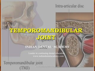
  • 1. TEMPOROMANDIBULARTEMPOROMANDIBULAR JOINTJOINT INDIAN DENTAL ACADEMY Leader in continuing dental education www.indiandentalacademy.com www.indiandentalacademy.comwww.indiandentalacademy.com
  • 2. CONTENTSCONTENTS  IntroductionIntroduction  Classification of the jointClassification of the joint  Development of the jointDevelopment of the joint  Special features of the jointSpecial features of the joint www.indiandentalacademy.comwww.indiandentalacademy.com
  • 3.  Anatomy of the jointAnatomy of the joint  Bony componentBony component  Articular discArticular disc  Articular cartilageArticular cartilage  Histology of articular surfacesHistology of articular surfaces  Joint capsuleJoint capsule  Synovial membraneSynovial membrane  Mechanism of lubricationMechanism of lubrication  Muscles involved in the jaw movementsMuscles involved in the jaw movements  LigamentsLigaments www.indiandentalacademy.comwww.indiandentalacademy.com
  • 4.  Innervation of the jointInnervation of the joint  Blood supply to the jointBlood supply to the joint  Biomechanics of the temporomandibular jointBiomechanics of the temporomandibular joint  Movements of the jointMovements of the joint  Age changes in the temporomandibular jointAge changes in the temporomandibular joint  Clinical examinationClinical examination  Functions of temporomandibular jointFunctions of temporomandibular joint  Functional and clinical considerationsFunctional and clinical considerations  Prosthodontic considerationsProsthodontic considerations  ConclusionConclusion  ReferencesReferences www.indiandentalacademy.comwww.indiandentalacademy.com
  • 5. INTRODUCTIONINTRODUCTION  Most human bones are joined with oneMost human bones are joined with one another. These connections of bones to eachanother. These connections of bones to each other are termed articulation’s or joints.other are termed articulation’s or joints. www.indiandentalacademy.comwww.indiandentalacademy.com
  • 6.  The articulation of the lower jaw with theThe articulation of the lower jaw with the cranium and upper facial skeleton involvescranium and upper facial skeleton involves two separate joints and the teeth when intwo separate joints and the teeth when in occlusion.occlusion.  The mandibular articulation, is a bilateralThe mandibular articulation, is a bilateral diarthrosis between the condyles of thediarthrosis between the condyles of the mandible and the articular eminences of themandible and the articular eminences of the temporal bone, anteriorly and madibular fossaetemporal bone, anteriorly and madibular fossae posteriorly.posteriorly. www.indiandentalacademy.comwww.indiandentalacademy.com
  • 7.  Temporomandibular joint (TMJ) is aTemporomandibular joint (TMJ) is a complex cranio mandibular articulationcomplex cranio mandibular articulation formed between the condyle of theformed between the condyle of the mandible and the glenoid fossamandible and the glenoid fossa www.indiandentalacademy.comwww.indiandentalacademy.com
  • 9.  The bones involved areThe bones involved are thethe MANDIBLEMANDIBLE and theand the OS TEMPORALEOS TEMPORALE andand the joint is thereforethe joint is therefore designated as thedesignated as the TemporomandibularTemporomandibular joint. – Other wisejoint. – Other wise called as ginglymo –called as ginglymo – arthroidal jointarthroidal joint www.indiandentalacademy.comwww.indiandentalacademy.com
  • 10.  It provides for hinging movement on one sideIt provides for hinging movement on one side and hence called a ginglymoid joint. At theand hence called a ginglymoid joint. At the other side it also provides for glidingother side it also provides for gliding movements, which classifies it as an arthroidalmovements, which classifies it as an arthroidal joint. Thus TMJ is considered ajoint. Thus TMJ is considered a ginglymoarthroidal joint.ginglymoarthroidal joint. www.indiandentalacademy.comwww.indiandentalacademy.com
  • 11. CLASSIFICATION OF THE JOINTSCLASSIFICATION OF THE JOINTS Joints are classified into three different kinds:Joints are classified into three different kinds:  FIBROUS JOINTFIBROUS JOINT  CARTILAGENOUS JOINTCARTILAGENOUS JOINT  SYNOVIAL JOINTSYNOVIAL JOINT www.indiandentalacademy.comwww.indiandentalacademy.com
  • 12. FIBROUS JOINTSFIBROUS JOINTS  In this type, two bonesIn this type, two bones are connected by fibrousare connected by fibrous tissue. Three types aretissue. Three types are described: The first is thedescribed: The first is the suture, a joint thatsuture, a joint that permits little or nopermits little or no movement.movement. www.indiandentalacademy.comwww.indiandentalacademy.com
  • 13.  Its function is to permit growth because itsIts function is to permit growth because its articulating surfaces are covered by anarticulating surfaces are covered by an osteogenetic layer responsible for new boneosteogenetic layer responsible for new bone formation to maintain the suture as the skullformation to maintain the suture as the skull bones which are separated by the expandingbones which are separated by the expanding brain.brain. www.indiandentalacademy.comwww.indiandentalacademy.com
  • 14.  The second type of fibrous joint is theThe second type of fibrous joint is the gomphosis, the socketed attachment of tooth togomphosis, the socketed attachment of tooth to bone by the fibrous periodontal ligament. Herebone by the fibrous periodontal ligament. Here functional movement is restricted to intrusionfunctional movement is restricted to intrusion and recovery in response to biting forces.and recovery in response to biting forces. www.indiandentalacademy.comwww.indiandentalacademy.com
  • 15.  The third type of fibrous type is theThe third type of fibrous type is the syndesmosis: examples of which are the jointssyndesmosis: examples of which are the joints between the fibula and the tibia and betweenbetween the fibula and the tibia and between the radius and the ulna.the radius and the ulna. www.indiandentalacademy.comwww.indiandentalacademy.com
  • 16.  Here the two bony components are someHere the two bony components are some distance apart but are joined by andistance apart but are joined by an interosseous ligament that permits limitedinterosseous ligament that permits limited movement.movement. www.indiandentalacademy.comwww.indiandentalacademy.com
  • 17. CARTILAGENOUS JOINTSCARTILAGENOUS JOINTS  In a primaryIn a primary cartilaginous joint,cartilaginous joint, bone and cartilagebone and cartilage are in directare in direct apposition.apposition. www.indiandentalacademy.comwww.indiandentalacademy.com
  • 18.  An example is the costochondral junction. In aAn example is the costochondral junction. In a secondary cartilaginous joint, the tissues of thesecondary cartilaginous joint, the tissues of the articulation occur in the sequence of bone-articulation occur in the sequence of bone- cartilage-fibrous tissue-cartilage-bone. Ancartilage-fibrous tissue-cartilage-bone. An example is the pubic symphysis.example is the pubic symphysis. www.indiandentalacademy.comwww.indiandentalacademy.com
  • 19. SYNOVIAL JOINTSSYNOVIAL JOINTS  In a synovial joint, twoIn a synovial joint, two bones are united andbones are united and surrounded by a capsule,surrounded by a capsule, thereby creating a jointthereby creating a joint cavity. This cavity iscavity. This cavity is filled with synovial fluidfilled with synovial fluid formed by a synovialformed by a synovial membrane that lines themembrane that lines the nonarticular surfaces.nonarticular surfaces. www.indiandentalacademy.comwww.indiandentalacademy.com
  • 20.  The cavity is dividedThe cavity is divided by an articular disk.by an articular disk. Various ligaments areVarious ligaments are associated with theassociated with the joints. These couldjoints. These could further be classifiedfurther be classified into uni-axial, biaxialinto uni-axial, biaxial and multi-axial.and multi-axial.  Example :Example : TemporomandibularTemporomandibular jointjoint www.indiandentalacademy.comwww.indiandentalacademy.com
  • 21. DEVELOPMENT OFDEVELOPMENT OF TEMPOROMANDIBULAR JOINTTEMPOROMANDIBULAR JOINT www.indiandentalacademy.comwww.indiandentalacademy.com
  • 22. EARLY DEVELOPMENT OF TMJEARLY DEVELOPMENT OF TMJ  In the first 8 weeks of intrauterineIn the first 8 weeks of intrauterine development, the mandible consists of adevelopment, the mandible consists of a cylindrical rod of cartilage, which iscylindrical rod of cartilage, which is surrounded by mesenchymal tissue.surrounded by mesenchymal tissue.  This cartilaginous rod provides the skeletalThis cartilaginous rod provides the skeletal support for the lower jaw.support for the lower jaw. www.indiandentalacademy.comwww.indiandentalacademy.com
  • 24.  It extends from the midline of the fusedIt extends from the midline of the fused mandibular processes, posteriorly, into themandibular processes, posteriorly, into the future middle ear cavity.future middle ear cavity.  At the posterior end, Meckel’s cartilage isAt the posterior end, Meckel’s cartilage is fused to the malleus cartilage.fused to the malleus cartilage. www.indiandentalacademy.comwww.indiandentalacademy.com
  • 26.  This round piece of cartilage articulates withThis round piece of cartilage articulates with another small cartilage termed the incus. Foranother small cartilage termed the incus. For the first 16 to 18 weeks of prenatal life, thethe first 16 to 18 weeks of prenatal life, the articulation fo the malleus and the incusarticulation fo the malleus and the incus functions as the primary temporomandibularfunctions as the primary temporomandibular joint.joint.  As far as is known, this is a hinge joint with noAs far as is known, this is a hinge joint with no lateral excursion. During this time, these twolateral excursion. During this time, these two cartilages become enclosed in the otic capsule.cartilages become enclosed in the otic capsule. www.indiandentalacademy.comwww.indiandentalacademy.com
  • 27.  Later they undergo endochondral boneLater they undergo endochondral bone formation to become middle ear bones.formation to become middle ear bones.  From the sixteenth to the eighteenth prenatalFrom the sixteenth to the eighteenth prenatal week, the secondary joint, consisting of theweek, the secondary joint, consisting of the head of the condyle, which articulates in thehead of the condyle, which articulates in the glenoid fossa, functions through out life byglenoid fossa, functions through out life by means of a sliding and rotating condylar head.means of a sliding and rotating condylar head. www.indiandentalacademy.comwww.indiandentalacademy.com
  • 30. CONDYLAR CARTILAGECONDYLAR CARTILAGE DEVELOPMENTDEVELOPMENT  Between the eigth and the twelfth week of theBetween the eigth and the twelfth week of the fetal life, the cartilagenous condyles developfetal life, the cartilagenous condyles develop anteriorly to the malleus and incus articulation.anteriorly to the malleus and incus articulation. The early cone – shaped cartilagenous condyleThe early cone – shaped cartilagenous condyle soon is altered by endochondral bonesoon is altered by endochondral bone formation and fuses with the posterior part offormation and fuses with the posterior part of the bony body of the mandible.the bony body of the mandible. www.indiandentalacademy.comwww.indiandentalacademy.com
  • 32.  At this same time, the lateral pterygoid muscleAt this same time, the lateral pterygoid muscle attaches to the medial surface of the condyle.attaches to the medial surface of the condyle.  Meckel’s cartilage, the malleus and the incus are seenMeckel’s cartilage, the malleus and the incus are seen medial to the developing mandible by now.medial to the developing mandible by now.  The secondary joint thus lies lateral to theThe secondary joint thus lies lateral to the cartilagenous rod and is anterior to the primary joint.cartilagenous rod and is anterior to the primary joint. The condyle is seen as a small condensation of theThe condyle is seen as a small condensation of the cartilage cells with a mesenchymal cap.cartilage cells with a mesenchymal cap. www.indiandentalacademy.comwww.indiandentalacademy.com
  • 33.  By the twelfth prenatal week, the condyleBy the twelfth prenatal week, the condyle consists of a large mass of hyaline cartilageconsists of a large mass of hyaline cartilage covered by a thin fibrous cap . In this, bonycovered by a thin fibrous cap . In this, bony ramus is outlined by connective tissue, and aramus is outlined by connective tissue, and a thin core of bone is seen. Meckel’s cartilage isthin core of bone is seen. Meckel’s cartilage is positioned medially to the ramus.positioned medially to the ramus. www.indiandentalacademy.comwww.indiandentalacademy.com
  • 35. ARTICULAR DISC FORMATIONARTICULAR DISC FORMATION  The first appearance of a temporomandibularThe first appearance of a temporomandibular joint cavity is seen in the 12- week old fetus.joint cavity is seen in the 12- week old fetus. The first of the two compartments to form isThe first of the two compartments to form is the inferior or mandibular compartment.the inferior or mandibular compartment.  A split first appears in the synovialA split first appears in the synovial mesenchyme overlying the condylar head andmesenchyme overlying the condylar head and extends into a small cleft.extends into a small cleft. www.indiandentalacademy.comwww.indiandentalacademy.com
  • 37.  The precise mechanism of cavitation stillThe precise mechanism of cavitation still remains unknown, although the split outlinesremains unknown, although the split outlines the head of the condyle.the head of the condyle.  The process probably is due to theThe process probably is due to the programmed cell death along the path of theprogrammed cell death along the path of the movement of the condyle and adjacentmovement of the condyle and adjacent connective tissue.connective tissue. www.indiandentalacademy.comwww.indiandentalacademy.com
  • 38.  Within another week or so, the superior orWithin another week or so, the superior or temporal compartment is formed by the sametemporal compartment is formed by the same process. The presence of both superior andprocess. The presence of both superior and inferior cavities thus creates the articular disc.inferior cavities thus creates the articular disc.  The primodium consists of dense, highlyThe primodium consists of dense, highly cellular mesenchyme pads.cellular mesenchyme pads. www.indiandentalacademy.comwww.indiandentalacademy.com
  • 40.  Each blends circumferentially into aEach blends circumferentially into a developing capsule at its borders.developing capsule at its borders.  With continued growth of the condyle andWith continued growth of the condyle and fossa, the articular disc becomes fibrous infossa, the articular disc becomes fibrous in character and assumes a typical shape, i.e. acharacter and assumes a typical shape, i.e. a very thin central zone and a much thickervery thin central zone and a much thicker peripheral area.peripheral area. www.indiandentalacademy.comwww.indiandentalacademy.com
  • 43. FATE OF MECKEL’SFATE OF MECKEL’S CARTILAGECARTILAGE  When the anterior aspect of Meckel’s cartilageWhen the anterior aspect of Meckel’s cartilage fuses in the medial wall of the mandible in thefuses in the medial wall of the mandible in the tenth prenatal week, the cartilage undergoestenth prenatal week, the cartilage undergoes endochondral bone formation along the medialendochondral bone formation along the medial aspect of the mandible.aspect of the mandible.  As the mandible enlarges, the remnants ofAs the mandible enlarges, the remnants of Meckel’s cartilage becomes relatively smaller.Meckel’s cartilage becomes relatively smaller. www.indiandentalacademy.comwww.indiandentalacademy.com
  • 45.  By the sixteenth prenatal week, the malleusBy the sixteenth prenatal week, the malleus and the incus have begun transformation intoand the incus have begun transformation into middle ear bones by endochondral bonemiddle ear bones by endochondral bone formation.formation.  As Meckel’s cartilage degenerates in the areaAs Meckel’s cartilage degenerates in the area anterior to the ear, two ligaments,anterior to the ear, two ligaments, the anteriorthe anterior malleus and sphenomandibular ,malleus and sphenomandibular , form withinform within its pathway.its pathway. www.indiandentalacademy.comwww.indiandentalacademy.com
  • 46.  When the secondary temporomandibular jointWhen the secondary temporomandibular joint becomes functional at 18 to 20 prenatal weeks,becomes functional at 18 to 20 prenatal weeks, Meckel’s cartilage loses its function andMeckel’s cartilage loses its function and disappears.disappears. www.indiandentalacademy.comwww.indiandentalacademy.com
  • 47. DIFFERENTIATION OF THE TMJDIFFERENTIATION OF THE TMJ  Once the component parts of the joint haveOnce the component parts of the joint have been established, at the fourteenth prenatalbeen established, at the fourteenth prenatal week, no major alterations occur, except inweek, no major alterations occur, except in differentiation of the joint tissues and thedifferentiation of the joint tissues and the increase in size of the joint.increase in size of the joint.  The growth of the condyle up to 14 prenatalThe growth of the condyle up to 14 prenatal weeks consists of both interstitial andweeks consists of both interstitial and appositional growth of the condylar cartilage.appositional growth of the condylar cartilage. www.indiandentalacademy.comwww.indiandentalacademy.com
  • 49.  From this time on, and continuing intoFrom this time on, and continuing into maturity in the middle twenties, thematurity in the middle twenties, the mandibular condlye increases in size bymandibular condlye increases in size by endochondral like bone formation.endochondral like bone formation.  This process of bone formation helps providedThis process of bone formation helps provided for the elongation of the ramus of thefor the elongation of the ramus of the mandible.mandible. www.indiandentalacademy.comwww.indiandentalacademy.com
  • 50.  The gradual formation of the temporal fossaThe gradual formation of the temporal fossa starts in the twelfth prenatal week, when thestarts in the twelfth prenatal week, when the synovial cavity outlines the condylar head.synovial cavity outlines the condylar head.  Then a heavy spicule of the temporal boneThen a heavy spicule of the temporal bone develops superior to the forming articular disc.develops superior to the forming articular disc. With continued intramembranous boneWith continued intramembranous bone formation, the small segments soon coalesce toformation, the small segments soon coalesce to form the glenoid fossa.form the glenoid fossa. www.indiandentalacademy.comwww.indiandentalacademy.com
  • 52.  The temporal bone as well as the condyleThe temporal bone as well as the condyle increases in size by 16 weeeks prenatally.increases in size by 16 weeeks prenatally.  There is further bone formation in the temporalThere is further bone formation in the temporal region, so that at 22 weeks prenatally, theregion, so that at 22 weeks prenatally, the glenoid fossa has developed a medial wall asglenoid fossa has developed a medial wall as well as extension of bone laterally.well as extension of bone laterally.  This joint becomes functional at 18 to 20This joint becomes functional at 18 to 20 weeks prenatally. At this time also, theweeks prenatally. At this time also, the articular eminence develops.articular eminence develops. www.indiandentalacademy.comwww.indiandentalacademy.com
  • 53.  The articular disc further takes on itsThe articular disc further takes on its characteristic shape during this period,characteristic shape during this period, appearing very thin centrally and thicker at itsappearing very thin centrally and thicker at its peripheral region where it blends into theperipheral region where it blends into the articular capsule.articular capsule.  The capsule is well differentiated at 26 weeksThe capsule is well differentiated at 26 weeks or 6 ½ months prenatally.or 6 ½ months prenatally. www.indiandentalacademy.comwww.indiandentalacademy.com
  • 55.  The disc now consists of a dense network ofThe disc now consists of a dense network of collagenous fibers. The fossa is formed bycollagenous fibers. The fossa is formed by typical endochondral bone.typical endochondral bone.  Thus, cartilage forms in the joint as well as inThus, cartilage forms in the joint as well as in the condylar head which articulates with it.the condylar head which articulates with it.  the true surface lining of both the upper andthe true surface lining of both the upper and the lower compartment is fibrous tissue orthe lower compartment is fibrous tissue or perichondrium. It is seen as the dark bandperichondrium. It is seen as the dark band covering the fossa and condyle.covering the fossa and condyle. www.indiandentalacademy.comwww.indiandentalacademy.com
  • 56. LATE PRENATALLATE PRENATAL DEVELOPMENTDEVELOPMENT  There are few major changes in theThere are few major changes in the temporomandibular joint after function beginstemporomandibular joint after function begins in the eighteenth to the twentieth prenatalin the eighteenth to the twentieth prenatal week.week.  The condylar bone increases in size andThe condylar bone increases in size and density, and the mandible undergoes changesdensity, and the mandible undergoes changes in shape and size associated within shape and size associated with differentiation and function of the muscles ofdifferentiation and function of the muscles of mastication.mastication. www.indiandentalacademy.comwww.indiandentalacademy.com
  • 57.  One noteworthy feature, occuring in the last 3One noteworthy feature, occuring in the last 3 prenatal months, is the appearance of clefts inprenatal months, is the appearance of clefts in the condylar head.the condylar head.  These connective tissue ingrowths originateThese connective tissue ingrowths originate from the fibrous perichondrium covering thefrom the fibrous perichondrium covering the cartilagenous condylar head.cartilagenous condylar head. www.indiandentalacademy.comwww.indiandentalacademy.com
  • 59.  They carry blood vessels into the rapidly growingThey carry blood vessels into the rapidly growing cartilage. This is unusual;other cartilages in thecartilage. This is unusual;other cartilages in the human body are considered to be avascular. Some ofhuman body are considered to be avascular. Some of the vascular ingrowths extend to the front of thethe vascular ingrowths extend to the front of the endochondral bone, forming the major end in theendochondral bone, forming the major end in the hyaline cartilage of the condyle. This feature ishyaline cartilage of the condyle. This feature is probably rekated to the rapid increase in size andprobably rekated to the rapid increase in size and function of the temporomandibular joint. Another latefunction of the temporomandibular joint. Another late prenatal change is the general thinning of the cartlageprenatal change is the general thinning of the cartlage on the condylar head.on the condylar head. www.indiandentalacademy.comwww.indiandentalacademy.com
  • 61.  During this period, endochondral boneDuring this period, endochondral bone replaces the cartilage more rapidly than thereplaces the cartilage more rapidly than the formation of new cartilage more rapidly thanformation of new cartilage more rapidly than the formation of new cartilage occurs on thethe formation of new cartilage occurs on the condylar surface.condylar surface.  A narrow band of cartilage does persist,A narrow band of cartilage does persist, however, on the head of the condyle untilhowever, on the head of the condyle until approximately the twenty – fifth postnatal yearapproximately the twenty – fifth postnatal year of life.of life. www.indiandentalacademy.comwww.indiandentalacademy.com
  • 62. SECONDARY GROWTHSECONDARY GROWTH CARTILAGESCARTILAGES  It is noteworthy that the bony mandibleIt is noteworthy that the bony mandible develops by both endochondral formation ofdevelops by both endochondral formation of the condyle and intra-membranousthe condyle and intra-membranous development of the mandibular body.development of the mandibular body.  There also appears several other cartilageThere also appears several other cartilage growth centres, known as secondary growthgrowth centres, known as secondary growth cartilages, in the mandible.cartilages, in the mandible. www.indiandentalacademy.comwww.indiandentalacademy.com
  • 63.  These includeThese include 1.1. The coronoid cartilagesThe coronoid cartilages 2.2. The tooth follicle cartilages andThe tooth follicle cartilages and 3.3. The symphseal cartilagesThe symphseal cartilages www.indiandentalacademy.comwww.indiandentalacademy.com
  • 65.  The carilage in the coronoid process is a smallThe carilage in the coronoid process is a small island that appears between the fourteenth andisland that appears between the fourteenth and sixteenth week and disappears by the twentiethsixteenth week and disappears by the twentieth week.week.  Tiny sites of cartilage appear near and aroundTiny sites of cartilage appear near and around early forming tooth buds, but these cartilageearly forming tooth buds, but these cartilage sites soon disappear. Only the symphysealsites soon disappear. Only the symphyseal cartilage persists until birth or longer.cartilage persists until birth or longer. www.indiandentalacademy.comwww.indiandentalacademy.com
  • 66.  Two bars of cartilage appear in theTwo bars of cartilage appear in the symphyseal region of the mandible by thesymphyseal region of the mandible by the twelfth prenatal week.twelfth prenatal week.  The two cartilages are separated by theThe two cartilages are separated by the perichondrium in the midline. On cessation ofperichondrium in the midline. On cessation of the growth of these cartilages, the suturethe growth of these cartilages, the suture persists at the midline.persists at the midline. www.indiandentalacademy.comwww.indiandentalacademy.com
  • 68. POST NATAL GROWTH ANDPOST NATAL GROWTH AND MATURATION OF TMJMATURATION OF TMJ  Further growth of the TMJ continues into theFurther growth of the TMJ continues into the second decade of postnatal life. The temporalsecond decade of postnatal life. The temporal fossa deepens as bone forms laterally on thefossa deepens as bone forms laterally on the skull and the articular tubercle enlarges byskull and the articular tubercle enlarges by further bone formation.further bone formation.  The articular surface of the fossa is covered byThe articular surface of the fossa is covered by fibrous tissue and a synovial membrane.fibrous tissue and a synovial membrane. www.indiandentalacademy.comwww.indiandentalacademy.com
  • 70.  The dense fibrous nature of the disc andThe dense fibrous nature of the disc and capsule is apparent.capsule is apparent.  The condyle continues to exhibit endochondralThe condyle continues to exhibit endochondral – like bone growth, however.– like bone growth, however.  The perichondrial covering of the condyleThe perichondrial covering of the condyle consists of two layers.consists of two layers. www.indiandentalacademy.comwww.indiandentalacademy.com
  • 72.  The portion lying next to the cartilage isThe portion lying next to the cartilage is highly cellular, and the outer layer is morehighly cellular, and the outer layer is more fibrous.fibrous.  The cartilaginous condyle consists of a numberThe cartilaginous condyle consists of a number of indistinct layers.of indistinct layers.  The outer articular surface is a highly cellularThe outer articular surface is a highly cellular zone cosisting of undifferentiated spindle –zone cosisting of undifferentiated spindle – shaped cells and fine collagen fibers and isshaped cells and fine collagen fibers and is termed thetermed the reserve or resting zonereserve or resting zone containingcontaining prechondroblasts.prechondroblasts. www.indiandentalacademy.comwww.indiandentalacademy.com
  • 73.  The next layer is theThe next layer is the multiplication zonemultiplication zone where dividing chondrocytes appear.where dividing chondrocytes appear.  TheThe maturation zonematuration zone is where theis where the chondrocytes begin to enlarge.chondrocytes begin to enlarge.  The next layer is theThe next layer is the hypertrophy zonehypertrophy zone wherewhere the large cells accumulate glycogen and fatthe large cells accumulate glycogen and fat droplets. As these cells die, the matrix arounddroplets. As these cells die, the matrix around them mineralizes, which forms thethem mineralizes, which forms the calcifiedcalcified matrix zone.matrix zone. www.indiandentalacademy.comwww.indiandentalacademy.com
  • 74.  TheThe resorption zoneresorption zone is next as osteoclastsis next as osteoclasts appear.appear.  Bone formationBone formation then occures on the calcifiedthen occures on the calcified cartilage trabeculae. As the bony trabeculaecartilage trabeculae. As the bony trabeculae fuse together and increase in size, vascularfuse together and increase in size, vascular sprouts are seen invading the resorbed areas.sprouts are seen invading the resorbed areas. www.indiandentalacademy.comwww.indiandentalacademy.com
  • 75.  The cycle continues with cartilageThe cycle continues with cartilage proliferation and bone formation which areproliferation and bone formation which are necessary for the ramus to grow.necessary for the ramus to grow.  During the second decade of life, there is aDuring the second decade of life, there is a reduction in the width of the proliferationreduction in the width of the proliferation layer, which indicates a slowing – growth ratelayer, which indicates a slowing – growth rate of the condylar head as well as of the ramus.of the condylar head as well as of the ramus. www.indiandentalacademy.comwww.indiandentalacademy.com
  • 77.  With time, there is a complete “sealing off” ofWith time, there is a complete “sealing off” of the cartilagenous region by bone. By the latterthe cartilagenous region by bone. By the latter part of the second decade, the cartilaginouspart of the second decade, the cartilaginous zone becomes mineralized and exhibitszone becomes mineralized and exhibits osteoclasts on its inner zone.osteoclasts on its inner zone.  At this time, the condyle has nearly reachedAt this time, the condyle has nearly reached maturity. The blood vessels are now limited tomaturity. The blood vessels are now limited to the nutrient canals of the adjacent bone.the nutrient canals of the adjacent bone. www.indiandentalacademy.comwww.indiandentalacademy.com
  • 78.  The cartilage of the condylar head varies inThe cartilage of the condylar head varies in several ways from that of the head of a largeseveral ways from that of the head of a large bone.bone.  First, the condyles do not develop secondaryFirst, the condyles do not develop secondary ossification centers, as do long bones, whichossification centers, as do long bones, which results in their developing cartilage in tworesults in their developing cartilage in two directions and forming an epiphyseal line.directions and forming an epiphyseal line. www.indiandentalacademy.comwww.indiandentalacademy.com
  • 79.  Each condyle forms a single front of cartilage andEach condyle forms a single front of cartilage and bone.bone.  Second, the chondrocytes in the condyles do not formSecond, the chondrocytes in the condyles do not form in rows, as do those in long bone growth..in rows, as do those in long bone growth..  Third, the condyles form a thick fibrousThird, the condyles form a thick fibrous perichondrium covering the cartilage and lining theperichondrium covering the cartilage and lining the joint cavity.joint cavity.  Fourth, vascular tracts enter the forming cartilageFourth, vascular tracts enter the forming cartilage which does not occur in other cartilages in our boides.which does not occur in other cartilages in our boides. www.indiandentalacademy.comwww.indiandentalacademy.com
  • 81.  Once the TMJ attains adult size, the articularOnce the TMJ attains adult size, the articular eminence becomes prominent and cartilageeminence becomes prominent and cartilage thins over the condylar head.thins over the condylar head.  During the second decade of post natal life, theDuring the second decade of post natal life, the reduction in the width of the proliferating layerreduction in the width of the proliferating layer causes the slowing growth rate of the condylarcauses the slowing growth rate of the condylar head as well as of the ramus.head as well as of the ramus. www.indiandentalacademy.comwww.indiandentalacademy.com
  • 91. CHRONOLOGICALCHRONOLOGICAL DEVELOPMENT OF THE TMJDEVELOPMENT OF THE TMJ  A. BirthA. Birth 1. The joint displays the structural components1. The joint displays the structural components of the adult joint, but it lies parallel with theof the adult joint, but it lies parallel with the occlusal plane instead of several mm superiorocclusal plane instead of several mm superior to it as in the adult.to it as in the adult. www.indiandentalacademy.comwww.indiandentalacademy.com
  • 92.  2. The articular eminence is inferiorly2. The articular eminence is inferiorly positioned, the fossa is relatively flat and thepositioned, the fossa is relatively flat and the joint functions as a pure hinge system with nojoint functions as a pure hinge system with no translation.translation. www.indiandentalacademy.comwww.indiandentalacademy.com
  • 93.  3. Throughout fetal life, the fibrous articular3. Throughout fetal life, the fibrous articular surfaces and interposing disc are vascularizedsurfaces and interposing disc are vascularized and innervated. This will disappear as functionand innervated. This will disappear as function induces compression of the disc between theinduces compression of the disc between the condyle and temporal bone.condyle and temporal bone. www.indiandentalacademy.comwww.indiandentalacademy.com
  • 94.  B. ChildhoodB. Childhood 1. With the occlusion of the primary dentition1. With the occlusion of the primary dentition at age three, pressure is placed on the jointat age three, pressure is placed on the joint which results in the change to nonvascularitywhich results in the change to nonvascularity of the articular surfaces. Before age three, aof the articular surfaces. Before age three, a fall or other trauma to the joint will oftenfall or other trauma to the joint will often result in a hemarthrosis and possible fibrousresult in a hemarthrosis and possible fibrous ankylosis.ankylosis. www.indiandentalacademy.comwww.indiandentalacademy.com
  • 95. SPECIAL FEATURES OF CRANIO -SPECIAL FEATURES OF CRANIO - MANDIBULAR ARTICULATIONMANDIBULAR ARTICULATION  A. Bilateral:A. Bilateral: also known as bipedalism. Thealso known as bipedalism. The craniomandibular articulation is composed ofcraniomandibular articulation is composed of two joints, a right and a lefttwo joints, a right and a left temporomandibular joint, which is atemporomandibular joint, which is a functioning unit. What affects one joint mustfunctioning unit. What affects one joint must affect the other.affect the other. www.indiandentalacademy.comwww.indiandentalacademy.com
  • 96.  B. Each of the joints functions as aB. Each of the joints functions as a compoundcompound jointjoint. For the human mandible, this is a. For the human mandible, this is a sliding-hinge joint which would imply threesliding-hinge joint which would imply three bones. Instead, the articular disc functions asbones. Instead, the articular disc functions as the third bone, supplying true articularthe third bone, supplying true articular surfaces superior and inferior.surfaces superior and inferior. www.indiandentalacademy.comwww.indiandentalacademy.com
  • 97.  The TMJ is a double joint composed of anThe TMJ is a double joint composed of an inferiorly positioned hinge joint and ainferiorly positioned hinge joint and a superiorly positioned sliding joint. The disc-superiorly positioned sliding joint. The disc- condyle complex is simple hinge joint and thecondyle complex is simple hinge joint and the superior joint is designed for slidingsuperior joint is designed for sliding movement in any direction, the range beingmovement in any direction, the range being limited by structural restraints.limited by structural restraints. www.indiandentalacademy.comwww.indiandentalacademy.com
  • 98.  C. The articular surfaces of theC. The articular surfaces of the temporomandibular joints are different fromtemporomandibular joints are different from other synovial joints in that they are notother synovial joints in that they are not composed ofcomposed of hyaline cartilage.hyaline cartilage. These surfacesThese surfaces are composed of nonvascularized andare composed of nonvascularized and noninnervated fibrous connective tissue.noninnervated fibrous connective tissue. www.indiandentalacademy.comwww.indiandentalacademy.com
  • 99.  The difference becomes important in theThe difference becomes important in the matter of the regenerative capabilities of thematter of the regenerative capabilities of the joint. In general, the temporomandibular jointsjoint. In general, the temporomandibular joints have somewhat greater potential for repair.have somewhat greater potential for repair. www.indiandentalacademy.comwww.indiandentalacademy.com
  • 100.  D. Joint position is determined byD. Joint position is determined by musclemuscle action until the moment of intercuspationaction until the moment of intercuspation,, when an irresistible force suddenly determineswhen an irresistible force suddenly determines that position (the occlusion). This is the onlythat position (the occlusion). This is the only joint in the body whose position is determinedjoint in the body whose position is determined by other hard structures.by other hard structures. www.indiandentalacademy.comwww.indiandentalacademy.com
  • 101.  There will frequently be muscle problemsThere will frequently be muscle problems associated with this joint, since the jointassociated with this joint, since the joint position is not determined by resting muscleposition is not determined by resting muscle tonus or postural position. When occlusaltonus or postural position. When occlusal position and muscular position are not inposition and muscular position are not in harmony, dysfunctional problems will occur.harmony, dysfunctional problems will occur. www.indiandentalacademy.comwww.indiandentalacademy.com
  • 102. ANATOMY OFANATOMY OF TEMPOROMANDIBULAR JOINTTEMPOROMANDIBULAR JOINT www.indiandentalacademy.comwww.indiandentalacademy.com
  • 103. BONES OF THE JOINTBONES OF THE JOINT  The bones of the temporomandibularThe bones of the temporomandibular articulation are thearticulation are the glenoid fossaglenoid fossa (on the(on the undersurface of the squmous part of theundersurface of the squmous part of the temporal bone) andtemporal bone) and the condylethe condyle (supported by(supported by the condylar process of the mandible).the condylar process of the mandible). www.indiandentalacademy.comwww.indiandentalacademy.com
  • 104.  The squamotympanic and petrotympanicThe squamotympanic and petrotympanic fissures limit the glenoid fossa posteriorly.fissures limit the glenoid fossa posteriorly.  Medially it is limited by the spine of sphenoid,Medially it is limited by the spine of sphenoid, and laterally by the root of zygomatic process ofand laterally by the root of zygomatic process of the temporal bone.the temporal bone. www.indiandentalacademy.comwww.indiandentalacademy.com
  • 105.  Anteriorly it is bounded by a ridge of boneAnteriorly it is bounded by a ridge of bone described as the articular eminence, alsodescribed as the articular eminence, also involved in the articulation. The middle part isinvolved in the articulation. The middle part is a fairly thin plate of bone whose upper surfacea fairly thin plate of bone whose upper surface forms the middle cranial fossa.forms the middle cranial fossa. www.indiandentalacademy.comwww.indiandentalacademy.com
  • 106. Temporal bone / Glenoid fossaTemporal bone / Glenoid fossa  The mandibular condyle articulates at the base of theThe mandibular condyle articulates at the base of the cranium, with the squamous portion of the temporalcranium, with the squamous portion of the temporal bone. The specific locations is on the posterior slopebone. The specific locations is on the posterior slope of articular eminence.of articular eminence.  This portion of the temporal bone is made up of aThis portion of the temporal bone is made up of a concave mandibular fossa, in which condyle isconcave mandibular fossa, in which condyle is situated and which has also been called the articularsituated and which has also been called the articular or glenoid fossa.or glenoid fossa. www.indiandentalacademy.comwww.indiandentalacademy.com
  • 107.  Anterior to the fossa is a convex bonyAnterior to the fossa is a convex bony prominence called the articular eminence.prominence called the articular eminence.  Posterior to mandibular fossa is thePosterior to mandibular fossa is the squamotympanic fissure.squamotympanic fissure.  Articular eminence is defined as a stronglyArticular eminence is defined as a strongly convex bony elevation on the root of theconvex bony elevation on the root of the zygomatic process.zygomatic process. www.indiandentalacademy.comwww.indiandentalacademy.com
  • 112. Mandibular condyleMandibular condyle  The mandible possesses two articularThe mandible possesses two articular surfaces a condyle located on the superiorsurfaces a condyle located on the superior extremity of each of the bilateral condylarextremity of each of the bilateral condylar processes.processes.  The condyle is the portion of the mandibleThe condyle is the portion of the mandible that articulates with the cranium, around whichthat articulates with the cranium, around which movement occurs.movement occurs. www.indiandentalacademy.comwww.indiandentalacademy.com
  • 114.  From the anterior view it has a medial and aFrom the anterior view it has a medial and a lateral projection called poles.lateral projection called poles.  The medial pole is generally moreThe medial pole is generally more prominent than the lateral.prominent than the lateral.  From above if a line drawn through theFrom above if a line drawn through the centres of the poles of the condyle will usuallycentres of the poles of the condyle will usually extend medially and posteriorly toward theextend medially and posteriorly toward the anterior border of the foramen magnum.anterior border of the foramen magnum. www.indiandentalacademy.comwww.indiandentalacademy.com
  • 115.  Each condyle articulates with a meniscusEach condyle articulates with a meniscus (disk) which is interposed between it and(disk) which is interposed between it and temporal bone.temporal bone.  The articulating surface of the condyle is quiteThe articulating surface of the condyle is quite convex anteroposteriorly and only slightlyconvex anteroposteriorly and only slightly convex mediolaterally.convex mediolaterally. www.indiandentalacademy.comwww.indiandentalacademy.com
  • 116.  The articular surface of the condyle is covered by aThe articular surface of the condyle is covered by a thick layer of fibrocartilage.thick layer of fibrocartilage.  The mediolateral length of the condyle is 15 to 20The mediolateral length of the condyle is 15 to 20 mm and the anteroposterior width is between 8 tomm and the anteroposterior width is between 8 to 10 mm.10 mm. www.indiandentalacademy.comwww.indiandentalacademy.com
  • 117. Different shapes of mandibularDifferent shapes of mandibular condylescondyles www.indiandentalacademy.comwww.indiandentalacademy.com
  • 118. 1 Mandibular condyle 2 Articular disk 3 Superior joint cavity 4 Articular eminence 5 External earwww.indiandentalacademy.comwww.indiandentalacademy.com
  • 119. ARTICULAR DISKARTICULAR DISK  A fibrous disk divides the joint cavity into twoA fibrous disk divides the joint cavity into two compartments and is a structure with ancompartments and is a structure with an important functional role: it provides, in effect,important functional role: it provides, in effect, an largely passive movable articular surfacean largely passive movable articular surface accommodating the translatory movementaccommodating the translatory movement made by the head of the condyle. It consists ofmade by the head of the condyle. It consists of dense fibrous tissue and its shape conforms todense fibrous tissue and its shape conforms to that of the opposed articular surfaces.that of the opposed articular surfaces. www.indiandentalacademy.comwww.indiandentalacademy.com
  • 120.  Its lower surface is concave and matches theIts lower surface is concave and matches the convex contour of the condyle. Its upperconvex contour of the condyle. Its upper surface is a concavo-convex.surface is a concavo-convex. AnteriorlyAnteriorly thethe disk divides into two lamellae-disk divides into two lamellae- www.indiandentalacademy.comwww.indiandentalacademy.com
  • 121.  upper one running forward to fuse with theupper one running forward to fuse with the capsule and periosteum on the anterior slopecapsule and periosteum on the anterior slope of the eminence, and a lower one runningof the eminence, and a lower one running down to attach to the front of the neck of thedown to attach to the front of the neck of the condyle. In between is the foot, merging eithercondyle. In between is the foot, merging either with the capsule or with the upper fiberswith the capsule or with the upper fibers constituting the superior head of the lateralconstituting the superior head of the lateral pterygoid muscle.pterygoid muscle. www.indiandentalacademy.comwww.indiandentalacademy.com
  • 122.  PosteriorlyPosteriorly the disk again divides into twothe disk again divides into two lamellae- an upper one, consisting of fibrouslamellae- an upper one, consisting of fibrous and elastic tissues, that fuses with the capsuleand elastic tissues, that fuses with the capsule and inserts into the squamotypanic fissure, andand inserts into the squamotypanic fissure, and a lower one consisting of collagen only.a lower one consisting of collagen only. www.indiandentalacademy.comwww.indiandentalacademy.com
  • 125.  The disc is avascular in the central region.The disc is avascular in the central region. During function it makes only relatively shortDuring function it makes only relatively short movements in a passive manner to fit best withmovements in a passive manner to fit best with the changing relationships of the condylarthe changing relationships of the condylar head and the glenoid fossa and articularhead and the glenoid fossa and articular eminence.eminence. www.indiandentalacademy.comwww.indiandentalacademy.com
  • 128. Disk positionDisk position  Normal position -12Normal position -12 o’clock positiono’clock position www.indiandentalacademy.comwww.indiandentalacademy.com
  • 133. CARTILAGE ASSOCIATED WITHCARTILAGE ASSOCIATED WITH THE JOINTTHE JOINT  The surface coverings of the joint consist ofThe surface coverings of the joint consist of fibro cartilage. A secondary growth cartilagefibro cartilage. A secondary growth cartilage associated with the developing TMJ formsassociated with the developing TMJ forms within the blastema the condylar cartilage.within the blastema the condylar cartilage. www.indiandentalacademy.comwww.indiandentalacademy.com
  • 134.  It consists essentially of a proliferating layer ofIt consists essentially of a proliferating layer of replicating cells that function as progenitorreplicating cells that function as progenitor cells for the growth cartilage. These cellscells for the growth cartilage. These cells become chondroblasts and elaborate an extrabecome chondroblasts and elaborate an extra cellular matrix consisting of proteoglycans andcellular matrix consisting of proteoglycans and type II collagen to form the extra cellulartype II collagen to form the extra cellular matrix of cartilage.matrix of cartilage. www.indiandentalacademy.comwww.indiandentalacademy.com
  • 135.  At the same time there is an increase in size ofAt the same time there is an increase in size of the chondroblasts (hypertrophy). Followingthe chondroblasts (hypertrophy). Following this cartilage production endochondralthis cartilage production endochondral ossification occurs involving mineralization ofossification occurs involving mineralization of the cartilage, vascular invasion, loss ofthe cartilage, vascular invasion, loss of chondrocytes and differentiation of osteoblastschondrocytes and differentiation of osteoblasts to produce bone on the mineralizedto produce bone on the mineralized cartilaginous framework.cartilaginous framework. www.indiandentalacademy.comwww.indiandentalacademy.com
  • 136.  In summary:In summary: Although fibrocartilage isAlthough fibrocartilage is associated with the temporomandibularassociated with the temporomandibular articulation, it does not form part of thearticulation, it does not form part of the articulation and has no formal functional rolearticulation and has no formal functional role to play in the everyday movements occurringto play in the everyday movements occurring between the two bones of the joint.between the two bones of the joint. www.indiandentalacademy.comwww.indiandentalacademy.com
  • 138. Histology of the articular surfacesHistology of the articular surfaces The articular surfaces of the mandibularThe articular surfaces of the mandibular condyle and fossa are composed of fourcondyle and fossa are composed of four distinct layers (or) zones.distinct layers (or) zones.  Articular zoneArticular zone  Proliferative zoneProliferative zone  Fibrocartilaginous zoneFibrocartilaginous zone  Calcified cartilage zoneCalcified cartilage zone www.indiandentalacademy.comwww.indiandentalacademy.com
  • 139.  Articular zone :Articular zone : the most superficial layer isthe most superficial layer is called the articular zone. It is found adjacent tocalled the articular zone. It is found adjacent to the joint cavity and forms the outermostthe joint cavity and forms the outermost functional surface. This articular layer is madefunctional surface. This articular layer is made up of dense fibrous connective tissue.up of dense fibrous connective tissue. www.indiandentalacademy.comwww.indiandentalacademy.com
  • 140.  The second zone is theThe second zone is the proliferative zone.proliferative zone. ThisThis is mainly cellular. This area hasis mainly cellular. This area has undifferentiated mesenchymal tissue. Thisundifferentiated mesenchymal tissue. This tissue is responsible for the proliferation oftissue is responsible for the proliferation of articular cartilage in response to the functionalarticular cartilage in response to the functional demands placed on it.demands placed on it. www.indiandentalacademy.comwww.indiandentalacademy.com
  • 141.  TheThe fibrocartilaginous zonefibrocartilaginous zone is the third zone.is the third zone. In this zone collagen fibrils are arranged inIn this zone collagen fibrils are arranged in bundles in a crossing pattern. They offerbundles in a crossing pattern. They offer resistance against compressive and lateralresistance against compressive and lateral forces.forces. www.indiandentalacademy.comwww.indiandentalacademy.com
  • 142.  TheThe calcified cartilage zonecalcified cartilage zone is the fourth andis the fourth and deepest zone. This zone is made up ofdeepest zone. This zone is made up of chondrocytes and chondroblasts distributedchondrocytes and chondroblasts distributed throughout the articular cartilage.throughout the articular cartilage. www.indiandentalacademy.comwww.indiandentalacademy.com
  • 145. CAPSULE, SYNOVIALCAPSULE, SYNOVIAL MEMBRANE AND DISK OF THEMEMBRANE AND DISK OF THE JOINTJOINT  The TMJ is divided into two compartments byThe TMJ is divided into two compartments by a disk and surrounded by a capsule lined witha disk and surrounded by a capsule lined with synovial membrane.synovial membrane. www.indiandentalacademy.comwww.indiandentalacademy.com
  • 146. CAPSULECAPSULE  The capsule, as it surrounds the joint, includes theThe capsule, as it surrounds the joint, includes the articualar eminence and consists of densearticualar eminence and consists of dense collagenous tissue, with its upper half (above thecollagenous tissue, with its upper half (above the articular disc) forming a loose envelope that isarticular disc) forming a loose envelope that is attached to the squamotympanic fissure behind, theattached to the squamotympanic fissure behind, the articular eminence in front, and the margins of thearticular eminence in front, and the margins of the glenoid fossa elsewhere.glenoid fossa elsewhere. www.indiandentalacademy.comwww.indiandentalacademy.com
  • 147.  It is attached at its medial and lateral marginsIt is attached at its medial and lateral margins to the articular disk. Below the disk it attachesto the articular disk. Below the disk it attaches tightly to the neck of the condyle.tightly to the neck of the condyle. www.indiandentalacademy.comwww.indiandentalacademy.com
  • 152. SYNOVIAL MEMBRANESYNOVIAL MEMBRANE  The capsule is lined on its inner surface by aThe capsule is lined on its inner surface by a synovial membrane, with folds or villi of thesynovial membrane, with folds or villi of the membrane protruding into the joint cavity,membrane protruding into the joint cavity, especially in its fornices and its upper posteriorespecially in its fornices and its upper posterior aspect.aspect. www.indiandentalacademy.comwww.indiandentalacademy.com
  • 153.  These folds increase in number with age andThese folds increase in number with age and are also more prominent in joints affected by aare also more prominent in joints affected by a pathologic process. Synovial membrane doespathologic process. Synovial membrane does not cover the articular surfaces of the joint ornot cover the articular surfaces of the joint or the disk, except for its bilaminar posteriorthe disk, except for its bilaminar posterior region.region. www.indiandentalacademy.comwww.indiandentalacademy.com
  • 154.  Histologically, it consists of two layers, a cellularHistologically, it consists of two layers, a cellular intima resting on a vascular subintima, which inintima resting on a vascular subintima, which in turn blends with the fibrous tissue of the capsule.turn blends with the fibrous tissue of the capsule. The subinitma is a loose connective tissueThe subinitma is a loose connective tissue containing vascular elements together withcontaining vascular elements together with scattered fibroblasts, macrophages, mast cells andscattered fibroblasts, macrophages, mast cells and fat cells.fat cells. www.indiandentalacademy.comwww.indiandentalacademy.com
  • 155.  The intima has one to four layers of synovialThe intima has one to four layers of synovial cells embedded in an amorphous fiber-freecells embedded in an amorphous fiber-free intercellular matrix. The cells are notintercellular matrix. The cells are not connected by junctional complexes and do notconnected by junctional complexes and do not rest on basement membrane.rest on basement membrane. www.indiandentalacademy.comwww.indiandentalacademy.com
  • 156.  The synovial membrane is responsible for theThe synovial membrane is responsible for the production of the synovial fluid, which isproduction of the synovial fluid, which is characterized by the physical properties ofcharacterized by the physical properties of viscosity, elasticity and plasticity.viscosity, elasticity and plasticity. www.indiandentalacademy.comwww.indiandentalacademy.com
  • 157.  The synovial fluid contains a small populationThe synovial fluid contains a small population of varying cell types such as monocytes,of varying cell types such as monocytes, lymphocytes, free synovial cells andlymphocytes, free synovial cells and occasionally polymorphonuclear leucocytes.occasionally polymorphonuclear leucocytes. The chemical composition is a dialysate ofThe chemical composition is a dialysate of plasma with some added protein and mucin.plasma with some added protein and mucin. www.indiandentalacademy.comwww.indiandentalacademy.com
  • 158. Functions of synovial fluidFunctions of synovial fluid  A liquid enivornment for the joint surfacesA liquid enivornment for the joint surfaces  Lubrication to increase efficiency and reduceLubrication to increase efficiency and reduce erosion.erosion. www.indiandentalacademy.comwww.indiandentalacademy.com
  • 159. 1 and 5 = Bone 2 = Cartilage 3 = Membrane 4 = Fluid www.indiandentalacademy.comwww.indiandentalacademy.com
  • 160. Mechanism of lubricationMechanism of lubrication  The first is called boundary lubrication, whichThe first is called boundary lubrication, which occurs when the joint is moved and theoccurs when the joint is moved and the synovial fluid is forced from one area of thesynovial fluid is forced from one area of the cavity into another. Boundary lubricationcavity into another. Boundary lubrication prevents friction in the moving joint.prevents friction in the moving joint. www.indiandentalacademy.comwww.indiandentalacademy.com
  • 161.  The second lubricating mechanism is calledThe second lubricating mechanism is called weeping lubrication. This refers to the abilityweeping lubrication. This refers to the ability of the articualr surfaces to absorb a smallof the articualr surfaces to absorb a small amount of synovial fluid. During function of aamount of synovial fluid. During function of a joint forces are created between the articularjoint forces are created between the articular surfaces.surfaces. www.indiandentalacademy.comwww.indiandentalacademy.com
  • 162.  These forces drive a small amount of synovialThese forces drive a small amount of synovial fluid in and out of the articular tissues.fluid in and out of the articular tissues.  Under compressive forces a small amount ofUnder compressive forces a small amount of synovial fluid is released.synovial fluid is released. www.indiandentalacademy.comwww.indiandentalacademy.com
  • 163.  This synovial fluid acts as a lubricant betweenThis synovial fluid acts as a lubricant between articular tissues to prevent sticking.articular tissues to prevent sticking.  Only a small amount of friction is eliminatedOnly a small amount of friction is eliminated as a result of weeping lubrication.as a result of weeping lubrication. www.indiandentalacademy.comwww.indiandentalacademy.com
  • 164.  Its function is to provideIts function is to provide 1.1. A liquid environment for the joint surfacesA liquid environment for the joint surfaces 2.2. Lubrication to increase efficiency and reduceLubrication to increase efficiency and reduce erosion.erosion. 3.3. Nutrition fro the disk and articular surfaces ofNutrition fro the disk and articular surfaces of the joint.the joint. 4.4. Phagocytic properties by removal ofPhagocytic properties by removal of extraneous material shed into the joint cavity.extraneous material shed into the joint cavity. www.indiandentalacademy.comwww.indiandentalacademy.com
  • 165. MUSCLES INVOLVED IN THEMUSCLES INVOLVED IN THE MOVEMENTMOVEMENT  Muscles involved are the muscles ofMuscles involved are the muscles of mastication namely the masseter, temporalis,mastication namely the masseter, temporalis, lateral pterygoid and the medial pterygoidlateral pterygoid and the medial pterygoid  Along with the digastric, geniohyoid and theAlong with the digastric, geniohyoid and the genioglossus.genioglossus. www.indiandentalacademy.comwww.indiandentalacademy.com
  • 166.  Muscles are contractile structures speciallyMuscles are contractile structures specially adapted to produce movement.adapted to produce movement. Three major categories.Three major categories.  –– Cardiac muscle.Cardiac muscle.  –– Smooth muscle (visceral,involuntary )Smooth muscle (visceral,involuntary )  –– Skeletal muscle (striated, voluntary).Skeletal muscle (striated, voluntary).  The masticatory muscles belong to the thirdThe masticatory muscles belong to the third categorycategory www.indiandentalacademy.comwww.indiandentalacademy.com
  • 167. Masticatory Muscles are otherwise called suprahyoidMasticatory Muscles are otherwise called suprahyoid group of musclesgroup of muscles  MasseterMasseter  TemporalTemporal  Lateral PterygoidLateral Pterygoid  Medial PterygoidMedial Pterygoid  -Less direct associations – Infrahyoid muscles-Less direct associations – Infrahyoid muscles  MylohyoidMylohyoid  Anterior belly of digastricAnterior belly of digastric  Tensor veli palatini and tensor veli tympaniTensor veli palatini and tensor veli tympani www.indiandentalacademy.comwww.indiandentalacademy.com
  • 169. EmbryologyEmbryology  Derived from the mesoderm of the firstDerived from the mesoderm of the first brachial arch.brachial arch.  Formed around ninth week in utero.Formed around ninth week in utero.  The mesenchymal condensations of theThe mesenchymal condensations of the mandibular arch forms the primitive cellsmandibular arch forms the primitive cells called myoblasts. These configure to formcalled myoblasts. These configure to form multinucleated myofibrils.multinucleated myofibrils.  These start forming much before the skeletalThese start forming much before the skeletal components to which they insert.components to which they insert. www.indiandentalacademy.comwww.indiandentalacademy.com
  • 170. Anatomy of MasseterAnatomy of Masseter  Quadrilateral muscle which covers the lateralQuadrilateral muscle which covers the lateral surface of the ramus of the mandible. Its fiberssurface of the ramus of the mandible. Its fibers are arranged in three layers.are arranged in three layers. www.indiandentalacademy.comwww.indiandentalacademy.com
  • 171. OriginOrigin  Superficial layer : from the anterior 2/3rd of the lowerSuperficial layer : from the anterior 2/3rd of the lower border of the zygomatic arch and from the zygomaticborder of the zygomatic arch and from the zygomatic process of the maxillaprocess of the maxilla  Middle layer : from anterior 2/3rd of deep surfaceMiddle layer : from anterior 2/3rd of deep surface and posterior 1/3rd of the lower border of theand posterior 1/3rd of the lower border of the zygomatic archzygomatic arch  Deep layer : from the deep surface of the zygomaticDeep layer : from the deep surface of the zygomatic arch.arch. www.indiandentalacademy.comwww.indiandentalacademy.com
  • 172. InsertionInsertion  Superficial fibers pass downwards andSuperficial fibers pass downwards and backwards at an angle of 45 degrees. They arebackwards at an angle of 45 degrees. They are inserted in the lower part of the lateral surfaceinserted in the lower part of the lateral surface of the ramus of the mandibleof the ramus of the mandible  Middle and deep fibers pass verticallyMiddle and deep fibers pass vertically downwards. Middle fiber is inserted in thedownwards. Middle fiber is inserted in the middle part of the ramus and into the coronoidmiddle part of the ramus and into the coronoid process.process. www.indiandentalacademy.comwww.indiandentalacademy.com
  • 173. RelationsRelations  Superficial : Skin, platysma, risorious,Superficial : Skin, platysma, risorious, zygomaticus major, parotid gland, parotidzygomaticus major, parotid gland, parotid duct, facial nerve, transverse facial vessels.duct, facial nerve, transverse facial vessels.  Medial : Temporalis and mandibular ramus.Medial : Temporalis and mandibular ramus. Fat separates it anteriorly from buccinator andFat separates it anteriorly from buccinator and the buccal nervethe buccal nerve  Posterior : margin overlap by parotid glandPosterior : margin overlap by parotid gland  Anterior : margin projects over buccinator andAnterior : margin projects over buccinator and is crossed below by the facial veinis crossed below by the facial vein www.indiandentalacademy.comwww.indiandentalacademy.com
  • 174.  Nerve Supply– Masseteric nerveNerve Supply– Masseteric nerve  Actions : elevates the mandible to close theActions : elevates the mandible to close the mouth and clenches teeth.mouth and clenches teeth. www.indiandentalacademy.comwww.indiandentalacademy.com
  • 178. TemporalisTemporalis  This muscle is fan shaped and fills theThis muscle is fan shaped and fills the temporal fossatemporal fossa www.indiandentalacademy.comwww.indiandentalacademy.com
  • 179. OriginOrigin  Temporal fossa excluding the zygomaticTemporal fossa excluding the zygomatic bonebone  ––Temporal fasciaTemporal fascia www.indiandentalacademy.comwww.indiandentalacademy.com
  • 180. InsertionInsertion  It converges and pass through the gap deep toIt converges and pass through the gap deep to the zygomatic archthe zygomatic arch  The margins and deep surface of coronoidThe margins and deep surface of coronoid process andprocess and  Anterior border of the ramus of the mandibleAnterior border of the ramus of the mandible www.indiandentalacademy.comwww.indiandentalacademy.com
  • 181. RelationsRelations  Superficial are skin, auricularis anterior and superior,Superficial are skin, auricularis anterior and superior, temporal fascia,superficial temporal vessels, auriculotemporal fascia,superficial temporal vessels, auriculo temporal nerve,temporal branches of facialtemporal nerve,temporal branches of facial nerve,zygomatico temporal nerve,zygomatic arch andnerve,zygomatico temporal nerve,zygomatic arch and massetermasseter  Medial : temporal fossa,lateral Pterygoid,superficialMedial : temporal fossa,lateral Pterygoid,superficial head of medial Pterygoid, a small part of buccinator,head of medial Pterygoid, a small part of buccinator, maxillary artery, deep temporal nerves,buccal nervemaxillary artery, deep temporal nerves,buccal nerve and vesselsand vessels  Posterior : Masseteric vessels and nerve,fat separatesPosterior : Masseteric vessels and nerve,fat separates its anterior border from zygomatic boneits anterior border from zygomatic bone www.indiandentalacademy.comwww.indiandentalacademy.com
  • 182.  Nerve SupplyNerve Supply –– Deep temporal branches from the anteriorDeep temporal branches from the anterior division of the mandibular nervedivision of the mandibular nerve www.indiandentalacademy.comwww.indiandentalacademy.com
  • 183. ActionAction  Elevates the mandibleElevates the mandible  Posterior fibers retract the protruded mandiblePosterior fibers retract the protruded mandible www.indiandentalacademy.comwww.indiandentalacademy.com
  • 188. Lateral PterygoidLateral Pterygoid  -This muscle has upper and lower heads-This muscle has upper and lower heads  Origin:Origin:  -Both heads arise from the sphenoid bone-Both heads arise from the sphenoid bone  -Upper head is small. It arises from the infra-Upper head is small. It arises from the infra temporal surface and crest of the greater wingtemporal surface and crest of the greater wing of the sphenoid bone.of the sphenoid bone.  -Lower head is large. It arises from the lateral-Lower head is large. It arises from the lateral Pterygoid plate(2).Pterygoid plate(2). www.indiandentalacademy.comwww.indiandentalacademy.com
  • 189. InsertionInsertion  Fibers run backwards and laterally andFibers run backwards and laterally and converge to be inserted into Pterygoid foveaconverge to be inserted into Pterygoid fovea on the anterior surface of the neck condyle ofon the anterior surface of the neck condyle of the mandiblethe mandible  Anterior margin of the articular disc andAnterior margin of the articular disc and capsule of the TMJcapsule of the TMJ www.indiandentalacademy.comwww.indiandentalacademy.com
  • 190. RelationsRelations  Superficial : Masseter, ramus of the mandible, tendonSuperficial : Masseter, ramus of the mandible, tendon of Temporalis, maxillary arteryof Temporalis, maxillary artery  Deep : Mandibular nerve, middle meningial artery,Deep : Mandibular nerve, middle meningial artery, sphenomandibular ligament, deep head of medialsphenomandibular ligament, deep head of medial PterygoidPterygoid  Structures emerging at the upper border : DeepStructures emerging at the upper border : Deep temporal nerves and Masseteric nervetemporal nerves and Masseteric nerve  Structures emerging at the lower border : inferiorStructures emerging at the lower border : inferior alveolar nerve and the middle meningial artery.alveolar nerve and the middle meningial artery.  Structures passing through the gap between the twoStructures passing through the gap between the two heads are maxillary artery,buccal branch ofheads are maxillary artery,buccal branch of mandibular nervemandibular nerve www.indiandentalacademy.comwww.indiandentalacademy.com
  • 191. ActionsActions  Depresses the mandible to open the mouthDepresses the mandible to open the mouth with supra hyoid muscleswith supra hyoid muscles  Lateral and medial Pterygoid muscles of bothLateral and medial Pterygoid muscles of both sides acting together protrude the mandiblesides acting together protrude the mandible  Medial and lateral Pterygoid muscles of theMedial and lateral Pterygoid muscles of the two sides contract alternately to produce sidetwo sides contract alternately to produce side to side movement of the mandible as into side movement of the mandible as in chewingchewing www.indiandentalacademy.comwww.indiandentalacademy.com
  • 192.  Nerve Supply : branch from the anteriorNerve Supply : branch from the anterior division of the mandibular nervedivision of the mandibular nerve www.indiandentalacademy.comwww.indiandentalacademy.com
  • 195. Medial PterygoidMedial Pterygoid  Quadrilateral muscle with two headsQuadrilateral muscle with two heads  OriginOrigin  -Superficial head (small) : from the tuberosity-Superficial head (small) : from the tuberosity of the maxilla and the adjoining bone.of the maxilla and the adjoining bone.  -Deep head from the medial surface of the-Deep head from the medial surface of the lateral Pterygoid plate and adjoining part oflateral Pterygoid plate and adjoining part of the palatine bone.the palatine bone. www.indiandentalacademy.comwww.indiandentalacademy.com
  • 196. InsertionInsertion  The fibers run backwards and downwards andThe fibers run backwards and downwards and laterally to be inserted into the roughened arealaterally to be inserted into the roughened area on the medial surface of angle and the ramuson the medial surface of angle and the ramus of the mandible i.e. below and behind theof the mandible i.e. below and behind the mandibular foramen and the mylohyhoidmandibular foramen and the mylohyhoid groovegroove www.indiandentalacademy.comwww.indiandentalacademy.com
  • 197. RelationRelation  Superficial : Lateral Pterygoid plate, lingualSuperficial : Lateral Pterygoid plate, lingual nerve, inferior alveolar nervenerve, inferior alveolar nerve  Deep : relations are tensor palati, superiorDeep : relations are tensor palati, superior constrictor of pharynx, styloglossus and styloconstrictor of pharynx, styloglossus and stylo phayngiousphayngious www.indiandentalacademy.comwww.indiandentalacademy.com
  • 198.  Nerve Supply : Nerve to medial Pterygoid :Nerve Supply : Nerve to medial Pterygoid : which is a branch of the main trunk of thewhich is a branch of the main trunk of the mandibular nervemandibular nerve www.indiandentalacademy.comwww.indiandentalacademy.com
  • 199. ActionsActions  ElevationElevation  ProtrusionProtrusion  Side to side movementSide to side movement www.indiandentalacademy.comwww.indiandentalacademy.com
  • 202. INFRAMANDIBULAR MUSCLESINFRAMANDIBULAR MUSCLES  DigastricDigastric  GeniohyoidGeniohyoid  MylohyoidMylohyoid  StylohyoidStylohyoid www.indiandentalacademy.comwww.indiandentalacademy.com
  • 203.  Their function is either to elevate hyoid boneTheir function is either to elevate hyoid bone or depress the mandibleor depress the mandible  If mandible is fixed the inframandibularIf mandible is fixed the inframandibular muscles elevate the hyoid bone and larynxmuscles elevate the hyoid bone and larynx  If these muscles contracts, the hyoid isIf these muscles contracts, the hyoid is stabilized, the mandible is depressed andstabilized, the mandible is depressed and retracts the lower jaw.retracts the lower jaw. www.indiandentalacademy.comwww.indiandentalacademy.com
  • 206. LIGAMENTS ASSOCIATED WITHLIGAMENTS ASSOCIATED WITH THE JOINTTHE JOINT Four ligaments have been identified:Four ligaments have been identified: 1.1. Capsular ligamentCapsular ligament 2.2. Temporomandibular ligamentTemporomandibular ligament 3.3. Sphenomandibular ligamentSphenomandibular ligament 4.4. Stylomandibular ligament.Stylomandibular ligament. www.indiandentalacademy.comwww.indiandentalacademy.com
  • 207. Capsular ligamentCapsular ligament  The entire TMJ is surrounded andThe entire TMJ is surrounded and encompassed by the capsular ligament. Theencompassed by the capsular ligament. The fibers of the capsular ligament are attachedfibers of the capsular ligament are attached superiorly to the temporal bone along thesuperiorly to the temporal bone along the borders of the articular surfaces of theborders of the articular surfaces of the mandibular fossa and articular eminence.mandibular fossa and articular eminence. www.indiandentalacademy.comwww.indiandentalacademy.com
  • 208.  Inferiorly, the fibers of the capsularInferiorly, the fibers of the capsular ligament attach to the neck of the condyle.ligament attach to the neck of the condyle.  Capsular ligament acts to resist any medial,Capsular ligament acts to resist any medial, lateral or inferior forces that tend to separate orlateral or inferior forces that tend to separate or dislocate the articular surfaces.dislocate the articular surfaces.  A significant function of the capsularA significant function of the capsular ligament is that as it encompass the joint, thusligament is that as it encompass the joint, thus retaining the synovial fluid.retaining the synovial fluid. www.indiandentalacademy.comwww.indiandentalacademy.com
  • 210.  TheThe temporomandibular ligamenttemporomandibular ligament is a fanis a fan shaped reinforcement of the lateral wall of theshaped reinforcement of the lateral wall of the capsule running obliquely backward andcapsule running obliquely backward and downward from the lateral aspect o thedownward from the lateral aspect o the articular eminence to the posterior aspect ofarticular eminence to the posterior aspect of the condylar neck.the condylar neck. www.indiandentalacademy.comwww.indiandentalacademy.com
  • 212.  Its functions in a similar way to collateralIts functions in a similar way to collateral ligaments of other joints because of theligaments of other joints because of the bilateral nature of the articulation. Thus bybilateral nature of the articulation. Thus by preventing lateral dislocation it also preventspreventing lateral dislocation it also prevents medial dislocation of the opposite joint.medial dislocation of the opposite joint. www.indiandentalacademy.comwww.indiandentalacademy.com
  • 213.  The ligament consists of two parts – anThe ligament consists of two parts – an oblique portion arising from the outer surfaceoblique portion arising from the outer surface of the articular eminence and extendingof the articular eminence and extending backward and downward to insert into thebackward and downward to insert into the outer surface of the condylar neck and in innerouter surface of the condylar neck and in inner horizontal portion with the same origin buthorizontal portion with the same origin but inserting into the lateral pole of the condyleinserting into the lateral pole of the condyle and the lateral margin of the disk.and the lateral margin of the disk. www.indiandentalacademy.comwww.indiandentalacademy.com
  • 214.  The position of the ligament is such that itsThe position of the ligament is such that its oblique portion limits the amount of inferioroblique portion limits the amount of inferior displacement that can occur whereas itsdisplacement that can occur whereas its horizontal portion prevents or at least limitshorizontal portion prevents or at least limits the posterior displacement of the condyle andthe posterior displacement of the condyle and disk.disk. www.indiandentalacademy.comwww.indiandentalacademy.com
  • 215.  TheThe sphenomandibular ligamentsphenomandibular ligament runningrunning from the linguala and shielding the opening offrom the linguala and shielding the opening of the inferior alveolar canal to the spine of thethe inferior alveolar canal to the spine of the sphenoid. It represents the residualsphenoid. It represents the residual perichondrium of the Meckel’s cartilage.perichondrium of the Meckel’s cartilage. www.indiandentalacademy.comwww.indiandentalacademy.com
  • 216.  TheThe stylomandibular ligamentstylomandibular ligament running fromrunning from the spine of the sphenoid to the angle of thethe spine of the sphenoid to the angle of the mandible. It represents the free border of themandible. It represents the free border of the deep cervical fascia.deep cervical fascia. www.indiandentalacademy.comwww.indiandentalacademy.com
  • 221.  Trigeminal nerveTrigeminal nerve provide the afferentprovide the afferent innervation to this joint.innervation to this joint.  Branches of the mandibular division of theBranches of the mandibular division of the fifth cranial nerve- the auriculotemporal, deepfifth cranial nerve- the auriculotemporal, deep temporal and masseteric supply the joint.temporal and masseteric supply the joint. INNERVATION OF THE JOINTINNERVATION OF THE JOINT www.indiandentalacademy.comwww.indiandentalacademy.com
  • 225. BLOOD SUPPLY TO THE JOINTBLOOD SUPPLY TO THE JOINT  The vascular supply the joint comes fromThe vascular supply the joint comes from branches of the superficial temporal, deepbranches of the superficial temporal, deep auricular, anterior tympanic and ascendingauricular, anterior tympanic and ascending pharyngeal arteries, all branches of thepharyngeal arteries, all branches of the external carotid.external carotid. www.indiandentalacademy.comwww.indiandentalacademy.com
  • 227. BIOMECHANICS OF THE JOINTBIOMECHANICS OF THE JOINT  The muscles act on the TMJ to achieveThe muscles act on the TMJ to achieve opening and closure of the jaw, protrusion andopening and closure of the jaw, protrusion and retrusion, and alternate lateral movements andretrusion, and alternate lateral movements and to provide stability.to provide stability. www.indiandentalacademy.comwww.indiandentalacademy.com
  • 228. Based on the anatomic configuration of theBased on the anatomic configuration of the muscles, and remembering that mostmuscles, and remembering that most movements of the joint involve both rotatorymovements of the joint involve both rotatory and translatory movement, we can now groupand translatory movement, we can now group muscle function as follows:-muscle function as follows:- www.indiandentalacademy.comwww.indiandentalacademy.com
  • 229.  Protrusion – Lateral and medial pterygoid muscles.Protrusion – Lateral and medial pterygoid muscles.  Retraction – Temporalis (posterior fibers), assisted byRetraction – Temporalis (posterior fibers), assisted by middle and deep parts of masseter, digastric andmiddle and deep parts of masseter, digastric and geniohyoid muscles.geniohyoid muscles.  Elevation – Temporalis, masseter, medial pterygoidElevation – Temporalis, masseter, medial pterygoid of both sides.of both sides.  Depression – Lateral pterygoidDepression – Lateral pterygoid  Lateral movements – Medial and lateral pterygoids ofLateral movements – Medial and lateral pterygoids of each side acting alternatively.each side acting alternatively. www.indiandentalacademy.comwww.indiandentalacademy.com
  • 236. MOVEMENTS OF THE JOINTMOVEMENTS OF THE JOINT  The mandible can be depressed or elevated,The mandible can be depressed or elevated, protruded or retracted since both joints alwaysprotruded or retracted since both joints always act together but may differ in actualact together but may differ in actual movement. Some rotation may occur. Thesemovement. Some rotation may occur. These actions involve gliding, spin, roll andactions involve gliding, spin, roll and angulation.angulation. www.indiandentalacademy.comwww.indiandentalacademy.com
  • 237.  In the position of rest upper and lower teethIn the position of rest upper and lower teeth are slightly apart; in closure they are apposed,are slightly apart; in closure they are apposed, thethe occlusal position.occlusal position. www.indiandentalacademy.comwww.indiandentalacademy.com
  • 238.  When theWhen the mouth opensmouth opens the mandible condylesthe mandible condyles rotate on a common horizontal axis and alsorotate on a common horizontal axis and also glide forwards and downwards on the inferiorglide forwards and downwards on the inferior surfaces of their articular discs, which slide insurfaces of their articular discs, which slide in the same direction on the temporal bones duethe same direction on the temporal bones due to their attachments to the mandibular hyeadsto their attachments to the mandibular hyeads and to contraction of the lateral pterygoidsand to contraction of the lateral pterygoids drawing the heads and to contraction of thedrawing the heads and to contraction of the lateral pterygoids drawing the heads and discslateral pterygoids drawing the heads and discs as the mouth opens fully.as the mouth opens fully. www.indiandentalacademy.comwww.indiandentalacademy.com
  • 239.  In closureIn closure the movements are reversed: eachthe movements are reversed: each head glides back and hinges on its disc, stillhead glides back and hinges on its disc, still held by the lateral pterygoid, which relaxes toheld by the lateral pterygoid, which relaxes to allow the disc to glide back and up into theallow the disc to glide back and up into the mandibular fossa.mandibular fossa. www.indiandentalacademy.comwww.indiandentalacademy.com
  • 241.  InIn protrusionprotrusion the teeth are parallel to thethe teeth are parallel to the occlusal plane but variably separated, theocclusal plane but variably separated, the lower carried forwards by both laterallower carried forwards by both lateral pterygoid muscles.pterygoid muscles.  InIn retractionretraction,, the mandible is returned to thethe mandible is returned to the position of rest.position of rest. www.indiandentalacademy.comwww.indiandentalacademy.com
  • 242.  In rotatory movements or mastication, oneIn rotatory movements or mastication, one head with its disc glides forwards, rotatinghead with its disc glides forwards, rotating around a vertical axis immediately behind thearound a vertical axis immediately behind the opposite head, then glides backwards rotatingopposite head, then glides backwards rotating in the opposite direction, as the opposite headin the opposite direction, as the opposite head comes forward in turn. This alteration swingscomes forward in turn. This alteration swings the mandible from side to side.the mandible from side to side. www.indiandentalacademy.comwww.indiandentalacademy.com
  • 245. FORCESFORCES  While chewing sudden waves of impact is sent fromWhile chewing sudden waves of impact is sent from articular surface to body of bony component.articular surface to body of bony component.  These waves are dissipated by gradual increase inThese waves are dissipated by gradual increase in resistance provided by the elegant design ofresistance provided by the elegant design of underlying architecture.underlying architecture.  These thrusts are first met by layer of maxillaryThese thrusts are first met by layer of maxillary flexibility in lubricating film and fibrous articularflexibility in lubricating film and fibrous articular covering.covering.  Then passed on to calcified cartilage – subchondralThen passed on to calcified cartilage – subchondral bone – bony trabeculae-in the end to the hard cortexbone – bony trabeculae-in the end to the hard cortex of body of bones.of body of bones. www.indiandentalacademy.comwww.indiandentalacademy.com
  • 246. POSTURESPOSTURES  Rest postureRest posture  Hinge postureHinge posture  Centric postureCentric posture  Occlusal postureOcclusal posture www.indiandentalacademy.comwww.indiandentalacademy.com
  • 247. RESTING POSTURERESTING POSTURE  Position of mandible when all the mandibularPosition of mandible when all the mandibular musculature are at restmusculature are at rest  To achieve it, the subject must stand / sit inTo achieve it, the subject must stand / sit in upright posture with head held so that the gazeupright posture with head held so that the gaze is toward the horizonis toward the horizon  If head is flexed, slightly forward soft tissuesIf head is flexed, slightly forward soft tissues between chin and neck push the mandiblebetween chin and neck push the mandible forward from resting posture.forward from resting posture. www.indiandentalacademy.comwww.indiandentalacademy.com
  • 248.  Teeth are not in occlusal contactTeeth are not in occlusal contact  Free way space / interocclusal clearance is 2-Free way space / interocclusal clearance is 2- 5mm, lips touch lightly5mm, lips touch lightly  Therefore, posture is independent of presenceTherefore, posture is independent of presence or absence of teethor absence of teeth  Dependant on resting tonus of muscles ofDependant on resting tonus of muscles of gravity.gravity.  Resting tonus means residual tension ofResting tonus means residual tension of muscle at rest.muscle at rest. www.indiandentalacademy.comwww.indiandentalacademy.com
  • 249. Clinical significanceClinical significance  If bite is raised upper and lower teeth touch andIf bite is raised upper and lower teeth touch and mandibular muscles are constantly stressed. So nomandibular muscles are constantly stressed. So no resting position.resting position.  This stimulate neural end organs in periodontiumThis stimulate neural end organs in periodontium which gives signal to the muscles of 5which gives signal to the muscles of 5thth cranial nervecranial nerve spreading impulses across the brain stem.spreading impulses across the brain stem.  So muscles go into spasm and causes trauma to teeth,So muscles go into spasm and causes trauma to teeth, supporting structures and joint structures resulting insupporting structures and joint structures resulting in pain and tissue destruction.pain and tissue destruction. www.indiandentalacademy.comwww.indiandentalacademy.com
  • 250. HINGE POSTUREHINGE POSTURE  Posture in which condyles stop at their mostPosture in which condyles stop at their most retrudable limits when teeth are free of occlusion.retrudable limits when teeth are free of occlusion.  Here condyles press against disks and retrodiscalHere condyles press against disks and retrodiscal pads are in articular fossa.pads are in articular fossa.  Hinge movements rotate around a transverse axis thatHinge movements rotate around a transverse axis that runs through the centre of condyle.runs through the centre of condyle.  In this hinge swing of mandible the incisal point ofIn this hinge swing of mandible the incisal point of lower central describes a short arc.lower central describes a short arc.  Resting posture is anterior to hinge posture.Resting posture is anterior to hinge posture. www.indiandentalacademy.comwww.indiandentalacademy.com
  • 251. CENTRIC OCCLUSAL POSTURECENTRIC OCCLUSAL POSTURE  Normal mandibular posture when the dentition isNormal mandibular posture when the dentition is occluded with all teeth fully interdigitated.occluded with all teeth fully interdigitated.  Condyles rotate slightly backward from restingCondyles rotate slightly backward from resting posture.posture.  Found in healthy young adult with normal occlusionFound in healthy young adult with normal occlusion  If teeth are free of contact mandible can be retrudedIf teeth are free of contact mandible can be retruded from this position to 0.5-1.0mm.from this position to 0.5-1.0mm.  This is a sign of normal balanced oral apparatus.This is a sign of normal balanced oral apparatus. www.indiandentalacademy.comwww.indiandentalacademy.com