Contenu connexe
Similaire à Noninvasive Face Mask Ventilation
Similaire à Noninvasive Face Mask Ventilation (20)
Noninvasive Face Mask Ventilation
- 1. Noninvasive face mask ventilation in patients with
acute respiratory failure.
G U Meduri, C C Conoscenti, P Menashe and S Nair
Chest 1989;95;865-870
DOI 10.1378/chest.95.4.865
The online version of this article, along with updated information and
services can be found online on the World Wide Web at:
http://chestjournal.chestpubs.org/content/95/4/865
CHEST is the official journal of the American College of Chest Physicians. It
has been published monthly since 1935. Copyright 1989 by the American
College of Chest Physicians, 3300 Dundee Road, Northbrook, IL 60062. All
rights reserved. No part of this article or PDF may be reproduced or distributed
without the prior written permission of the copyright holder.
(http://chestjournal.chestpubs.org/site/misc/reprints.xhtml) ISSN:0012-3692
Downloaded from chestjournal.chestpubs.org by guest on October 14, 2009
© 1989 American College of Chest Physicians
- 2. clinical investigations in critical care
Noninvasive Face Mask Ventilation in
Patients with Acute Respiratory Failure*
Gianfranco Umber-to Meduri, lti. D. , F. C. C ;t . P
Craig C. Conoscenti, M.D.; tPhillip Menashe, Ttf.D.;
and SreedharNair, M.D., F.C.C.PII
Noninvasive face mask ventilation has been used success- or to improve oxygen exchange; three eventually required
fully in patients with paralytic respiratory failure. This endotracheal intubation. The mask was generally well
study evaluated whether noninvasive face mask ventilation tolerated. All patients had a nasogastric tube placed on
can be used for patients with acute respiratory failure due suction, and none vomited or aspirated. The mean duration
to intrinsic lung disease. Six patients with hypercapnia and of treatment was 33 h (range, 3 to 88). The physiologic
four with hypoxemic acute respiratory failure met clinical response was considered similar to that which would have
and objective criteria for mechanical ventilation, which was been achieved with conventionally delivered ventilation.
delivered with pressure control and pressure support via a Noninvasive face mask ventilation may have a role in
tightly strapped, clear face mask. No patient terminated managing respiratory failure. (Chest 1989; 95:865-70)
the study because ofinability to deliver adequate ventilation
N oninvasive mechanical ventilation is used success- patients with acute respiratory failure from intrinsic
fully in patients with acute or chronic paralytic lung disease.
respiratory failure. Many patients with only moderate MATERIAL AND METHOD
respiratory muscle weakness during the daytime de- Patients with acute hypoxemic or hypercapneic respiratory failure
velop severe respiratory failure at night and require nonselectively entered the study from April 1987 to June 1988 if
ventilatory support. In recent years, noninvasive pos- they met clinical and physiologic parameters indicating the need
itive-pressure ventilation via the mouth or nose has for mechanical ventilation. The protocol was approved by the
Institutional Review Board.
partially replaced tracheostomy or the use of negative-
Diagnostic criteria for hypercapnic respiratory failure were severe
pressure ventilation for this group of patients. Nonin- difficulty in breathing as expressed by the patient, hypercapnia,
vasive positive-pressure ventilation with a face mask acute respiratory acidosis with a respiratory rate more than 30
or mouth seal is effective in supporting ventilation for breaths/mm, and signs of increased respiratory work such as
years; however, it can be associated with complications intercostal and suprasternal retraction. Intubation was considered
for those patients with COPD only after they failed conventional
such as aerophagia, dehydration ofthe oropharynx, or
aggressive treatment with inhaled and systemic bronchodilators and
bite deformities from the mouth seal.’2 Nose inter- IV steroids.
mittent ventilation administered via a nasal continuous Diagnostic criteria for hypoxemic respiratory failure included
positive-airway pressure (CPAP) mask and a fitted difficulty in breathing spontaneously as expressed by the patient,
foani rubber piece have been equally effective and respiratory rate more than 30 breaths/mm, PaO2:F1o2 less than 200,
and signs of increased work of breathing, such as use of accessory
better tolerated in patients with neuromuscular dis-
muscles of respiration or Pco2 retention.
ease, severe kyphoscoliosis, or central hypoventila- Criteria for excluding patients from the study included hemody-
tion.3” namic and ECG instability (patients with pulmonary edema without
We initiated this study to evaluate whether nonin- hypotension were included), need for endotracheal intubation to
vasive ventilation via a face mask can be used for protect the airways or to manage respiratory secretions, and inability
to properly fit the face mask. Criteria for leaving the study included
*From the Hinds Center for Respiratory Research, Norwalk Hos- patient’s request, hemodynamic or ECC instability, need for intu-
bation to protect the airways or manage secretions, inability to
pital, Yale University School of Medicine, Norwalk, CT
tAssistant Professor of Medicine, Director of Respiratory Services, improve alveolar ventilation or oxygen exchange, and failure to
University oflennessee Medical Center University of Tennessee, improve mental status of patients who were lethargic from CO2
Memphis; Former Associate Director, Pulmonary Fellowship Pro- retention or agitated from hypoxemia before initiating noninvasive
gram, Norwalk Hospital, Yale University School of Medicine.
jAttending, Pulmonary Section, Norwalk Hospital. face mask ventilation.
§Senior Pulmonary Fellov; Norwalk Hospital. Each patient used one of two masks: the Snuggy anesthesia and
liChief, Section of Pulmonary Medicine, Norwalk Hospital; Clinical respiratory mask (No. 8887, Hospitak mc) incorporated a large,
Professor of Medicine, Yale University School of Medicine. high-compliance, low-pressure inflatable cuff for facial sealing, and
Presented in abstract form at the 54th Annual Scientific Assembly,
the Downs CPAP mask (9000, Vital Signs Corp) allowed for a larger
American College ofChest Physicians, Anaheim, Oct 3-7, 1988.
Manuscript received October 31; revision accepted December 9. mask surface area. A nasogastric tube was inserted before initiating
Reprint requests: Dr Meduri, 956 Court Avenue, Memphis 38163 face mask ventilation and placed on suction.
CHEST I 95 I 4 I APRIL 1989 865
Downloaded from chestjournal.chestpubs.org by guest on October 14, 2009
© 1989 American College of Chest Physicians
- 3. Table 1 -Diagnosis, Outcome, and Baaeline Blood Gas Values oflO Patients with Acute Respiratory Failure0
Patient Pco,, Po,, PEEP, RB,
No. Diagnosis Age, yr Sex Outcome Survival H pH mm Hg mm Hg Flo, cm H2O breaths/min
1 COPD/RTI 58 F Excellent S 43 7.10 83 114 0.28 0 36
2 COPD 78 F Seizure/Intub. S 3 7.25 84 60 0.35 0 44
3 COPD/CHF 70 M Refused to NS 88 7.21 93 46 0.32 0 28
continue
4 COPD/RTI 68 M MI/Died NS 16 7.22 92 47 0.24 0 13
5 COPD/RTI 84 F Excellent S 29 7.39 59 66 0.28 0 40
6 COPD/RTL’OD 62 F Excellent S 7 7.25 85 59 0.35 0 40
7 ARDS/Sepsis 32 F Excellent S 71 7.33 52 87 0.70 18 50
8 ARDS/AIDS-PCP 40 F Excellent S 48 7.42 42 74 0.90 10 40
9 Cardiac edema 48 F Not tolerated, S 20 7.34 34 51 0.50 0 40
intubated
10 Cardiac edema 77 M Excellent S 4 7.25 59 73 1.00 0 34
*H = Duration of face mask ventilation; PEEP positive end-expiratory pressure; BE respiratory rate; RTI respiratory tract infection;
OD = overdose; PCP = Pneumocystts carinii pneumonia; S survivor; NS nonsurvivor.
All patients received mechanical ventilation using a Servo 900C obtained 10 years before this admission. The mean
Ventilator (Siemens-Elema Corporation). During mechanical venti-
age was 70 (range, 58 to 84 years). The cause of the
lation, all lung volumes, including exhaled minute ventilation (VE)
and exhaled tidal volume (VT) were measured using the standard acute decompensation was believed to be a respiratory
Servo flow monitoring system. Blood gases were measured with a tract infection in patients 1, 4, and 5; congestive heart
Radiometer ABL 30 Acid-Base Analyzer (Radiometer Corp). Meas- failure in patient 3; a combination of bronchitis and
urements were made following daily calibration of the acid-base narcotic overdose in patient 6; and not known in
analyzer using standard testing solutions. Patients received contin-
patient 2.
uous ECG and oximetric monitoring.
The following data were collected: use of the accessory muscles
All patients were in severe respiratory distress, four
of respiration, presence of paradoxic abdominal motion, exhaled were using the accessory muscles of respiration, and
volumes, vital signs, baseline pulmonary function testing, duration all but one (patient 4) were tachypneic, with a mean
of face mask ventilation, causes of exit from the study, and possible respiratory rate of 34 breaths/mm. All patients failed
complications.
to improve despite aggressive treatment with systemic
RESULTS
and inhaled bronchodilators and IY steroids. Three
patients (2, 4, and 6) were lethargic but became alert
Ten patients with acute respiratory failure entered
and responsive soon after beginning ventilation.
the study (Table 1). Six had COPD with acute venti-
The arterial blood gas drawn before initiating face
latory failure, and four had refractory hypoxemia of
mask ventilation showed a pH ranging from 7. 10 to
different etiologies. Two patients who met the entrance
7.39, with a mean of 7.23, and PCO2 ranging from 58
criteria were unable to participate in the study because
to 93 mm Hg, with a mean of 82.5. Based on their
of the inability to find a mask that properly fit their
clinical or physiologic status, all would have required
facial contour.
intubation and mechanical ventilation. The single
patient with a normal pH (patient 5) was in severe
Hypercapnic Respiratory Failure
distress, and it was thought that decompensation was
Six patients had acute exacerbation ofsevere COPD imminent. Two patients (3 and 6) expressed the will
with a mean baseline FEY1 of 577 ml. Pulmonary not to be intubated.
function test results for the four women and two men Pressure control ranging from 20 to 30 cm H2O
are shown in Table 2. Results for patients 1 and 3 were delivered a TY from 340 to 790 ml with a VE between
Table 2-Pulmonary Function Testing in Patients with COPD0
Patient FVC, ml FEV1, ml FEV1/FVC, Dsb, m/C&min/mm Hg
No. (%) (%) (%) (%) RV!FLC, %
it 1,700 (52) 900 (36) 53 NA NA
2 945 (40) 591 (37) 62 NA 65
3t 1,031 (23) 486 (14) 47 9.8 (32) 75
4 1,592 (38)1: 694 (24) 44 9.3 (32) 68
5 760(30) 430(23) 56 NA NA
6 1,074 (38) 752 (36) 70 10.3 (41) 63
*NA = not available.
tPFT obtained 10 years before this admission.
15% increase in FVC following inhaled bronchodilator.
N. Face Mask Ventilation in Patients with ARF (Modurietal)
Downloaded from chestjournal.chestpubs.org by guest on October 14, 2009
© 1989 American College of Chest Physicians
- 4. Changes in PaCO2 in Six Patients
with Hypercapnic Respiratory Failure
o-o#1 D-DH4
‘--O#2 #{163}--A5
#{163}s#{149}#{149}t#3 #{149}-#{149}#6
100
*0
I 90
#{163}&.
. ..*
E 80
70
0
0
0 60
0
50
40
.7,’
1216203040 60 80 FIGURE 1. Changes in Pco, over time in the group with
Time (Hours) hypercapnic respiratory failure.
6.3 and 9. 1 . Acceptance ofthe face mask was excellent, (respiratory rate, 13 breaths/mm). He responded
and immediate improvement in the sensation of dysp- quickly and well to face mask ventilation with clinical
nea was recorded in four patients. and physiologic improvement. Sixteen hours later he
Clinical and physiologic improvements were seen developed acute bradycardia and electromechanical
shortly after beginning face mask ventilation (eg, a dissociation. He was intubated but failed to respond
mean fall in respiratory rate of 18 breaths/mm). An to resuscitation efforts. An arterial blood gas analysis
arterial blood gas determination within 1 h of initiating 30 mm before the arrest showed a pH of 7.29, PCO2
face masks ventilation showed a mean fall in PCO2 of of 66 mm Hg, and Po2 of 69 mm Hg. Yital signs and
18 mm Hg (Fig 1). By the sixth hour, the PCO2 had oximetry results before the event were satisfactory.
fallen by 24 mm Hg and pH had risen by 0.145. Face (Postmortem study limited to the heart showed a
mask ventilation lasted from 3 to 88 h (mean, 31 h). myocardial infarction.)
The mask was removed for briefperiods ofrest for the One other patient (2) required intubation after she
patients to receive nebulizer treatment with a developed generalized seizure, which is thought to be
agonist agent, to drink water, to expectorate secretions, secondary to hyponatremia and acute respiratory
or to speak. alkalosis in patients with a previous history of seizure.
Patients 1, 5, and 6 had an excellent response and Arterial pH rose from 7.25 to 7.50 with face mask
an uncomplicated course. The death oftwo patients (3 ventilation and did not correct itselfdespite a decrease
and 4) was not due to a failure offace mask ventilation. in ventilatory rate.
Patient 3, a 70-year-old man with severe COPD, was
a chronic CO2 retainer who received oxygen therapy Hypoxemic Respiratory Failure
at home and had a history ofcongestive heart failure. The second group consisted offour patients (patients
After successful resuscitation at home following car- 7 to 10) with acute hypoxemic respiratory failure, two
diopulmonary arrest, he was admitted intubated. The with adult respiratory distress syndrome (ARDS) and
following day the patient seif-extubated and expressed two with cardiogenic pulmonary edema. Patient 7 had
the will not to be reintubated. Twenty-four hours later, noncardiogenic pulmonary edema from sepsis. She
he developed severe respiratory difficulties and aci- was neutropenic and thrombocytopenic 10 days follow-
dosis. For four days he tolerated face mask ventilation ing chemotherapy for Wilms’ tumor; the source of
well and showed clinical and physiologic improve- infection was not identified. Patient 8 had AIDS and
ment. On day 5, after several unsuccessful weaning developed noncardiogenic pulmonary edema follow-
trials, he decided not to continue the therapy; 15 hours ing an open lung biopsy examination that diagnosed
later he died. the presence of Pneumocystis carinii pneumonia.
Patient 4, a 68-year-old man with severe COPD, Patients 9 and 10 had acute cardiogenic pulmonary
was receiving nocturnal oxygen at home and had a edema. Myocardial infarction was suspected in one
history of coronary artery disease. He was admitted and diagnosed in the other.
with an acute exacerbation and theophylline intoxica- Their mean age was 40 years (range, 32 to 77), and
tion (Wml level 51.5). Two days after entering the the female-to-male ratio was 3:1. Three patients were
hospital, he developed severe respiratory difficulties in severe respiratory distress; patient 10 was described
and lethargy Arterial pH fell from 7.36 to 7.22 as in moderate distress. All four patients were tachy-
CHE5T/95/4/APRIL 1989 867
Downloaded from chestjournal.chestpubs.org by guest on October 14, 2009
© 1989 American College of Chest Physicians
- 5. Table 3-Initial Ventilatory Settings infour Ibtienta with Hypoxemic Respiratory Failure
Respiratory Bate, VE, Positive End-Expiratory
Patient Pressure* breaths/mm VT, ml (Llmin) Flo, Pressure, cm H,O
7 PS10 46 349 16.1 .70 18
8 PC1O 2U118 290 9.4 1.00 10
9 P520 20 895 17 1.00 5
10 PC18 12/6 835 15 1.00 0
*PS = pressure support; PC pressure control predetermined rate/additional spontaneous rate.
pneic, with a mean respiratory rate of 40 breaths/mm The mean PaO2:F1o2 before initiating face mask
(range, 34 to 50). The decision to begin mechanical ventilation was 95 mm Hg and increased to 192 mm
ventilation in each patient was based on the following Hg at the first hour and to 261 mm Hg after 6 h, with
information: a mean increase of 97 and 167 mm Hg, respectively
Patient 7 had ventilatory fatigue with respiratory (Fig 2). The mean duration of face mask ventilation
acidosis (Pco2, 52 mm Hg; respiratory rate, 50 breaths/ was 36 h (range, 4 to 71). After initiating face mask
mm) and acute change in mental status (agitation and ventilation, the respiratory rate halved in the subgroup
disorientation) while receiving CPAP of 18 cm H2O by with cardiogenic edema, from a mean of 38 to of 19.
face mask. In the subgroup with ARDS, the respiratory rate did
Patient 8 had refractory hypoxemia (Po2 74 mm Hg not change, which may have resulted from our inability
on 90 percent FIo2 and CPAP 10 cm H20) and clinical to deliver a higher tidal volume or from intrinsic lung
signs of imminent ventilatory decompensation with disease. Significant oxygen desaturation was seen in
rising PC02. these two patients if the mask was removed for even a
Patient 9 had a marked increase in an effort to brief period, while they were receiving high positive
breathe soon after extubation and two days following end-expiratory pressure (PEEP).
cardiac arrest. The only observed complication was the develop-
Patient 10 had resistant hypoxemia (Po2, 73 mm Hg ment of the nasal bridge abrasion at the site of mask
on 100 percent F1o2 by nonrebreathing face mask) . application in patients 7 and 8. The abrasion healed
and acute ventilatory decompensation (pH, 7.25; PCO2 spontaneously in a few days.
50 mm Hg; respiratory rate, 34 breaths/mm, using the
accessory muscles of respiration).
DISCUSSION
The initial ventilatory settings in this group are
given in Table 3. Patient 8 initially experienced a Mechanical ventilation is a lifesaving support meas-
moderate degree of anxiety from having a tight-fitting ure for patients with respiratory failure. Its primary
mask; the anxiety slowly improved. Patient 9 tolerated purposes are to achieve adequate alveolar ventilation
the mask well initially but after 20 h became uncom- and to improve oxygen exchange. Traditionally, insert-
fortable and was finally intubated, despite adequate ing an endotracheal tube is needed to deliver the
oxygen tension (Po2, 122 mm Hg on 40 percent FIo). mechanical tidal breath. Complications can result from
This was the only patient who required intubation the intubation procedure, either while the tube is in
because of an inability to tolerate the face mask. place or after ti”2
Pa02/Fj02 in Four Patients
With Hypoxemic Respiratory Failure
700
600 .,. O-O#7 ‘‘4#9
500 (10)
(5) ‘ . . , , O---’# 8 o-D# 10
400
300 I-..-. : t(5)
‘_% I
c,,1 200
o)
I A
0 1 80 (10) ,,‘(io)
La
1 60 - ---.
1 40
0
0 1 20 (.
C-
1 00
(6)
90
80#{149}
70
60
012345678910 15 25 35 45 55
Ficun 2. Change in Po,:FIo, over time in the group
with hypoxemic respiratory failure. lime ( Hours)
N. Face Mask %ntIIatIon m PatIents with ARF (M.dud it a!)
Downloaded from chestjournal.chestpubs.org by guest on October 14, 2009
© 1989 American College of Chest Physicians
- 6. Placement of a tube in the oral cavity and the
inability of patients to verbalize are associated with a
great degree of discomfort and anxiety that can neces-
sitate the use of sedatives or paralyzing agents to
control them . In recent years, new noninvasive mo-
dalities have been developed to improve alveolar
ventilation and oxygenation in patients with respira-
tory failure. Patients with respiratory failure from
neuromuscular disorders and without significant in-
trinsic lung disease have been successfully ventilated
nonmnvasively through a face mask’2 or a nose mask,’#{176}
thereby avoiding or postponing for years the need for
tracheostomy.
CPAP applied noninvasively through a face or nose
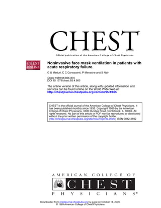
mask has been used successfully to correct refractory FIGURE 3. View of the face mask and ventilator.
hypoxemia in patients with cardiogenic’3 and noncar-
diogenic pulmonary 46 Retention of CO2 has showed a myocardial infarction.
occasionally been a limiting factor for this technique, The physiologic response to face mask ventilation
however. was considered satisfactory in all patients (Fig 3). In
Ten patients nonselectively entered this study. Six the hypercapnic group, mean Pco, was reduced by 18
had severe COPD with acute hypercapnic respiratory and 24 mm Hg at the first and sixth hours of ventilation,
failure and four had hypoxemic respiratory failure respectively, while the mean respiratory rate fell by
from cardiogenic and noncardiogenic pulmonary 18 breaths/mm. Patients also experienced a marked
edema. All patients met objective or clinical criteria amelioration of dyspnea and decreased effort to
indicating the need for mechanical ventilation. Face breathe. Overcorrection of respiratory acidosis (from
mask ventilation rapidly ameliorated dyspnea and pH 7.25 to 7.50) was, in part, responsible for the
reduced respiratory rates. development ofgeneralized seizures in patient 2, who
The use of a tight-fitting mask was well tolerated; had hyponatremia.
only one patient stopped using the mask after 20 h The patients with COPD and hypercapnia had the
because of discomfort. Only two patients developed advantage of being able to remove the mask for brief
abrasions on the nasal bridge, and they healed spon- periods (10 to 15 mm) during which they could drink
taneously; this was the only obvious complication noted fluids, verbalize, receive nebulized 3-agonist agents,
in this group. A nasogastric tube was inserted in all or expectorate secretions. Weaning from face mask
patients before beginning face mask ventilation. This ventilation was achieved by prolonging the periods off
tube resulted in leaking air where the tube exited the mask, while the patients received additional F1o2
from the mask, but no patient developed abdominal by nasal cannula or a Yenturi mask. Noninvasive face
distention secondary to aerophagia or gaseous insuffla- mask mechanical ventilation was successful in achiev-
lion, and no vomiting occurred. We did not observe ing adequate alveolar ventilation and correcting the
obvious aspiration clinically or radiographically. respiratory acidemia (mean rise in pH at 6 h was
Three patients, lethargic from CO2 retention (pa- 0. 145) while allowing the respiratory muscles to rest.
tients 2, 4, and 6), became alert and oriented after Compared with mechanical ventilation delivered via
beginning face mask ventilation. One patient (7) with an endotracheal tube, face mask ventilation had the
refractory hypoxemia was acutely agitated and disori- advantage of being noninvasive and more comfortable
ented before treatment and became comfortable and to the patients. It can be used also in an earlier stage
oriented soon after beginning ventilation. of ventilatory decompensation while medical manage-
The mean duration of face mask ventilation was ment is initiated. Because of the risk of aspiration, we
33 h (range, 3 to 88 h). No patient left the study think that face mask ventilation should not be contin-
because of failure to achieve adequate oxygenation or ued in patients who are lethargic and whose mental
ventilation. Three patients eventually required endo- status fails to improve after beginning ventilation.
tracheal intubation: patient 2 after generalized sei- In the group with hypoxemia and low lung compli-
zures, patient 4 during cardiopulmonary resuscitation, ance, face mask ventilation satisfactorily improved gas
and patient 9 because of discomfort from the tight- exchange and effective alveolar ventilation. Patients 7
fitting mask. Two patients died. Patient 3 refused to and 8 had ARDS with refractory hypoxemia and
continue after four days of successful ventilation and ventilatory insufficiency (tachypnea and PCO, reten-
died 15 h after discontinuing ventilation. Patient 4 had tion) while receiving CPAP delivered by a face mask.
a sudden, unexpected cardiac arrest, and an autopsy In patient 7, tachypnea and the sensation of dyspnea
CHEST I 95 I 4 I APRIL, 1989 869
Downloaded from chestjournal.chestpubs.org by guest on October 14, 2009
© 1989 American College of Chest Physicians
- 7. were corrected by instituting face mask ventilation; nelles. Paris: Universite R Descartes, 1984:1-124
4 Rideau Y. Management of the wheelchair muscular dystrophy
however, the PaO2:F1o2 ratio did not improve for
patient: prevention ofdeath [Abstract]. Los Angeles: 4th Inter-
24 h. Patient 8 experienced a rapid improvement in national Congress on Neuromuscular Diseases, 1986
oxygen exchange that permitted a reduction in FIo2 5 Ellis ER, Bye PTP, Bruderer JW, Sullivan CE. Treatment of
from 0.9 to 0.5 in 13 h, while the PEEP remained respiratory failure during sleep in patients with neuromuscular
constant at 10 cm H2O. A significant and rapid im- disease: positive-pressure ventilation through a nose mask. Am
Rev Respir Dis 1987; 135:148-52
provement in PaO2:FIO2 ratio was also seen in the two
6 Ellis ER, McCauley VB, Mellis C, Sullivan CE. Treatment of
patients with cardiogenic pulmonary edema.
alveolar hypoventilation in a six-year-old girl with intermittent
Face mask ventilation was used in a small hetero- positive pressure ventilation through a nose mask. Am Rev
geneous group ofpatients with acute respiratory failure Respir Dis 1987; 136:188-91
due to intrinsic lung disease. This procedure appears 7 Bach JR. Alba A, Mosher R, Delaubier A. Intermittent positive
pressure ventilation via nasal access in the management of
to produce physiologic improvements similar to those
respiratory 1987; 92:168-70
insafficiency. Chest
in intubated patients receiving mechanical ventilation.
8 Kerby CR, Mayer IS, Pingleton SK. Nocturnal positive pressure
The mask was well accepted and tolerated for pro- ventilation via nasal mask. Am Rev Respir Dis 1987; 135:738-40
longed periods of time without significant complica- 9 Carroll N, Branthwaite MA. Intermittent positive pressure
tions. While this preliminary report on a new mode ventilation by nasal mask: technique and applications. Intensive
Care Med 1988; 14:115-17
of noninvasive mechanical ventilation is encouraging,
10 Segall D. Noninvasive nasal mask-assisted ventilation in respi-
a much larger study is necessary before firm conclu- ratory failure of Duchenne muscular dystrophy. Chest 1988; 93:
sions and recommendations can be reached. 1298-1300
11 Zwiuich CW, Pierson DJ, Creagh CD, Sutton FD, Schatz E,
ACKNOWLEDGMENT: The authors wish to thank the respiratory Petty U. Complications of assisted
ventilation: a prospective
therapists and the Pulmonary attendings of Norwalk Hospital for
study of354 consecutive episodes. Am J Med 1974; 57:161-70
their dedication and enthusiasm that have made this study possible.
We would like to acknowledge the assistance of Dr. Norman Soskel 12 Stanifer JL, Silvestri RC. Complications of endotracheal intu-
and Dr. David Armbruster in editing the manuscript, Nancy Smith bation, tracheostomy, and artificial airways. Respir Care 1982;
and Vicky Franke for secretarial support, and Denis Selmont, PhD, 27:417-34
for organizing Tables 1-3.
13 Vaisanen IT, Rasanen J. Continuous positive airway pressure and
supplemental oxygen in the treatment ofcardiogenic pulmonary
edema. Chest 1987; 92:481-85
REFERENCES
14 Naver L, Walter 5, GlowinskiJ. Pulmonary fat embolism treated
1 Bach JR. O’Brien J, Krotenberg R, Alba AS. Management of by intermittent continuous positive airway pressure given by
end stage respiratory failure in Duchenne muscular dystrophy. face mask. Br Med J 1980; 14:1413-14
Muscle Nerve 1987; 10:177-82 15 Smith RA, Kirby RB, Gooding JM, Civetta JM. Continuous
2 Bach JR. Alba AS, Bohatiuk G, Saporito L, Lee M. Mouth positive airway pressure (CPAP) by face mask. Crit Care Med
intermittent positive pressure ventilation in the management of 1980; 8:483-85
postpolio respiratory insufilciency. Chest 1987; 91:859-64 16 Hurst JM, DeHaven CB, Branson RD. Use of CPAP mask as
3 Delaubier A. Traitement de l’insufllsance respiratoire chronique the sole mode of ventilatory support in trauma patients with
dans les dystrophies musculaires. In: Memoires de certificat mild to moderate respiratory insufficiency. Trauma 1985; 25:
d’#{233}tudes
superieures de reeducation et readaptation fonction- 1065-68
870 Face Mask Venthation in Patents with ARF (Medun at a!)
Downloaded from chestjournal.chestpubs.org by guest on October 14, 2009
© 1989 American College of Chest Physicians
- 8. Noninvasive face mask ventilation in patients with acute respiratory failure.
G U Meduri, C C Conoscenti, P Menashe and S Nair
Chest 1989;95; 865-870
DOI 10.1378/chest.95.4.865
This information is current as of October 14, 2009
Updated Information Updated Information and services, including high-resolution
& Services figures, can be found at:
http://chestjournal.chestpubs.org/content/95/4/865
Citations This article has been cited by 20 HighWire-hosted articles:
http://chestjournal.chestpubs.org/content/95/4/865#related-
urls
Open Access Freely available online through CHEST open access option
Permissions & Licensing Information about reproducing this article in parts (figures,
tables) or in its entirety can be found online at:
http://www.chestjournal.org/site/misc/reprints.xhtml
Reprints Information about ordering reprints can be found online:
http://www.chestjournal.org/site/misc/reprints.xhtml
Email alerting service Receive free email alerts when new articles cite this article. Sign
up in the box at the top right corner of the online article.
Images in PowerPoint Figures that appear in CHEST articles can be downloaded for
format teaching purposes in PowerPoint slide format. See any online
article figure for directions
Downloaded from chestjournal.chestpubs.org by guest on October 14, 2009
© 1989 American College of Chest Physicians