SlideShare une entreprise Scribd logo
1  sur  44
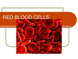
RED BLOOD CELLS
Three Phases of an Erythrocyte Life

    A) Erythropoiesis or Erythrocyte
                 Production

    B) Release from Bone Marrow to
                  Circulation

     C) Destruction and Death
RBC
    Materials needed for Red Blood
                 Production
     a) Metals e.g. Copper, Iron, Cobalt, Zinc
                b) Vitamins e.g. Thiamine, Folate
                  Cyanocobalamin
                      c) Amino Acids e.g.
     Glutamic, Lysine,
                  Leucine
                         d) Hormones e.g.
     Erythropoietin,
                 Androgen
RBC

  Erythrokinetic Evaluation states that Red
   Cell Production is equal to Red Cell
   Destruction
  Erythrocyte Kinetics ( M = P x S)
                   M - Total Erythrocyte
   Mass                               P–
   Production ( Number of New RBC
            produced per day)
                           S – RBC Life
   Span
Erythrocyte Kinetics
    Ex: A 70 kg man with 2 liters of Erythrocyte
     must produced 20 ml of new erythrocytes each
     day to replace the 20 ml. normally lost RBC
     due to cell senecent 2000 (M) = 20 ml /day (P)

                         100 days (S)
 If Survival time is decreased by half, the BM
    must double its production to maintain the man
    of 2000 ml of RBC 2000 ml(M)= 40 ml/day (P)

                       50 days(S)
Tests to Measure Erythropoiesis

    A) M/E Ratio ( 3:1) or E:G Ratio (1:3)

    B) Fecal Urobilinogen

    C) PITR ( Plasma Iron Turnover Rate)
Two Types of Erythropoiesis
  A) Effective Erythropoiesis
                    1) Reticulocyte count
                               2) Red Cell
   Utilization of Iron
   3) Red Cell Survival Time
  B) Ineffective Erythropoiesis
               Two groups demonstrating
   I.E.                              1)
   Hypoproliferative anemias
                     2) Anemias secondary
   to maturation
Factors Involved in the Release of
               RBC
   1)   Hypoxia
   2)   Presence of Erythropoietin
   3)   Thyroid Hormone
   4)   Androgenic Hormone
   5)   Growth Hormone
   6)   Prostaglandins
Normal red cells
   Normal red cells or
    erythrocytes show only slight
    variation in size and shape.
    The blood film should be
    examined in the area where
    the red cells are touching but
    not often overlapping. In this
    area many red cells have an
    area of central pallor which
    may be up to a third of the
    diameter of the cell. This is
    consequent on the shape of a
    normal red cell, which
    resembles a disc that is
    thinner in the centre.
Variation of RBC ( Anisocytosis)
   Anisocytosis
   Anisocytosis refers to
    increased variation in
    the size of red cells. This
    image also shows
    anisochromasia, i.e. an
    increased variation in
    the staining of red cells.
    Anisochromasia
    indicates variation in the
    haemoglobin
    concentration between
    different cells.
Macrocytosis

   Macrocytosis.
    Macrocytosis refers to
    an increase in the
    average size of red
    cells. A large red cell is
    referred to a macrocyte.
    This blood film is from a
    patient with
    macrocytosis caused by
    liver disease. The MCV
    was 105 fl (normal range
    82-98).
Macro-ovalocyte

   An ovalocyte which is
    appreciably larger than a
    normal erythrocyte is
    referred to as a macro-
    ovalocyte [arrow].
    Macro-ovalocytes are a
    feature of megaloblastic
    anaemia, congenital
    dyserythropoietic
    anaemias and South-
    East Asian ovalocytosis
Microcytosis

   The term microcytosis means
    that red cells are smaller than
    normal. Small red cells are
    referred to as microcytes.
    This blood film is from a
    blood sample with a mean
    cell volume (MCV) of 72 fl
    (normal range 82-98). There
    is also hypochromia, i.e. the
    cells have an increased area
    of central pallor.
Microspherocytes

   Microspherocytes
    resemble spherocytes in
    that they lack central
    pallor but differ in that
    they are smaller than
    normal discocytes. They
    result from red cell
    fragmentation. This
    photograph also shows
    large red cells, which
    are likely to be young
    cells, and a nucleated
    red cell or erythroblast
Variation of RBC (Poikilocytosis)
    Poikilocytosis in a baby with
     hereditary elliptocytosis and
     transient neonatal
     poikilocytosis. Poikilocytosis
     means an increased
     variability in red cell shape. A
     poikilocyte is a red cell of
     abnormal shape. An
     individual may have a
     predominance of a particular
     type of poikilocyte or there
     may be cells of a variety of
     shapes.
Classification of poikilocytes

   Poikilocytes can be
    classified as shown.
Acanthocyte

   An acanthocyte is an
    erythrocyte that is
    irregular in shape and is
    covered with a small
    number of spicules
    (2-20) which vary in
    length and thickness.
    The spicules are
    irregularly distributed
    over the surface of the
    red cell. The presence of
    acanthocytes is referred
    to as acanthocytosis.
Severe Liver Disease (Acanthocytosis
– Spur Cell Anemia)
    Acanthocyte are            Scanning electron
     irregularly shape and       micrograph of
     is covered with a           acanthocyte
     small spicules (2 to
     20)
Echinocyte
   An echinocyte is an erythrocyte
    with a large number of short
    blunt spicules (10-30), regularly
    distributed over the surface of
    the cell. The presence of
    echinocytes is referred to as
    echinocytosis. In this case the
    abnormality was caused by
    chronic renal failure but much
    more often echinocytes
    represent a storage artefact
    indicating that EDTA-
    anticoagulated blood has been
    stored for too long before the
    blood film was made.
Elliptocytes in iron deficiency

   Elliptocytes are quite
    common in iron deficiency.
    They are usually particularly
    long and thin and for this
    reason are sometimes
    referred to as 'pencil cells'.
    This film also shows
    anaemia, mild anisocytosis,
    hypochromia and
    microcytosis.
Spherocyte

   Spherocytes in
    hereditary
    spherocytosis. The
    spherocytes are the
    cells that lack central
    pallor. Cells which have
    rounded up to become
    spherocytes have a
    reduced diameter in
    comparison with normal
    discocytes but the cell
    size is normal.
Sickle cell and boat-shaped cell

    Sickle cell and boat-shaped
     erythrocytes in sickle cell
     anaemia. Sickle cells [dark
     red arrow] are sickle- or
     crescent-shaped red cells
     with both ends being pointed.
     Boat-shaped cells [dark blue
     arrows] are pointed at one or
     both ends but are not curved.
     Sickle cells are
     pathognomonic for sickle cell
     disease whereas boat-
     shaped cells are suggestive
     but not pathognomonic.
Stomatocytes

 Stomatocytes are red cells
  that, on a stained blood film,
  appear to have a slit-like
  mouth or stoma. In three
  dimensions they are cup-
  shaped.
 Common causes of
  stomatocytosis are liver
  disease and alcohol excess.
 Less common causes include
  myelodysplastic syndromes
  and hydroxyurea therapy.
 Congenital stomatocytosis is
  a rare cause of
  stomatocytosis.
Target cell
   Target cells are erythrocytes
    that, in stained blood films,
    resemble a target. They have
    an area of increased staining
    in the centre of the normal
    area of central pallor. In three
    dimensions, target cells are
    found to be bell-shaped.
    Target cells are a feature of
    hyposplenism, obstructive
    jaundice, liver disease and
    various
    haemoglobinopathies.
Teardrop poikilocyte

   Teardrop poikilocytes or
    dacrocytes are pear-
    shaped or teardrop-
    shaped red cells. They
    are a feature of
    idiopathic myelofibrosis
    (this case), secondary
    myelofibrosis,
    megaloblastic anaemia
    and congenital or
    acquired
    dyserythropoiesis
Keratocyte

   Keratocytes or horned cells
    are erythrocytes with a pair of
    spicules or 'horns'
    surrounding a gap in the cell
    outline. There may be a
    single pair of spicules [red
    arrow] or two pairs of
    spicules [blue arrow].
    Keratocytes can result from
    removal of a Heinz body from
    a cell (this case) or from red
    cell fragmentation.
Elliptocytes in hereditary elliptocytosis


    Elliptocytes are cells of
     elliptical shape. This film
     also shows ovalocytes
     which have an oval
     outline and are
     intermediate in shape
     between elliptocytes and
     discocytes.
RBC INCLUSIONS (alpha Chain
        precipitates)

   When synthesis of b globin
    chain is reduced excess
    alpha chains may precipitate
    in erythroblasts or mature
    erythrocytes. a chain
    precipitates [blue arrow] have
    the same staining
    characteristics as
    haemoglobin and appear as
    an irregular mass within a
    hypochromic red cell. They
    are seen in patients with b
    thalassaemia major.
Basophilic stippling

   Basophilic stippling [arrows]
    or punctate basophilia refers
    to the presence of numerous
    small basophilic inclusions
    scattered through red cells.
    They differ from
    Pappenheimer bodies in
    being present in larger
    numbers within an individual
    red cell and being scattered
    through the cytoplasm rather
    than being peripherally sited
Haemoglobin C crystal

   Haemoglobin C is prone
    to crystallize, forming
    crystals with straight
    parallel edges [arrows].
    They are usually
    rhomboidal or
    hexagonal. Usually all
    the haemoglobin in the
    cell crystallizes so that
    the cell appears
    otherwise empty of
    haemoglobin.
Heinz body

   Heinz bodies are red cell
    inclusions that are seen only
    after a specific vital stain, a
    Heinz body stain, has been
    performed. They stain pale pink
    or purple, are attached to the red
    cell membrane and sometimes
    protrude through the membrane.
    Heinz bodies cannot be seen on
    a routinely stained blood film.
    However when they are present
    the routine blood film usually
    shows irregularly contracted cells
    and keratocytes. Sometimes
    there is a protrusion of the red
    cell membrane
Glucose-6-phosphate dehydrogenase
         (G6PD) deficiency

   The commonest cause of
    congenital haemolytic anaemia is
    glucose-6-phosphate
    dehydrogenase (G6PD)
    deficiency which affects millions
    of people world-wide. Most
    individuals with G6PD deficiency
    suffer only intermittent
    haemolysis. The blood film then
    shows irregularly contracted cells
    [deep red arrows] and
    sometimes hemighosts [deep
    blue arrow] in which all the
    haemoglobin appears to have
    retracted to one side of the
    erythrocyte.
Howell-Jolly body

   Howell-Jolly bodies
    [arrow] are small nuclear
    fragments within
    erythrocytes. Their
    staining characteristics
    resemble those of the
    nucleus of a mature
    erythrocyte. They are
    round in shape and are
    sited closer to the edge
    of the cell than the
    centre.
Irregularly contracted cells

   Irregularly contracted
    cells are rounded
    cells lacking central
    pallor but with
    irregular outlines.
    This image also
    shows large cells
    that are likely to be
    young red cells.
Acquired haemoglobin H disease

    Acquired haemoglobin H
     disease occurs in
     myelodysplastic
     syndromes and
     erythroleukaemia. This
     patient also has
     sideroblastic
     erythropoiesis.
    The film is dimorphic
     and shows target cells,
     other poikilocytes,
     hypochromia and
     Pappenheimer bodies.
Pappenheimer bodies
   Pappenheimer bodies
    [arrows] are small
    basophilic inclusions
    sited near the periphery
    of red cells. They are
    smaller than Howell-
    Jolly bodies, stain deep
    blue rather than purple
    and are more likely to be
    multiple..
Siderotic granules (Perls' stain)

   Siderotic granules
    [arrow] are cytoplasmic
    granules composed of
    haemosiderin. The term
    'siderotic granule' is
    used when an iron stain
    is employed.
   The same red cell
    inclusions on a routinely
    stained film are referred
    to as 'Pappenheimer
    bodies'.
Variation of RBC (Anisochromasia)
    Anisochromasia
    This film from an iron
     deficient patient shows
     anisochromasia, i.e.
     there is increased
     variation in staining from
     cell to cell. Some cells
     are normally
     haemoglobinized while
     others show only a thin
     rim of haemoglobinized
     cytoplasm.
Dimorphic            blood film

 The term dimorphic indicates
  that there are two distinct
  populations of red cells. Most
  often the term is used, as in
  this case, to refer to a mixture
  of normal cells and
  hypochromic microcytes.
 This film is from a
  heterozygous carrier of
  sideroblastic anaemia. The
  photograph also shows three
  cells of abnormal shape.
Variation of RBC (Hypochromic)
    Hypochromia
    Hypochromia refers to
     reduced staining of red
     cells, indicative of a
     reduced haemoglobin
     concentration. Cells that
     are hypochromic are
     often also microcytic.
    In this film, from a
     patient with
     haemoglobin H disease,
     the great majority of
     cells are hypochromic.
Hyperchromia caused by spherocytosis

 Hyperchromia. The term
  hyperchromia can be used to
  indicate the presence of
  increased numbers of cells
  lacking central pallor.
  However it is more useful to
  state specifically why cells
  lack central pallor.
 This film shows hyperchromia
  consequent on
  spherocytosis, i.e. the
  presence of cells which are
  more spherical than normal
  cells. In this case they are
  consequent on autoimmune
  haemolytic anaemia.
Rouleaux and increased background staining


  Rouleaux and increased
   background staining often
   occur together since both are
   consequent on increased
   concentration of plasma
   proteins.
  Rouleaux are stacks of red
   cells, arranged like a pile of
   pennies.
  Increased background
   staining means that there is a
   basophilic background
   between the red cells.
Red cell agglutination

 Red cell agglutination is the
  clumping of red cells into
  irregular masses. It is caused
  by the presence of an
  antibody, usually a cold
  antibody, which bridges red
  cells.
 Agglutination can be
  distinguished from rouleaux
  formation by the irregularity
  of the clumps of cells, in
  contrast with the stacks of
  red cells in rouleaux.
Red cell rosetting

   Red cell rosetting is
    an uncommon
    phenomenon of
    uncertain
    significance in which
    erythrocytes are
    rosetted around
    leucocytes.

Contenu connexe

Tendances

Peripheral Blood Smear Examination
Peripheral Blood Smear ExaminationPeripheral Blood Smear Examination
Peripheral Blood Smear ExaminationDr Snehal Kosale
 
Rbc morphology
Rbc morphologyRbc morphology
Rbc morphologyPrince Avi
 
2. blood cells morphology
2. blood cells morphology2. blood cells morphology
2. blood cells morphologyaungkyawmyint26
 
Blood cell morphology
Blood cell morphologyBlood cell morphology
Blood cell morphologyTasmiaZeb1
 
Hb electrophoresis (principle materials and procedure)
Hb electrophoresis (principle materials and procedure)Hb electrophoresis (principle materials and procedure)
Hb electrophoresis (principle materials and procedure)hussainshahid55
 
iron staining.
iron staining.iron staining.
iron staining.milaya23
 
RBC morphology and Disease that may be associated with abnormal morphologies.
RBC morphology and Disease that may be associated with abnormal morphologies.RBC morphology and Disease that may be associated with abnormal morphologies.
RBC morphology and Disease that may be associated with abnormal morphologies.Faheem Javed
 
Benign White blood cell (WBC) Disorders
Benign White blood cell (WBC) DisordersBenign White blood cell (WBC) Disorders
Benign White blood cell (WBC) DisordersDr. Varughese George
 
Platelet Function Tests
Platelet Function TestsPlatelet Function Tests
Platelet Function TestsAhmed Makboul
 
Abnormal morphology of rbc notes
Abnormal morphology of rbc notesAbnormal morphology of rbc notes
Abnormal morphology of rbc notesVamsi kumar
 
Leukopiesis and agranulositosis 2011
Leukopiesis and agranulositosis 2011Leukopiesis and agranulositosis 2011
Leukopiesis and agranulositosis 2011wayan sugiritama
 
Morphology of white blood cells
Morphology of white blood cellsMorphology of white blood cells
Morphology of white blood cellsPrince Lokwani
 
Abnormalities of WBC
Abnormalities of WBCAbnormalities of WBC
Abnormalities of WBCSUNIL SHAH
 
Sideroblastic anemia - Etiopathogenesis, Clinical features, Advances in Manag...
Sideroblastic anemia - Etiopathogenesis, Clinical features, Advances in Manag...Sideroblastic anemia - Etiopathogenesis, Clinical features, Advances in Manag...
Sideroblastic anemia - Etiopathogenesis, Clinical features, Advances in Manag...Chetan Ganteppanavar
 

Tendances (20)

Peripheral Blood Smear Examination
Peripheral Blood Smear ExaminationPeripheral Blood Smear Examination
Peripheral Blood Smear Examination
 
Rbc morphology
Rbc morphologyRbc morphology
Rbc morphology
 
2. blood cells morphology
2. blood cells morphology2. blood cells morphology
2. blood cells morphology
 
Blood cell morphology
Blood cell morphologyBlood cell morphology
Blood cell morphology
 
RBC Inclusions
RBC InclusionsRBC Inclusions
RBC Inclusions
 
Hb electrophoresis (principle materials and procedure)
Hb electrophoresis (principle materials and procedure)Hb electrophoresis (principle materials and procedure)
Hb electrophoresis (principle materials and procedure)
 
Reticulocyte count
Reticulocyte countReticulocyte count
Reticulocyte count
 
iron staining.
iron staining.iron staining.
iron staining.
 
Rbc indices
Rbc indicesRbc indices
Rbc indices
 
Hematocrit determination
Hematocrit determinationHematocrit determination
Hematocrit determination
 
RBC morphology and Disease that may be associated with abnormal morphologies.
RBC morphology and Disease that may be associated with abnormal morphologies.RBC morphology and Disease that may be associated with abnormal morphologies.
RBC morphology and Disease that may be associated with abnormal morphologies.
 
Benign White blood cell (WBC) Disorders
Benign White blood cell (WBC) DisordersBenign White blood cell (WBC) Disorders
Benign White blood cell (WBC) Disorders
 
Laboratory hematology
Laboratory hematologyLaboratory hematology
Laboratory hematology
 
Platelet Function Tests
Platelet Function TestsPlatelet Function Tests
Platelet Function Tests
 
Abnormal morphology of rbc notes
Abnormal morphology of rbc notesAbnormal morphology of rbc notes
Abnormal morphology of rbc notes
 
Leukopiesis and agranulositosis 2011
Leukopiesis and agranulositosis 2011Leukopiesis and agranulositosis 2011
Leukopiesis and agranulositosis 2011
 
morphology of red blood cells
morphology of red blood cellsmorphology of red blood cells
morphology of red blood cells
 
Morphology of white blood cells
Morphology of white blood cellsMorphology of white blood cells
Morphology of white blood cells
 
Abnormalities of WBC
Abnormalities of WBCAbnormalities of WBC
Abnormalities of WBC
 
Sideroblastic anemia - Etiopathogenesis, Clinical features, Advances in Manag...
Sideroblastic anemia - Etiopathogenesis, Clinical features, Advances in Manag...Sideroblastic anemia - Etiopathogenesis, Clinical features, Advances in Manag...
Sideroblastic anemia - Etiopathogenesis, Clinical features, Advances in Manag...
 

En vedette

En vedette (20)

The erythrocyte
The erythrocyteThe erythrocyte
The erythrocyte
 
The erythrocyte lecture 2
The erythrocyte lecture 2The erythrocyte lecture 2
The erythrocyte lecture 2
 
normal and abnormalities in red blood cell
normal and abnormalities in red blood cellnormal and abnormalities in red blood cell
normal and abnormalities in red blood cell
 
Red cell membrane
Red cell membraneRed cell membrane
Red cell membrane
 
Rbc membrane
Rbc membraneRbc membrane
Rbc membrane
 
Red blood cell
Red blood cellRed blood cell
Red blood cell
 
Red blood cells in Blood
Red blood cells in BloodRed blood cells in Blood
Red blood cells in Blood
 
Structure and function of erythropoietic tissue
Structure and function of erythropoietic tissueStructure and function of erythropoietic tissue
Structure and function of erythropoietic tissue
 
Resealed erythrocytes
Resealed erythrocytesResealed erythrocytes
Resealed erythrocytes
 
Rbc
RbcRbc
Rbc
 
Megaloblastic Anaemia - Vit B12 deficiency
Megaloblastic Anaemia - Vit B12 deficiencyMegaloblastic Anaemia - Vit B12 deficiency
Megaloblastic Anaemia - Vit B12 deficiency
 
BLOOD:facts and fun
BLOOD:facts and funBLOOD:facts and fun
BLOOD:facts and fun
 
Megaloblastic anemia
Megaloblastic anemiaMegaloblastic anemia
Megaloblastic anemia
 
Peripheral blood smear examination
Peripheral blood smear examinationPeripheral blood smear examination
Peripheral blood smear examination
 
Blood
BloodBlood
Blood
 
Blood Physiology - Ppt
Blood Physiology - PptBlood Physiology - Ppt
Blood Physiology - Ppt
 
Hematology review '04
Hematology review '04Hematology review '04
Hematology review '04
 
Circulatory system
Circulatory system Circulatory system
Circulatory system
 
Category of blood variation in shape "POIKILOCYTOSIS"
Category of blood variation in shape "POIKILOCYTOSIS"Category of blood variation in shape "POIKILOCYTOSIS"
Category of blood variation in shape "POIKILOCYTOSIS"
 
Cell types
Cell typesCell types
Cell types
 

Similaire à Erythocyte

Red blood cells
Red blood cellsRed blood cells
Red blood cellsRaghu Veer
 
Morphology of erythrocytes
Morphology of erythrocytes Morphology of erythrocytes
Morphology of erythrocytes Amira Shehata
 
BLOOD FILM COMMENT part one.pptx
BLOOD FILM COMMENT part one.pptxBLOOD FILM COMMENT part one.pptx
BLOOD FILM COMMENT part one.pptxjnrkwesi
 
2 Hematopoiesis.pptgisgwvjshhsvffghvvgsz
2 Hematopoiesis.pptgisgwvjshhsvffghvvgsz2 Hematopoiesis.pptgisgwvjshhsvffghvvgsz
2 Hematopoiesis.pptgisgwvjshhsvffghvvgszsiddarthsaini007
 
Rbcmorphology 140722103459-phpapp02 - copy
Rbcmorphology 140722103459-phpapp02 - copyRbcmorphology 140722103459-phpapp02 - copy
Rbcmorphology 140722103459-phpapp02 - copyebtihal babekir
 
Hema II Chapter 2_RBC morphology study_AT.ppt
Hema II Chapter 2_RBC morphology study_AT.pptHema II Chapter 2_RBC morphology study_AT.ppt
Hema II Chapter 2_RBC morphology study_AT.pptaadillkhan1995
 
Hema II Chapter 2_RBC morphology study_AT.ppt
Hema II Chapter 2_RBC morphology study_AT.pptHema II Chapter 2_RBC morphology study_AT.ppt
Hema II Chapter 2_RBC morphology study_AT.pptAfeedaRazik
 
Hema II Chapter 2_RBC morphology study_AT.ppt
Hema II Chapter 2_RBC morphology study_AT.pptHema II Chapter 2_RBC morphology study_AT.ppt
Hema II Chapter 2_RBC morphology study_AT.pptssuser29c92d
 
Interpreting the c.b.c differential blood film Examination(part 1)
Interpreting the c.b.c  differential blood film Examination(part 1)Interpreting the c.b.c  differential blood film Examination(part 1)
Interpreting the c.b.c differential blood film Examination(part 1)Ahmed Redwan
 
Normal Hematopoiesis
Normal HematopoiesisNormal Hematopoiesis
Normal Hematopoiesisbalsan
 
hemopoiesisabbasa-170303103105 (1).pdf
hemopoiesisabbasa-170303103105 (1).pdfhemopoiesisabbasa-170303103105 (1).pdf
hemopoiesisabbasa-170303103105 (1).pdfMariumNSiddiqui
 
Chapter 2. Hematology(1).pptx
Chapter 2. Hematology(1).pptxChapter 2. Hematology(1).pptx
Chapter 2. Hematology(1).pptxSuraMAn2
 

Similaire à Erythocyte (20)

Red blood cells
Red blood cellsRed blood cells
Red blood cells
 
Morphology of erythrocytes
Morphology of erythrocytes Morphology of erythrocytes
Morphology of erythrocytes
 
RBC Morphology.pdf
RBC Morphology.pdfRBC Morphology.pdf
RBC Morphology.pdf
 
BLOOD FILM COMMENT part one.pptx
BLOOD FILM COMMENT part one.pptxBLOOD FILM COMMENT part one.pptx
BLOOD FILM COMMENT part one.pptx
 
hematology
hematologyhematology
hematology
 
My Presentation.pptx
My Presentation.pptxMy Presentation.pptx
My Presentation.pptx
 
2 Hematopoiesis.pptgisgwvjshhsvffghvvgsz
2 Hematopoiesis.pptgisgwvjshhsvffghvvgsz2 Hematopoiesis.pptgisgwvjshhsvffghvvgsz
2 Hematopoiesis.pptgisgwvjshhsvffghvvgsz
 
Hema intro anemia
Hema intro anemiaHema intro anemia
Hema intro anemia
 
erythropoieses
erythropoieseserythropoieses
erythropoieses
 
Rbcmorphology 140722103459-phpapp02 - copy
Rbcmorphology 140722103459-phpapp02 - copyRbcmorphology 140722103459-phpapp02 - copy
Rbcmorphology 140722103459-phpapp02 - copy
 
Hema II Chapter 2_RBC morphology study_AT.ppt
Hema II Chapter 2_RBC morphology study_AT.pptHema II Chapter 2_RBC morphology study_AT.ppt
Hema II Chapter 2_RBC morphology study_AT.ppt
 
Hema II Chapter 2_RBC morphology study_AT.ppt
Hema II Chapter 2_RBC morphology study_AT.pptHema II Chapter 2_RBC morphology study_AT.ppt
Hema II Chapter 2_RBC morphology study_AT.ppt
 
Hema II Chapter 2_RBC morphology study_AT.ppt
Hema II Chapter 2_RBC morphology study_AT.pptHema II Chapter 2_RBC morphology study_AT.ppt
Hema II Chapter 2_RBC morphology study_AT.ppt
 
Interpreting the c.b.c differential blood film Examination(part 1)
Interpreting the c.b.c  differential blood film Examination(part 1)Interpreting the c.b.c  differential blood film Examination(part 1)
Interpreting the c.b.c differential blood film Examination(part 1)
 
Blood
BloodBlood
Blood
 
Normal Hematopoiesis
Normal HematopoiesisNormal Hematopoiesis
Normal Hematopoiesis
 
hemopoiesisabbasa-170303103105 (1).pdf
hemopoiesisabbasa-170303103105 (1).pdfhemopoiesisabbasa-170303103105 (1).pdf
hemopoiesisabbasa-170303103105 (1).pdf
 
Chapter 2. Hematology(1).pptx
Chapter 2. Hematology(1).pptxChapter 2. Hematology(1).pptx
Chapter 2. Hematology(1).pptx
 
Anaemia
AnaemiaAnaemia
Anaemia
 
Blood.pptx
Blood.pptxBlood.pptx
Blood.pptx
 

Plus de jinx11

Tyrosine kinase report
Tyrosine kinase reportTyrosine kinase report
Tyrosine kinase reportjinx11
 
Acanthamoeba
AcanthamoebaAcanthamoeba
Acanthamoebajinx11
 
Apoptosis
ApoptosisApoptosis
Apoptosisjinx11
 
Microbe human interaction
Microbe human interactionMicrobe human interaction
Microbe human interactionjinx11
 
Epidemiology
EpidemiologyEpidemiology
Epidemiologyjinx11
 
Laboratory procedure of bacterial inhibition assay
Laboratory procedure of bacterial inhibition assayLaboratory procedure of bacterial inhibition assay
Laboratory procedure of bacterial inhibition assayjinx11
 

Plus de jinx11 (6)

Tyrosine kinase report
Tyrosine kinase reportTyrosine kinase report
Tyrosine kinase report
 
Acanthamoeba
AcanthamoebaAcanthamoeba
Acanthamoeba
 
Apoptosis
ApoptosisApoptosis
Apoptosis
 
Microbe human interaction
Microbe human interactionMicrobe human interaction
Microbe human interaction
 
Epidemiology
EpidemiologyEpidemiology
Epidemiology
 
Laboratory procedure of bacterial inhibition assay
Laboratory procedure of bacterial inhibition assayLaboratory procedure of bacterial inhibition assay
Laboratory procedure of bacterial inhibition assay
 

Dernier

"LLMs for Python Engineers: Advanced Data Analysis and Semantic Kernel",Oleks...
"LLMs for Python Engineers: Advanced Data Analysis and Semantic Kernel",Oleks..."LLMs for Python Engineers: Advanced Data Analysis and Semantic Kernel",Oleks...
"LLMs for Python Engineers: Advanced Data Analysis and Semantic Kernel",Oleks...Fwdays
 
Vertex AI Gemini Prompt Engineering Tips
Vertex AI Gemini Prompt Engineering TipsVertex AI Gemini Prompt Engineering Tips
Vertex AI Gemini Prompt Engineering TipsMiki Katsuragi
 
Nell’iperspazio con Rocket: il Framework Web di Rust!
Nell’iperspazio con Rocket: il Framework Web di Rust!Nell’iperspazio con Rocket: il Framework Web di Rust!
Nell’iperspazio con Rocket: il Framework Web di Rust!Commit University
 
Vector Databases 101 - An introduction to the world of Vector Databases
Vector Databases 101 - An introduction to the world of Vector DatabasesVector Databases 101 - An introduction to the world of Vector Databases
Vector Databases 101 - An introduction to the world of Vector DatabasesZilliz
 
Scanning the Internet for External Cloud Exposures via SSL Certs
Scanning the Internet for External Cloud Exposures via SSL CertsScanning the Internet for External Cloud Exposures via SSL Certs
Scanning the Internet for External Cloud Exposures via SSL CertsRizwan Syed
 
Powerpoint exploring the locations used in television show Time Clash
Powerpoint exploring the locations used in television show Time ClashPowerpoint exploring the locations used in television show Time Clash
Powerpoint exploring the locations used in television show Time Clashcharlottematthew16
 
My INSURER PTE LTD - Insurtech Innovation Award 2024
My INSURER PTE LTD - Insurtech Innovation Award 2024My INSURER PTE LTD - Insurtech Innovation Award 2024
My INSURER PTE LTD - Insurtech Innovation Award 2024The Digital Insurer
 
"ML in Production",Oleksandr Bagan
"ML in Production",Oleksandr Bagan"ML in Production",Oleksandr Bagan
"ML in Production",Oleksandr BaganFwdays
 
Connect Wave/ connectwave Pitch Deck Presentation
Connect Wave/ connectwave Pitch Deck PresentationConnect Wave/ connectwave Pitch Deck Presentation
Connect Wave/ connectwave Pitch Deck PresentationSlibray Presentation
 
DevoxxFR 2024 Reproducible Builds with Apache Maven
DevoxxFR 2024 Reproducible Builds with Apache MavenDevoxxFR 2024 Reproducible Builds with Apache Maven
DevoxxFR 2024 Reproducible Builds with Apache MavenHervé Boutemy
 
Integration and Automation in Practice: CI/CD in Mule Integration and Automat...
Integration and Automation in Practice: CI/CD in Mule Integration and Automat...Integration and Automation in Practice: CI/CD in Mule Integration and Automat...
Integration and Automation in Practice: CI/CD in Mule Integration and Automat...Patryk Bandurski
 
Commit 2024 - Secret Management made easy
Commit 2024 - Secret Management made easyCommit 2024 - Secret Management made easy
Commit 2024 - Secret Management made easyAlfredo García Lavilla
 
My Hashitalk Indonesia April 2024 Presentation
My Hashitalk Indonesia April 2024 PresentationMy Hashitalk Indonesia April 2024 Presentation
My Hashitalk Indonesia April 2024 PresentationRidwan Fadjar
 
"Debugging python applications inside k8s environment", Andrii Soldatenko
"Debugging python applications inside k8s environment", Andrii Soldatenko"Debugging python applications inside k8s environment", Andrii Soldatenko
"Debugging python applications inside k8s environment", Andrii SoldatenkoFwdays
 
"Subclassing and Composition – A Pythonic Tour of Trade-Offs", Hynek Schlawack
"Subclassing and Composition – A Pythonic Tour of Trade-Offs", Hynek Schlawack"Subclassing and Composition – A Pythonic Tour of Trade-Offs", Hynek Schlawack
"Subclassing and Composition – A Pythonic Tour of Trade-Offs", Hynek SchlawackFwdays
 
SAP Build Work Zone - Overview L2-L3.pptx
SAP Build Work Zone - Overview L2-L3.pptxSAP Build Work Zone - Overview L2-L3.pptx
SAP Build Work Zone - Overview L2-L3.pptxNavinnSomaal
 
Bun (KitWorks Team Study 노별마루 발표 2024.4.22)
Bun (KitWorks Team Study 노별마루 발표 2024.4.22)Bun (KitWorks Team Study 노별마루 발표 2024.4.22)
Bun (KitWorks Team Study 노별마루 발표 2024.4.22)Wonjun Hwang
 
Leverage Zilliz Serverless - Up to 50X Saving for Your Vector Storage Cost
Leverage Zilliz Serverless - Up to 50X Saving for Your Vector Storage CostLeverage Zilliz Serverless - Up to 50X Saving for Your Vector Storage Cost
Leverage Zilliz Serverless - Up to 50X Saving for Your Vector Storage CostZilliz
 
Story boards and shot lists for my a level piece
Story boards and shot lists for my a level pieceStory boards and shot lists for my a level piece
Story boards and shot lists for my a level piececharlottematthew16
 
Advanced Test Driven-Development @ php[tek] 2024
Advanced Test Driven-Development @ php[tek] 2024Advanced Test Driven-Development @ php[tek] 2024
Advanced Test Driven-Development @ php[tek] 2024Scott Keck-Warren
 

Dernier (20)

"LLMs for Python Engineers: Advanced Data Analysis and Semantic Kernel",Oleks...
"LLMs for Python Engineers: Advanced Data Analysis and Semantic Kernel",Oleks..."LLMs for Python Engineers: Advanced Data Analysis and Semantic Kernel",Oleks...
"LLMs for Python Engineers: Advanced Data Analysis and Semantic Kernel",Oleks...
 
Vertex AI Gemini Prompt Engineering Tips
Vertex AI Gemini Prompt Engineering TipsVertex AI Gemini Prompt Engineering Tips
Vertex AI Gemini Prompt Engineering Tips
 
Nell’iperspazio con Rocket: il Framework Web di Rust!
Nell’iperspazio con Rocket: il Framework Web di Rust!Nell’iperspazio con Rocket: il Framework Web di Rust!
Nell’iperspazio con Rocket: il Framework Web di Rust!
 
Vector Databases 101 - An introduction to the world of Vector Databases
Vector Databases 101 - An introduction to the world of Vector DatabasesVector Databases 101 - An introduction to the world of Vector Databases
Vector Databases 101 - An introduction to the world of Vector Databases
 
Scanning the Internet for External Cloud Exposures via SSL Certs
Scanning the Internet for External Cloud Exposures via SSL CertsScanning the Internet for External Cloud Exposures via SSL Certs
Scanning the Internet for External Cloud Exposures via SSL Certs
 
Powerpoint exploring the locations used in television show Time Clash
Powerpoint exploring the locations used in television show Time ClashPowerpoint exploring the locations used in television show Time Clash
Powerpoint exploring the locations used in television show Time Clash
 
My INSURER PTE LTD - Insurtech Innovation Award 2024
My INSURER PTE LTD - Insurtech Innovation Award 2024My INSURER PTE LTD - Insurtech Innovation Award 2024
My INSURER PTE LTD - Insurtech Innovation Award 2024
 
"ML in Production",Oleksandr Bagan
"ML in Production",Oleksandr Bagan"ML in Production",Oleksandr Bagan
"ML in Production",Oleksandr Bagan
 
Connect Wave/ connectwave Pitch Deck Presentation
Connect Wave/ connectwave Pitch Deck PresentationConnect Wave/ connectwave Pitch Deck Presentation
Connect Wave/ connectwave Pitch Deck Presentation
 
DevoxxFR 2024 Reproducible Builds with Apache Maven
DevoxxFR 2024 Reproducible Builds with Apache MavenDevoxxFR 2024 Reproducible Builds with Apache Maven
DevoxxFR 2024 Reproducible Builds with Apache Maven
 
Integration and Automation in Practice: CI/CD in Mule Integration and Automat...
Integration and Automation in Practice: CI/CD in Mule Integration and Automat...Integration and Automation in Practice: CI/CD in Mule Integration and Automat...
Integration and Automation in Practice: CI/CD in Mule Integration and Automat...
 
Commit 2024 - Secret Management made easy
Commit 2024 - Secret Management made easyCommit 2024 - Secret Management made easy
Commit 2024 - Secret Management made easy
 
My Hashitalk Indonesia April 2024 Presentation
My Hashitalk Indonesia April 2024 PresentationMy Hashitalk Indonesia April 2024 Presentation
My Hashitalk Indonesia April 2024 Presentation
 
"Debugging python applications inside k8s environment", Andrii Soldatenko
"Debugging python applications inside k8s environment", Andrii Soldatenko"Debugging python applications inside k8s environment", Andrii Soldatenko
"Debugging python applications inside k8s environment", Andrii Soldatenko
 
"Subclassing and Composition – A Pythonic Tour of Trade-Offs", Hynek Schlawack
"Subclassing and Composition – A Pythonic Tour of Trade-Offs", Hynek Schlawack"Subclassing and Composition – A Pythonic Tour of Trade-Offs", Hynek Schlawack
"Subclassing and Composition – A Pythonic Tour of Trade-Offs", Hynek Schlawack
 
SAP Build Work Zone - Overview L2-L3.pptx
SAP Build Work Zone - Overview L2-L3.pptxSAP Build Work Zone - Overview L2-L3.pptx
SAP Build Work Zone - Overview L2-L3.pptx
 
Bun (KitWorks Team Study 노별마루 발표 2024.4.22)
Bun (KitWorks Team Study 노별마루 발표 2024.4.22)Bun (KitWorks Team Study 노별마루 발표 2024.4.22)
Bun (KitWorks Team Study 노별마루 발표 2024.4.22)
 
Leverage Zilliz Serverless - Up to 50X Saving for Your Vector Storage Cost
Leverage Zilliz Serverless - Up to 50X Saving for Your Vector Storage CostLeverage Zilliz Serverless - Up to 50X Saving for Your Vector Storage Cost
Leverage Zilliz Serverless - Up to 50X Saving for Your Vector Storage Cost
 
Story boards and shot lists for my a level piece
Story boards and shot lists for my a level pieceStory boards and shot lists for my a level piece
Story boards and shot lists for my a level piece
 
Advanced Test Driven-Development @ php[tek] 2024
Advanced Test Driven-Development @ php[tek] 2024Advanced Test Driven-Development @ php[tek] 2024
Advanced Test Driven-Development @ php[tek] 2024
 

Erythocyte

  • 2. Three Phases of an Erythrocyte Life  A) Erythropoiesis or Erythrocyte Production  B) Release from Bone Marrow to Circulation C) Destruction and Death
  • 3. RBC  Materials needed for Red Blood Production a) Metals e.g. Copper, Iron, Cobalt, Zinc b) Vitamins e.g. Thiamine, Folate Cyanocobalamin c) Amino Acids e.g. Glutamic, Lysine, Leucine d) Hormones e.g. Erythropoietin, Androgen
  • 4. RBC  Erythrokinetic Evaluation states that Red Cell Production is equal to Red Cell Destruction  Erythrocyte Kinetics ( M = P x S) M - Total Erythrocyte Mass P– Production ( Number of New RBC produced per day) S – RBC Life Span
  • 5. Erythrocyte Kinetics  Ex: A 70 kg man with 2 liters of Erythrocyte must produced 20 ml of new erythrocytes each day to replace the 20 ml. normally lost RBC due to cell senecent 2000 (M) = 20 ml /day (P) 100 days (S) If Survival time is decreased by half, the BM must double its production to maintain the man of 2000 ml of RBC 2000 ml(M)= 40 ml/day (P) 50 days(S)
  • 6. Tests to Measure Erythropoiesis  A) M/E Ratio ( 3:1) or E:G Ratio (1:3)  B) Fecal Urobilinogen  C) PITR ( Plasma Iron Turnover Rate)
  • 7. Two Types of Erythropoiesis  A) Effective Erythropoiesis 1) Reticulocyte count 2) Red Cell Utilization of Iron 3) Red Cell Survival Time  B) Ineffective Erythropoiesis Two groups demonstrating I.E. 1) Hypoproliferative anemias 2) Anemias secondary to maturation
  • 8. Factors Involved in the Release of RBC 1) Hypoxia 2) Presence of Erythropoietin 3) Thyroid Hormone 4) Androgenic Hormone 5) Growth Hormone 6) Prostaglandins
  • 9. Normal red cells  Normal red cells or erythrocytes show only slight variation in size and shape. The blood film should be examined in the area where the red cells are touching but not often overlapping. In this area many red cells have an area of central pallor which may be up to a third of the diameter of the cell. This is consequent on the shape of a normal red cell, which resembles a disc that is thinner in the centre.
  • 10. Variation of RBC ( Anisocytosis)  Anisocytosis  Anisocytosis refers to increased variation in the size of red cells. This image also shows anisochromasia, i.e. an increased variation in the staining of red cells. Anisochromasia indicates variation in the haemoglobin concentration between different cells.
  • 11. Macrocytosis  Macrocytosis. Macrocytosis refers to an increase in the average size of red cells. A large red cell is referred to a macrocyte. This blood film is from a patient with macrocytosis caused by liver disease. The MCV was 105 fl (normal range 82-98).
  • 12. Macro-ovalocyte  An ovalocyte which is appreciably larger than a normal erythrocyte is referred to as a macro- ovalocyte [arrow]. Macro-ovalocytes are a feature of megaloblastic anaemia, congenital dyserythropoietic anaemias and South- East Asian ovalocytosis
  • 13. Microcytosis  The term microcytosis means that red cells are smaller than normal. Small red cells are referred to as microcytes. This blood film is from a blood sample with a mean cell volume (MCV) of 72 fl (normal range 82-98). There is also hypochromia, i.e. the cells have an increased area of central pallor.
  • 14. Microspherocytes  Microspherocytes resemble spherocytes in that they lack central pallor but differ in that they are smaller than normal discocytes. They result from red cell fragmentation. This photograph also shows large red cells, which are likely to be young cells, and a nucleated red cell or erythroblast
  • 15. Variation of RBC (Poikilocytosis)  Poikilocytosis in a baby with hereditary elliptocytosis and transient neonatal poikilocytosis. Poikilocytosis means an increased variability in red cell shape. A poikilocyte is a red cell of abnormal shape. An individual may have a predominance of a particular type of poikilocyte or there may be cells of a variety of shapes.
  • 16. Classification of poikilocytes  Poikilocytes can be classified as shown.
  • 17. Acanthocyte  An acanthocyte is an erythrocyte that is irregular in shape and is covered with a small number of spicules (2-20) which vary in length and thickness. The spicules are irregularly distributed over the surface of the red cell. The presence of acanthocytes is referred to as acanthocytosis.
  • 18. Severe Liver Disease (Acanthocytosis – Spur Cell Anemia)  Acanthocyte are  Scanning electron irregularly shape and micrograph of is covered with a acanthocyte small spicules (2 to 20)
  • 19. Echinocyte  An echinocyte is an erythrocyte with a large number of short blunt spicules (10-30), regularly distributed over the surface of the cell. The presence of echinocytes is referred to as echinocytosis. In this case the abnormality was caused by chronic renal failure but much more often echinocytes represent a storage artefact indicating that EDTA- anticoagulated blood has been stored for too long before the blood film was made.
  • 20. Elliptocytes in iron deficiency  Elliptocytes are quite common in iron deficiency. They are usually particularly long and thin and for this reason are sometimes referred to as 'pencil cells'. This film also shows anaemia, mild anisocytosis, hypochromia and microcytosis.
  • 21. Spherocyte  Spherocytes in hereditary spherocytosis. The spherocytes are the cells that lack central pallor. Cells which have rounded up to become spherocytes have a reduced diameter in comparison with normal discocytes but the cell size is normal.
  • 22. Sickle cell and boat-shaped cell  Sickle cell and boat-shaped erythrocytes in sickle cell anaemia. Sickle cells [dark red arrow] are sickle- or crescent-shaped red cells with both ends being pointed. Boat-shaped cells [dark blue arrows] are pointed at one or both ends but are not curved. Sickle cells are pathognomonic for sickle cell disease whereas boat- shaped cells are suggestive but not pathognomonic.
  • 23. Stomatocytes  Stomatocytes are red cells that, on a stained blood film, appear to have a slit-like mouth or stoma. In three dimensions they are cup- shaped.  Common causes of stomatocytosis are liver disease and alcohol excess.  Less common causes include myelodysplastic syndromes and hydroxyurea therapy.  Congenital stomatocytosis is a rare cause of stomatocytosis.
  • 24. Target cell  Target cells are erythrocytes that, in stained blood films, resemble a target. They have an area of increased staining in the centre of the normal area of central pallor. In three dimensions, target cells are found to be bell-shaped. Target cells are a feature of hyposplenism, obstructive jaundice, liver disease and various haemoglobinopathies.
  • 25. Teardrop poikilocyte  Teardrop poikilocytes or dacrocytes are pear- shaped or teardrop- shaped red cells. They are a feature of idiopathic myelofibrosis (this case), secondary myelofibrosis, megaloblastic anaemia and congenital or acquired dyserythropoiesis
  • 26. Keratocyte  Keratocytes or horned cells are erythrocytes with a pair of spicules or 'horns' surrounding a gap in the cell outline. There may be a single pair of spicules [red arrow] or two pairs of spicules [blue arrow]. Keratocytes can result from removal of a Heinz body from a cell (this case) or from red cell fragmentation.
  • 27. Elliptocytes in hereditary elliptocytosis  Elliptocytes are cells of elliptical shape. This film also shows ovalocytes which have an oval outline and are intermediate in shape between elliptocytes and discocytes.
  • 28. RBC INCLUSIONS (alpha Chain precipitates)  When synthesis of b globin chain is reduced excess alpha chains may precipitate in erythroblasts or mature erythrocytes. a chain precipitates [blue arrow] have the same staining characteristics as haemoglobin and appear as an irregular mass within a hypochromic red cell. They are seen in patients with b thalassaemia major.
  • 29. Basophilic stippling  Basophilic stippling [arrows] or punctate basophilia refers to the presence of numerous small basophilic inclusions scattered through red cells. They differ from Pappenheimer bodies in being present in larger numbers within an individual red cell and being scattered through the cytoplasm rather than being peripherally sited
  • 30. Haemoglobin C crystal  Haemoglobin C is prone to crystallize, forming crystals with straight parallel edges [arrows]. They are usually rhomboidal or hexagonal. Usually all the haemoglobin in the cell crystallizes so that the cell appears otherwise empty of haemoglobin.
  • 31. Heinz body  Heinz bodies are red cell inclusions that are seen only after a specific vital stain, a Heinz body stain, has been performed. They stain pale pink or purple, are attached to the red cell membrane and sometimes protrude through the membrane. Heinz bodies cannot be seen on a routinely stained blood film. However when they are present the routine blood film usually shows irregularly contracted cells and keratocytes. Sometimes there is a protrusion of the red cell membrane
  • 32. Glucose-6-phosphate dehydrogenase (G6PD) deficiency  The commonest cause of congenital haemolytic anaemia is glucose-6-phosphate dehydrogenase (G6PD) deficiency which affects millions of people world-wide. Most individuals with G6PD deficiency suffer only intermittent haemolysis. The blood film then shows irregularly contracted cells [deep red arrows] and sometimes hemighosts [deep blue arrow] in which all the haemoglobin appears to have retracted to one side of the erythrocyte.
  • 33. Howell-Jolly body  Howell-Jolly bodies [arrow] are small nuclear fragments within erythrocytes. Their staining characteristics resemble those of the nucleus of a mature erythrocyte. They are round in shape and are sited closer to the edge of the cell than the centre.
  • 34. Irregularly contracted cells  Irregularly contracted cells are rounded cells lacking central pallor but with irregular outlines. This image also shows large cells that are likely to be young red cells.
  • 35. Acquired haemoglobin H disease  Acquired haemoglobin H disease occurs in myelodysplastic syndromes and erythroleukaemia. This patient also has sideroblastic erythropoiesis.  The film is dimorphic and shows target cells, other poikilocytes, hypochromia and Pappenheimer bodies.
  • 36. Pappenheimer bodies  Pappenheimer bodies [arrows] are small basophilic inclusions sited near the periphery of red cells. They are smaller than Howell- Jolly bodies, stain deep blue rather than purple and are more likely to be multiple..
  • 37. Siderotic granules (Perls' stain)  Siderotic granules [arrow] are cytoplasmic granules composed of haemosiderin. The term 'siderotic granule' is used when an iron stain is employed.  The same red cell inclusions on a routinely stained film are referred to as 'Pappenheimer bodies'.
  • 38. Variation of RBC (Anisochromasia)  Anisochromasia  This film from an iron deficient patient shows anisochromasia, i.e. there is increased variation in staining from cell to cell. Some cells are normally haemoglobinized while others show only a thin rim of haemoglobinized cytoplasm.
  • 39. Dimorphic blood film  The term dimorphic indicates that there are two distinct populations of red cells. Most often the term is used, as in this case, to refer to a mixture of normal cells and hypochromic microcytes.  This film is from a heterozygous carrier of sideroblastic anaemia. The photograph also shows three cells of abnormal shape.
  • 40. Variation of RBC (Hypochromic)  Hypochromia  Hypochromia refers to reduced staining of red cells, indicative of a reduced haemoglobin concentration. Cells that are hypochromic are often also microcytic.  In this film, from a patient with haemoglobin H disease, the great majority of cells are hypochromic.
  • 41. Hyperchromia caused by spherocytosis  Hyperchromia. The term hyperchromia can be used to indicate the presence of increased numbers of cells lacking central pallor. However it is more useful to state specifically why cells lack central pallor.  This film shows hyperchromia consequent on spherocytosis, i.e. the presence of cells which are more spherical than normal cells. In this case they are consequent on autoimmune haemolytic anaemia.
  • 42. Rouleaux and increased background staining  Rouleaux and increased background staining often occur together since both are consequent on increased concentration of plasma proteins.  Rouleaux are stacks of red cells, arranged like a pile of pennies.  Increased background staining means that there is a basophilic background between the red cells.
  • 43. Red cell agglutination  Red cell agglutination is the clumping of red cells into irregular masses. It is caused by the presence of an antibody, usually a cold antibody, which bridges red cells.  Agglutination can be distinguished from rouleaux formation by the irregularity of the clumps of cells, in contrast with the stacks of red cells in rouleaux.
  • 44. Red cell rosetting  Red cell rosetting is an uncommon phenomenon of uncertain significance in which erythrocytes are rosetted around leucocytes.