Cestodes and trematodes
•Télécharger en tant que PPTX, PDF•
26 j'aime•17,896 vues

Parasitology

Recommandé
Contenu connexe
Tendances
Tendances (20)
En vedette
En vedette (19)
Similaire à Cestodes and trematodes
Similaire à Cestodes and trematodes (20)
Dernier
Dernier (20)
Cestodes and trematodes
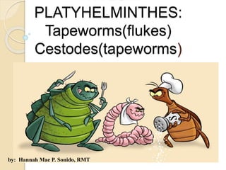
- 1. PHYLUM PLATYHELMINTHES: Tapeworms(flukes) Cestodes(tapeworms) by: Hannah Mae P. Sonido, RMT
- 2. Phylum Platyhelminthes (Flatworms) Bilaterally symmetrical Compressed dorso-ventrally Has a definite anteroposterior axis They lack a circulatory system Some species are hermaphroditic or monoecious Some are diecious
- 3. Possess a bilaterally symmetrical excretory system, collecting tubules, and capillaries which terminate in “flame cells”.
- 4. Two classes under Phylum Platyhelminthes which are of medical importance: a. Class Cestoidea b. Class Trematoda
- 5. Class Cestoidea Cestodes or tapeworms inhabit the small intestinal tract of vertebrates while the larva parasitize the tissues of vertebrates and invertebrates. Adult cestodes are usually ribbon or tape-like segmented parasites varying in size from a few millimeters to several meters. The body consists of 3 distinct regions: Proglottids or segment: a. Immmature- no well developed organs b. Mature- w/ well developed organ c. Gravid/ripe- filled with eggs
- 7. The life cycle of cestodes include: 1. the egg with a hexacanth embryo or oncosphere 2. the larval stage (cysticercus, cysticercoid larvae, or coracidium, procercoid and plerocercoid larvae) 3. adult stage All cestodes usually require an intermediate host although in some species the definitive host can serve as intermediate host.
- 8. Two orders of tapeworms with medical importance: Order PSEUDOPHYLLIDEA Order CYCLOPHYLLIDEA False tapeworm True tapeworm example D. latum All except D. latum scolex Spoon-shaped with slit-like sucking grooves called bothria With 4 cup-like suckers strobila Anapolytic (don’t shed segments) Apolytic (shed segments) ova Operculated, immature when laid Non-operculated, embryonated Larval stages 1st: coracidium 2nd: procercoid 3rd: plerocercoid (IS) 1st: cysticercus 2nd: cysticercoid 3rd: hydatid Intermediate host 2 1 only
- 9. Class Trematoda Leaf-like and unsegmented Adult trematodes are provided with an oral sucker and a ventral sucker called acetabulum. A third sucker called genital sucker or gonotyl is observed only among the heterophyids
- 10. The life cycle of trematodes includes: 1. The egg stage 2. The larval stages (miracidium, sporocyst, redia, cercaria, metacercariae) 3. Adult The definitive host (man) harbors the adult worm Intermediate host usually a freshwater snail or mollusk harbors the larval stage 2nd intermediate host (fish, crab, or another snail) is required for encystment
- 11. Phylum Platyhelminthes Class: Cestoidea (tapeworms) Subclass: Cestodes Order: Pseudophyllidea – scolex spatulate with bothria 1. Diphyllobothrium latum Order: Cyclophyllidea- scolex globular with 4 cuplike suckers A. Species which requires vertebrate intermediate hosts 1. Taenia solium 2. Taenia saginata B. Species which require invertebrate intermediate hosts 1. Dipyllidium caninum 2. Hymenolepis diminuta C. Species which may or may not require intermediate host 1. Hymenolepis nana D. Species which may infect man in their larval stages 1. Echinococcus granulosus 2. Echinococcus multilocularis
- 12. Class: Trematoda (flukes) A. Species which inhabit the portal blood stream of vertebrates 1. Schistosoma japonicum 2. Schistosoma mansoni 3. Schistosoma haematobium B. Species which inhabit the liver of vertebrates 1. Fasciola hepatica 2. Clonorchis sinensis 3. Opistorchis felineus C. Species which inhabit the small intestines of vertebrates 1. Fasciolopsis buski 2. Echinostoma ilocanum 3. Heterophyid group D. Species which inhabit the lungs of vertebrates 1. Paragonimus westermani
- 13. CESTODES
- 14. Intestinal Cestodes Taenia saginata Taenia solium Common name Beef tapeworm Pork tapeworm Intermediate host cattle Pig scolex No rostellum Armed rostellum length <25 m <7 m # of uterine branches 15 or more 13 or less # of proglottids 1000 to 2000 <1000 Gravid proglottid Tree-like ( dichomotous) Finger-like (dendritic) eggs Spherical and striated, inside is an embryo or oncosphere with 6 hooklets larva Cysticercus bovis Cysticercus cellulosae Infective stage Cysticercus bovis Cysticercus cellulosae Pathogenesis Taeniasis saginata Taeniasis solium, cysticercosis
- 16. Taenia solium Taenia saginata
- 17. Taenia species The eggs of the two species cannot be differentiated. Differentiation is usually done by examination of gravid proglottids that have been injected with India ink to reveal the lateral uterine branches. The eggs are spherical, yellow-brown measuring 31-43 mm. The shell is thick and radially striated, giving it a "prismatic" appearance. The egg also contains a six-hooked embryo called an oncosphere.
- 18. Hymenolepis Hymenolepis nana Hymenolepis diminuta Commin name Dwarf tapeworm (smallest tapeworm) Rat tapeworm Scolex Armed rostellum Unarmed rostellum eggs With 4 to 8 polar filaments Without filaments Infective stage Eggs (direct) Cysticercoid larvae (indirect) Cysticercoid larvae Intermediate host Fleas (Ctenocephalides canis, Xenopsylla cheopis, Pulex iritans) Flour beetles (Tenebrio molitor, T. obscurus, Tribolium confusum) Insects Final host man Norway rats
- 21. Dipylidium caninum Dog tapeworm or double-pored tapeworm Producing dipylidiasis very common intestinal parasites of dogs and cats Scolex is small w/ four deeply cupped suckers an protrusible rostellum Armed w/ 1 to 7 rows of rose-thorn shaped hooklets
- 22. The gravid proglottids have the size and shape of the pumpkin seed and are filled with capsules or packets of about 8 to 15 eggs enclosed in a embryonic membrane. D. caninum egg packet, containing 8 visible eggs, in a wet mount
- 23. Diphyllobothrium latum Fish tapeworm/ broad tapeworm Producing dyphillobothriasis Scolex is spatulate and measures 2-3mm in length by 1mm in diameter It has two bothria or sucking grooves
- 24. The dark, rosette-like, coiled uterus located in the middle of the gravid proglottid Ova are usually yellowish brown, w/ a moderately thick shell and an inconspicuous operculum.
- 25. Extraintestinal Cestodes Echinococcus sp. Belong to the Family Taeniidae, Order Cyclophyllidea Echinococcus granulosus Cause cystic echinococcosis Echinococcus multilocularis Cause alveolar echinococcosis
- 26. Echinococcus granulosus Hydatid worm Causes hydatid disease The smallest tapeworm (consisting only of a pyriform scolex, neck and three segments) Three segments: one immature, one mature, and one gravid The scolex bears a prominent rostellum and has four acetabula
- 27. Definitive host: dogs and other canines Intermediate host: goats, horses, camels, and sheep Larval stage: hydatid cyst - numerous protoscolices may be found within the cyst - cyst has outer laminated layer and inner nucleated germinal layer When protoscolices and broad capsules lie free in the cyst, these are referred to as “hydatid sand”.
- 28. Pathogenesis and Clinical Manifestations Liver (right lobe) – the most common and most important site of involvement Unilocular hydatid cyst – cyst of E. granulosus Alveolar cysts – cyst of E. multilocularis Osseous hydatid cyst- Unilocular cyst when located in the bone E. granulosus has led to down regulation of inflammatory cytokines leading to a local immunosuppression Hepatic cyst – mostly found in the inferior right lobe and may cause obstructive jaundice Abdominal cyst – may cause discomfort when the cyst are large enough
- 29. The rupture of hepatic cyst produces a characteristic triad: intermittent jaundice, fever, and eosinophilia. Sputum may contain frothy blood, mucus, hydatid fluid and bits of membrane. Involvement of the brain may cause increased intracranial pressure and Jacksonian epilepsy. Jacksonian seizures are partial seizures that begin in one part of the body such as the side of the face, the toes on one foot, or the fingers on one hand. The jerking movements then spread to other muscles on the same side of the body. This type of seizure is associated with a lesion or defect in the area of the cerebral cortex that controls voluntary movement
- 30. It shows multiple destructive cystic lesions in the proximal of the tibia and distal of the femur which some of them show extra-osseous extension. Enlargement of the head in a 5-year-old male who was finally diagnosed to have brain hydatid disease. Extra –osseus extension in the knee
- 31. Diagnosis 1. Roentgenogram 2. Exploratory cyst puncture 3. Immunologic tests (intradermal, precipitin, complement fixation, hemagglutination, and bentonite flocculation latex slide agglutination and fluorescent antibody tests) 4. Serologic tests ( IHA, IFA, EIA) Positive cases will have to undergo a gel diffusion assay that would demonstrate the echinococcal “Arc 5” for confirmation.
- 32. Sparganosis Refers to the larval infection with the plerocercoid larvae also known as spargana of pseudophyllidean tapeowrms falling under the Genus Spirometra. Common in man: Spirometra mansoni Spirometra erinacei Spirometra ranarum
- 33. Trematodes (Flukes) All FLUKES Schistosomes shape Flat and leaf-shaped Elongated and cylindrical sexes hermaphroditic Separate sexes egg operculated Nonoperculated transmission ingestion Skin penetration Infective stage metarcercaria Cercaria Intermediate host 2 1 •Attaches to host by means of 2 suckers: oral sucker and ventral sucker (acetabulum) •Heterophyes has 3 suckers, the 3rd one is the genital sucker (gonotyle)
- 34. Life cycle: 1. egg stage 2. larval stage a. 1st intermediate: miracidium-sporocyst-redia-cercaria b. 2nd intermediate host: cercaria-metacercaria 3. adult stage Requires 2 intermediate hosts (except for Schistosomes) 1st IH: snail 2nd IH: a. fish – H. heterophyes, C. sinensis, O. felineus b. crab – P. westermanni c. plant/vegetation – F. hepatica, F. gigantica, F. buski d. snail: E. ilocanum
- 35. Lung Fluke Paragonimus westermanni Common name: Oriental lung fluke Causing : paragonimiasis, lung fluke disease, pulmonary distomiasis, endemic hemoptysis, parasitic hemoptysis Two other species: P. philippinensis P. siamensis (only in cats)
- 36. adult: reddish-brown resembles a coffee bean ovary: has six long unbranched lobes
- 37. egg: oval yellowish-brown thick-shelled has a flattened operculum, opposite is a thickened abopercular
- 38. First intermediate host: Antemelania asperata Antemelania dactylus (former name: Brotia asperata) Second intermediate host: mountain crab – Sundathelphusa philippina (former name: Parathelphusa grapsoides)
- 39. Pathogenesis and Clinical Manifestations Heavy infections: from dry cough and later produce bloodstained or rust- colored sputum with foul fish odor, most pronounced in the morning. Elevated levels of IL-5 Migration in the brain may cause: Jacksonian epilepsy, cerebral hemorrhage, edema, visual disturbances, or meningits Often misdiagnosed as PTB
- 40. Diagnosis Radiographs Complement fixation (CF) – standard serological test Enzyme Immunoassay (EIA) Immunoblot (IB) Based on the detection of characteristic eggs in the stool
- 41. Intestinal Flukes Fasciolopsis buski Common name: Giant intestinal fluke Causing: fasciolopsiasis *The largest fluke parasitizing man Adult: -does not have cephalic cone
- 42. egg: -ellipsoidal, rounded at both ends -has a thin shell and a delicate operculum
- 43. First intermediate host: -snails belonging to the genus Segmentina or Hippeutis Second intermediate host: Trapa bicornis (water caltrop) Eliocharis tuberosa (water chesnut) Ipomea obscura (water morning glory) Nymphaea lotus (lotus) Definitive host: Humans Pigs
- 44. Pathogenesis and Clinical Manifestations Pathological changes are traumatic, obstructive, and toxic. Inflammation and ulceration Heavy infections: intestinal obstruction Toxic and allergic symptoms: edema in the face, abdominal wall, and lower limbs Profound intoxication can result to death Diagnosis Detection of parasite in stool
- 45. Echinostoma ilocanum Artyfechinostomum malayanum The echinomastids are digenetic trematodes characterized by a collar of spines around their oral suckers. A. Malayanum adult - Has rounded posterior end - 43-45 collar spines - Two testes: large w/ six to nine lobes arranged in tandem A. Malayanum egg - Larger - golden brown in color - operculated
- 46. E. ilocanum adult - Reddish-gray - Tapered posterior end - Has 49 to 51 collar spines - testes: deeply bilobed, arranged in tandem E. ilocanum egg - straw-colored - operculated Echinostoma ilocanum: Common name: Garrison’s fluke Causing: echinostomiasis
- 47. E. ilocanum First snail intermediate host: Gyraulus convexiusculus Hippeutis umbilicalis Second snail IH: Pila luzonica (kuhol) Vivipara angularis (susong pampang) A. Malayanum Second snail IH: Lymnaea(Bullastra) cumingiana(birabid)
- 48. Pathogenesis and Clinical Manifestations Ulceration Diarrhea (sometimes bloody) Abdominal pain Diagnosis Detection of eggs in the stool
- 49. Heterophyid Flukes Major species: Heterophyes heterophyes Metagonimus yokogawai Haplorchis taichuia Haplorchis yokogawai
- 50. Heterophyes heterophyes Common name: Von Siebod’s fluke *smallest fluke of man, yet deadliest. Adult: - elongated, oval or pyriform - Tegument has fine scale-like spines Egg: - Light brown in color - ovoid
- 51. Pathogenesis and Clinical Manifestations Peptic Ulcer Disease (PUD) Acid Peptic Disease (APD) Diagnosis Kato-katz
- 52. Blood Fluke Schistosomes: S. japonicum – aka as Oriental Blood fluke - causes Katayama’s disease - habitat: mesentric veins of small intestines - IH: Oncomelania quadrasi
- 53. S. mansoni – smallest blood fluke - habitat: mesentric veins of colon, rectum - IH: Biophalaria and Australorbis S. haematobium – aka as Vesical blood fluke - causes: Bilharziasis or uninary schistosomiasis (bloody urine) - habitat: veins of urinary bladder - IH: Bulinus and Physopsis
- 54. S. japonicum & S. mansoni - parasites of branches of the portal vein - both cause primarily hepato-intestinal schistomiasis S. haematobium - inhabits the veins of the urinary bladder - causes urinary schistosomiasis
- 56. Ovum S. japonicum - ovoidal, rounded or pear-shaped - when recovered in stool, the eggs are pale yellow - on the side near one of the polar ends is a curved hook or spine.
- 57. S. mansoni - a prominent lateral spine near the posterior end. - the anterior end is tapered and slightly curved.
- 58. S. haematobium - bear a conspicuous terminal spine.
- 60. Miracidium -morphological features includes: apical papilla epidermal plates covered w/ cilia pair of cephalic unicellular penetration glands two pairs of flame cells & germinal cells
- 61. Miracidium - are phototactic and swim actively in surface water - factors which influence the infection of snail by miracidia: - age of snails & miracidia - number of miracidia per snail - length of contact time - water flow & turbulence
- 62. Intramolluscan Developmental Stages - the miracidium becomes the first generation or mother sporocyst soon after penetration of its snail host. - in 96 hours, it is an elongated sac filled with germinal cells. - on the 8th day, germ cells budded off and develop into daughter sporocyst.
- 63. Cercariae - mature cercariae emerge from daughter sporocysts and escape from the snail into surrounding waters. - the cercariae has a body and a forked tail, oral and ventral sucker. - elongated body: 100 to 500 um in length 40 to 60 um in diameter - tail trunk: 140 to 150 um by 20 to 35 um - fork: 50 to 70 um long - cercarial penetration is mediated by lytic enzymes secreted by cephalic glands aided by muscular activity.
- 65. Schistosomule - after skin penetration,the cercaria is transformed into a schistosomule, which has different physiological adaptations and requirements.
- 66. Adult flukes - unlike other trematodes, schistosomes have separate sexes. - adults: large sucker capping the anterior end ventral sucker gonophore - the suckers aid in the movement and enable the flukes to maintain their position inside the veins. - male: shorter but sturdier sex - 12-20 mm in length by 0.4 o -0.5 mm in diameter - female: measures 15 to 26 mm by 0.3 mm
- 67. male: -also has a gynecophoral canal where the longer and more slender female is held.
- 68. Pathogenesis and Clinical Manifestations Early Schistosomiasis - chills, fever or non-productive cough Colonic Schistosomiasis - ulcerations result in dysentery or diarrhea Hepatosplenic Disease - the most serious consequence of chronic schistomiasis - characterized by hepatosplenomegaly, ascites, and collateral circulation
- 69. Pulmonary Schistosomiasis - cor pulmonale resulting from the obstruction of the pulmonary vasculature Cerebral Schistosomiasis - meningoencephalitis with fever, headache, confusion, lethargy, and coma.
- 72. Hepatosplenic schistosomiasis Collateral circulation Patients infected with a large load of parasites are more likely to produce disease in the liver. The eggs of the parasite tend to migrate and settle in the portal vein, causing inflammation and obstruction of the passage of blood by fibrosis. As all the blood coming from gastrointestinal system passes through the portal vein to the liver before going to the rest of the body, an obstruction in this region causes a huge "traffic jam" of blood, which leads to what we call portal hypertension. If no blood reaches the liver, it has to find other ways to get to the rest of the body, forming a collateral circulation. Portal hypertension is responsible for complications of hepatosplenic schistosomiasis, among them, ascites, splenomegaly (enlarged spleen) and esophageal varices. The esophageal varices are a feared complication of portal hypertension, as they may rupture causing severe gastrointestinal bleeding and bloody vomiting.
- 73. Liver Flukes - these are digenetic trematode species belong to family Fasciolidae. - they are parasites found in the liver and biliary passages of humans and herbivorous mammals, especially ruminants. - in tropical countries, fascioliasis is considered the most important helminth infection of cattle.
- 74. First intermediate host: - belongs to the family Lymnaeidae - L. philippinensis & L. auricularia rubiginosa Second intermediate host: - aquatic plants: Ipomea obscura (kangkong) Nasturtium officinale (water cress)
- 75. Fasciola hepatica - common name: sheep liver fluke - causes: sheep liver rot, fascioliasis hepatica - prevalent in sheep raising countries Fasciola gigantica - common name: Giant liver fluke - infects cattles in the Philippines
- 77. egg -large, ovoidal, operculated and yellowish to brownish in color -measures: 140- 180 um by 80- 100 um in size -released from the worm still immature
- 79. Pathogenesis and Clinical Manifestations -two clinical stages: 1. Acute stage - coincides with larval migration and worm maturation in hepatic tissue. 2. Chronic stage - coincides with the persistence of Fasciola worms in the biliary ducts
- 80. Hepatic fascioliasis: -can be asymptomatic or may produce fever -right upper quadrant abdominal pain -hypereosinophilia Fibrosis when the worm reached the bile ducts Pharyngeal fascioliasis or Halzuon (suffocation) has been reported in Mediterranean countries. This has been attributed to ingestion of raw liver containing the parasites w/c attach to the pharyngeal mucosa causing asphyxiation.
- 81. Clonorchis sinensis Opisthorchis felineus Opisthorchis viverrini - belong to family Opistorchiidae - parasites of the bile duct and gallbladder of humans and fish-eating mammals
- 82. Clonorchis sinensis -common name: Chinese liver fluke Oriental liver fluke -causes: clonorchiasis -most important liver fluke in man -eggs are like old fashioned electric light bulb Opisthorchis felineus -common name: cat liver fluke -causes: opistorchiasis felineus
- 83. First snail intermediate host of C. sinensis: - belong to the genera Parafossarulus P. manchouricus, P. anomaloispralis, P. stratulus - genera Bulimus (B. striatulus) - genera Semisulcospira - genera Alcinma (A. longicornis) - genera Thiara ( T. granifera) - genera Melanoides (M. tuberculatus) First snail intermediate host of O. felineus and O. viverrini: - genus Bithynia
- 84. Second intermediate host: - fish species that belongs to family Cyprinidae - freshwater shrimp
- 85. Adult worms - leaf like in shape with transparent tegument
- 86. egg - yellowish brown - ovoid - there is a distinctly convex operculum