
Recommandé
Recommandé
Contenu connexe
Tendances
Tendances (20)
En vedette
En vedette (20)
Similaire à Hepato-renal effects of cypermethrin
Similaire à Hepato-renal effects of cypermethrin (20)
Dernier
Dernier (20)
Hepato-renal effects of cypermethrin
- 1. INTERNATIONAL JOURNAL OF AGRICULTURE & BIOLOGY ISSN Print: 1560–8530; ISSN Online: 1814–9596 11–414/ARL/2011/13–6–865–872 http://www.fspublishers.org Full Length Article Cypermethrin Induced Biochemical and Hepato-renal Pathological Changes in Rabbits LATIF AHMAD, AHRAR KHAN1 AND MUHAMMAD ZARGHAM KHAN Department of Pathology, Faculty of Veterinary Science, University of Agriculture, Faisalabad, Pakistan 1 Corresponding author’s e-mails: ahrar1122@yahoo.com; ahrar1122@gmail.com ABSTRACT The effects of intra-peritoneal administration of cypermethrin (CY) on biochemistry and histology of liver and kidneys in rabbits were studied. Male rabbits (n = 10 x 4 = 40) in groups B, C and D received low (50 mg.kg-1 body weight), medium (100 mg.kg-1 body weight) and high (150 mg.kg-1 body weight) CY doses, respectively in mustard oil at weekly interval up to day 71. Group A served as control and each animal in this group received equivalent volume of mustard oil. Blood samples without anticoagulant from all animals were collected prior to start of experiment (day 0) and after every treatment, which were used for extraction of serum. The serum was used for analysis of proteins (serum total proteins/STP, serum albumin, serum globulins), aminotransferases (AST & ALT), alkaline phosphatase (ALP), urea and creatinine. Two animals from each group were euthanized fortnightly for histopathological studies. Increases in AST level in sera of CY-treated rabbits were accompanied by histological lesions in liver (different stages of degeneration & bile duct hyperplasia). Increased urea and creatinine concentrations and decreased STP, albumin and globulins in sera of CY-treated rabbits could be due to renal damage. The renal damage appeared histologically in the form of different lesions (pyknotic nuclei, necrosis, sloughed tubular epithelium, cast deposition & increased urinary space) in CY-treated rabbits. It was concluded that cypermethrin at various doses administered during the study produced moderate histological lesions in liver and kidneys along-with biochemical alterations in serum samples. © 2011 Friends Science Publishers Key Words: Cypermethrin; Liver enzymes; Proteins; Kidneys; Creatinine; Histopathpology INTRODUCTION Although hepato-renal consequences have been addressed in a few studies in animals, yet present study is A wide range of pests including those challenging peculiar in regard to long term repeated treatments and the public health are being killed/inactivated with follow up. Changes produced in the pyrethroid exposed cypermethrin (CY), which is very forceful synthetic animals can be regarded as immediate, since pyrethroids are pyrethroid pesticide. It is extensively used as insecticidal rapidly metabolized in the body (Sayim et al., 2005). sprays on cotton, vegetables and other crops. Likewise, However, it was realized that frequent stress, debility and some veterinary products are based on CY (e.g., residue accumulation etc. with repeated CY exposure could Ecofleece), which are popularly used for dipping or reveal more pronounced lesions than single exposure. The showering of food animals (Shah et al., 2007). half-life of orally administered cis-isomer of CY in fat, liver Pyrethroids have less mammalian toxicity among the and kidneys has been reported to be about 12 days, although pesticides but have been reported to affect physiological residues in liver and kidneys were much lower as compared activities and produce pathological entities in animals to fat at a given time (Crawford et al., 1981). On the other (Khan et al., 2009). Among pyrethroids, CY is rapidly hand, the in vivo half-life of trans-isomer of CY has been absorbed in the body and exhibit clinical signs of reported to be 3.4 days (Sayim et al., 2005). So, in vivo half- neurotoxicity including loss of coordination, muscular life was one of the factors considered, while establishing twitching and death due to respiratory failure (Sharaf et weekly interval between intraperitoneal injections in the al., 2010). Due to being lipid soluble and of smaller size present study. Furthermore, almost weekly agricultural (V = 536.40 Å3), CY easily passes through cell membrane spraying practices and anti-parasite application in animals and may damage DNA by causing destabilization and also required the weekly interval to make contrast of the unwinding of the DNA helix and chromosomal injuries study to animal exposures under field conditions. (Saxena et al., 2005). The DNA damage has led to When exogenous and endogenous toxic products enter different consequences in different body systems (Sharaf the body, their detoxification is carried out with active et al., 2010). participation of liver. So, there is a greater risk of hepatocyte To cite this paper: Ahmad, L., A. Khan and M.Z. Khan, 2011. Cypermethrin induced biochemical and hepato-renal pathological changes in rabbits. Int. J. Agric. Biol., 13: 865–872
- 2. AHMAD et al. / Int. J. Agric. Biol., Vol. 13, No. 6, 2011 injury (Wight, 1982). Serum enzyme levels are considered peritonealy. Since CY is not soluble in water, it was indicators of overall health status of an individual especially dissolved in mustard oil for intra-peritoneal (IP) injection. hepatocyte injury and related stress (Khan et al., 2009). By Dose to be injected was adjusted according to body weight. processing blood plasma and excreting urine, the kidneys Groups B, C and D received low, medium and high (50, 100 play a vital role in the clearance and excretion of & 150 mg.kg-1 body weight) CY doses, respectively at xenobiotics including drugs and drug-products from the weekly interval up to day 71. Group A served as control and body. Valuable facts about the nephrotic health can be each animal in this group received equivalent volume of known from the estimation of several waste products of mustard oil. metabolism (such as urea & creatinine), which are excreted Sample collection and analysis: Blood samples from all completely through kidneys (Garba et al., 2007). Urea and animals were collected prior to first treatment (d 0) and then creatinine are waste products of protein metabolism and are after every treatment and subjected to extraction of serum. withheld in blood in cellular damage (Aslam et al., 2010). The serum samples were stored in aliquots at -20°C for Therefore, these are the most sensitive biochemical markers biochemical studies. Serum aspartate aminotransferase for the diagnosis of renal damage (Garba et al., 2007). The (AST), alanine aminotransferase (ALT) and alkaline major reasons for decreased proteins in serum include: renal phosphatase (ALP) activities were measured with and intestinal protein loss, hemorrhages, malabsorption and commercially available colorimetric kits (AMP liver failure (Khan, 2008). So determination of proteins in Medizintechnic GmbH, Austria Cat # BR0415, BR061 & CY-treated animals might yield interesting information in BR0202, respectively). The principles for the leakage connection with hepato-renal toxicity. enzymes (AST & ALT) measurements were to monitor the Numerous pesticides are in use for the control of concentration of L-lactate or L-Malate with α-ketoglutarate agricultural pests and animal disease causing vectors in at wavelength of 340 nm, while that for ALP was to monitor Pakistan. Despite the fact that the pyrethroids (including the concentration of p-nitrophenol formed with water at CY) have potential adverse health effects, they are still wavelength of 405 nm with a spectrophotometer. Urea and being widely used in agriculture and animal husbandry creatinine levels in serum were measured with (Shah et al., 2007). During the previous decade, pyrethroid commercially available colorimetric kits (AMP application has increased tremendously (Ahmad et al., Medizintechnic GmbH, Austria Cat # BR04006 & BR2810, 2011). Although, many studies are available addressing respectively) using spectro-photometer. The principle for hepato-renal pathology in pyretroid exposed animals, yet enzymatic determination of urea was measurement of L- present study is peculiar with regard to duration and more glutamate with water and α-ketoglutarate, while that for clearly correlates the biochemical alterations with renal and creatinine determination was to measure the speed of hepatic histopathology. production of colored complex between creatinine and alkaline picrate using spectrophotometer at wavelengths of MATERIALS AND METHODS 340 and 500 nm, respectively. The serum total proteins (STP) were estimated by Synopsis of this experiment was tailored keeping in Biuret method and serum albumin by bromocresol green view all the national legislations and research ethics laid (BCG) dye binding method (Khan et al., 2011) using down by the Ethics Committee about the animal welfare and spectrophotometer. The serum globulin was calculated by following the strategies and guidelines of the Advanced subtracting albumin from STP (Javed et al., 2010). Studies and Research Board (ASRB) of the University. Two animals randomly selected from each group were Before implementation, the experimental proposal was euthanized fortnightly (on experimental days 15, 29, 43, 57 approved by the ASRB. & 71). The liver and kidneys from each animal were Experimental animals and protocol: Apparently healthy carefully dissected and processed for histopathology New Zealand white adult male rabbits (Oryctolagus (Awaad et al., 2010; Khan et al., 2010). Briefly, about 5 cuniculus) (n = 40) almost of the same age (about 6 months) mm thick pieces of the morbid organs were fixed in 10% and weight (990±50 g), were procured from the local market buffered formalin and later processed for histopathological and were kept under similar management conditions. The studies using routine methods of dehydration, paraffin animal room temperature was maintained at 25-27°C with embedding, sectioning (4-5 µm) and staining (H & E). 45-70% humidity and 12-h light–dark cycle throughout the During the microscopic examination of slides of each group study. Drinking water was available ad libitum. The green at a particular period, histological lesions were scored on a fodder Berseem (Trifolium alexandrinum) was offered in scale of ---- to ++++. From this a cumulative lesion score the morning and evening. After 5 days acclimatization, the was derived for the overall severity of pathology in a rabbits were randomly divided into four equal groups i.e., particular group. A-D. Cypermethrin (92%) used in the study was gifted by Data analysis: Randomized Complete Block Design was M/S Pak-China Chemicals, Lahore. Oral LD50 of CY in used and serum parameters’ data collected were subjected to rabbits is 2400 mg.kg-1 body weight (Hartley & Kidd, analysis of variance using Minitab statistical software 1990), 1/48th, 1/24th and 1/16th of it were administered intra- package on personal computer. Mean values of various 866
- 3. HEPATO-RENAL PATHOLOGY IN RABBITS / Int. J. Agric. Biol., Vol. 13, No. 6, 2011 treatments were compared by Duncan’s Multiple Range II). Significantly (P≤0.05) higher serum creatinine was Test (DMR) at P<0.05. recorded in all treated groups at day 64, in groups C and D at days 1, 29 and 71 and in group D at day 43. RESULTS A dose and time dependent decreasing trend was observed in the concentration of STP in all treatment groups Biochemical parameters: Significantly (P≤0.05) higher (Fig. 1). Significantly (P≤0.05) lower STP were recorded in AST was recorded in all treated groups at day 43 and in all treated groups at days 8 and 57, in groups B at day 15, in group D (150 mg.kg-1 body weight) at days 36, 57 and 71 group C at day 36, in group D at day 15 and in groups C and (Table I). Significantly (P≤0.05) lower ALT was recorded D at days 50, 64 and 71 (Fig. 1). Significantly (P≤0.05) in all treated groups at day 71, while significantly higher lower serum albumin was recorded in all treated groups at ALP at day 15 only in group B (Table I). A dose and time day 8 and in group B at day 15 (Table II). Significantly dependent increasing trend was observed in the (P≤0.05) lower serum globulins were recorded in all treated concentration of urea (Fig. 1) and creatinine concentration groups at day 57, in groups C and D at days 64 and 71 and (Table II) in all treatment groups Significantly (P≤0.05) in group B at day 8 (Table II). lower serum urea was recorded in all treated groups at day Gross and histopathology: In group A (control) 1. Contrarily, significantly (P≤0.05) higher serum urea was throughout the course of experiment, the liver and kidneys recorded in all treatment groups at day 64 and 71, in groups did not exhibit any gross morphological alteration. All C and D at days 43 and 57 and in group D at days 29 (Table treated groups were having dose and time related frequency Table I: Serum aspartate transaminase, alanine transaminase and alkaline phosphatase concentrations (IU) in cypermethrin treated rabbits Parameters/Experimental Group (CY dose: mg.kg-1 body weight) P-Value Days A (0) B (50) C (100) D (150) Aspartate transaminase 0 95.2 ± 13.4 91.3 ± 17.5 90.3 ± 14.4 94.7 ± 20.7 0.945 1 88.0 ± 17.9 84.8 ± 38.2 116.3 ± 70.9 95.7 ± 28.0 0.602 8 93.2 ± 20.2 171.7 ± 56.0 119.0 ± 36.8 115.3 ± 71.2 0.098 15 112.6 ± 48.8 155.2 ± 50.3 127.7 ± 24.0 126.7 ± 32.9 0.367 22 97.6 ± 36.6 160.2 ± 35.0 135.8 ± 45.8 146.2 ± 56.9 0.184 29 95.8 ± 61.7 129.6 ± 46.5 177.0 ± 86.0 144.8 ± 99.2 0.465 36 75.8 ± 15.7 118.0 ± 46.3 149.5 ± 13.2 178.0 ± 72.2* 0.036 43 80.0 ± 8.7 149.5 ± 7.4* 172.5 ± 31.3* 157.3 ± 47.5* 0.003 50 104.3 ± 16.0 95.0 ± 21.9 139.0 ± 35.7 162.7 ± 65.4 0.213 57 90.3 ± 18.5 52.0 ± 12.1 118.0 ± 45.0 277.0 ± 114.0* 0.010 64 94.0 ± 49.5 91.0 ± 32.5 114.5 ± 46.0 154.0 ± 59.2 0.400 71 91.0 ± 11.3 138.0 ± 28.3 149.5 ± 20.5 207.5 ± 21.9* 0.025 Alanine transaminase 0 114.3 ± 6.0 131.2 ± 6.5 113.17 ± 26.06 128.2 ± 60.7 0.709 1 144.0 ± 34.3 135.7 ± 70.5 106.00 ± 69.13 154.5 ± 72.8 0.597 8 140.6 ± 85.1 121.2 ± 54.4 172.17 ± 56.92 99.3 ± 31.9 0.207 15 156.4 ± 41.9 140.8 ± 30.6 112.67 ± 20.92 158.2 ± 34.6 0.093 22 163.2 ± 47.8 125.7 ± 31.1 191.17 ± 90.66 201.2 ± 94.5 0.302 29 148.6 ± 25.4 105.0 ± 47.5 157.60 ± 91.75 205.4 ± 57.0 0.113 36 152.0 ± 66.5 125.6 ± 52.9 114.75 ± 10.87 115.4 ± 27.8 0.577 43 164.7 ± 53.4 116.0 ± 33.1 104.25 ± 16.78 123.8 ± 36.8 0.170 50 162.7 ± 144.0 88.3 ± 16.7 99.00 ± 42.29 86.3 ± 48.8 0.501 57 116.0 ± 12.5 116.0 ± 12.0 145.00 ± 12.00 87.7 ± 48.7 0.150 64 159.0 ± 14.0 97.0 ± 21.0 153.67 ± 62.50 79.0 ± 49.0 0.109 71 155.0 ± 25.9 80.0 ± 19.4* 69.00 ± 17.57* 45.0 ± 14.2* 0.000 Alkaline phosphatase 0 273.0 ± 165.5 284.0 ± 104.5 326.2 ± 164.7 239.3 ± 99.9 0.749 1 220.2 ± 74.8 386.7 ± 227.0 311.3 ± 114.0 290.7 ± 46.0 0.278 8 239.2 ± 39.6 291.2 ± 106.0 309.3 ± 87.8 254.2 ± 72.9 0.471 15 232.2 ± 31.1 364.3 ± 79.4* 275.2 ± 35.7 307.7 ± 59.3 0.007 22 267.2 ± 29.3 262.8 ± 29.7 245.8 ± 47.0 272.8 ± 53.3 0.755 29 226.8 ± 43.6 182.2 ± 64.9 174.0 ± 44.2 243.6 ± 112.0 0.421 36 252.7 ± 32.9 285.0 ± 98.9 247.3 ± 66.9 244.8 ± 69.1 0.840 43 231.8 ± 81.4 343.0 ± 84.1 317.8 ± 133.1 208.0 ± 150.2 0.330 50 200.7 ± 22.5 333.7 ± 44.5 281.0 ± 140.0 284.0 ± 82.9 0.353 57 235.0 ± 12.0 324.0 ± 16.0 222.0 ± 19.0 261.7 ± 110.4 0.201 64 239.5 ± 67.2 274.0 ± 32.5 331.0 ± 79.2 255.0 ± 12.7 0.457 71 227.5 ± 61.5 256.5 ± 26.2 362.0 ± 168.3 243.5 ± 34.7 0.525 The values (mean ± SD) bearing asterisks in a row differ significantly (P≤0.05) 867
- 4. AHMAD et al. / Int. J. Agric. Biol., Vol. 13, No. 6, 2011 Fig. 1: Serum urea (upper) and total proteins (lower) concentrations (mean ± SD) in cypermethrin treated rabbits. Table II: Serum creatinine, albumin and globulin concentrations in cypermethrin treated rabbits Parameters/Experimental days Group (CY dose: mg.kg-1 body weight) P-Value A (0) B (50) C (100) D (150) Creatinine (mg.dl-1) 0 1.45 ± 0.51 1.35 ± 0.77 1.84 ± 0.34 1.57 ± 1.15 0.711 1 1.62 ± 0.38 1.91 ± 0.42 2.88 ± 0.55* 2.77 ± 0.60* 0.000 8 2.32 ± 0.90 2.45 ± 0.36 1.04 ± 0.22 0.95 ± 0.71 0.533 15 1.37 ± 0.31 1.53 ± 0.22 2.02 ± 0.49 2.76 ± 0.33 0.528 22 1.21 ± 0.72 1.34 ± 0.57 4.95 ± 0.47 4.03 ± 0.23 0.072 29 1.04 ± 0.27 1.04 ± 0.54 4.21 ± 0.42* 5.39 ± 0.12* 0.011 36 1.04 ± 0.36 4.11 ± 3.12 3.30 ± 2.19 3.99 ± 2.24 0.150 43 1.00 ± 0.148 3.27 ± 1.76 3.08 ± 1.69 5.52 ± 1.66* 0.008 50 1.53 ± 0.66 2.49 ± 2.17 1.56 ± 1.53 2.75 ± 2.07 0.851 57 1.27 ± 0.26 2.75 ± 2.69 1.62 ± 0.08 2.87 ± 0.20 0.419 64 1.88 ± 0.77 4.90 ± 0.85* 5.15 ± 0.50* 6.47 ± 0.26* 0.008 71 2.28 ± 1.01 4.65 ± 0.93 5.93 ± 1.96* 6.95 ± 1.58* 0.004 Albumin (g.dl-1) 0 2.61 ± 0.38 2.88 ±0.46 2.75 ± 0.20 3.00 ± 0.24 0.242 1 2.63 ± 0.45 2.55 ±0.59 1.94 ± 0.55 2.07 ± 0.69 0.140 8 3.60 ± 0.09 3.30 ± 0.07* 2.92 ± 0.20* 2.98 ± 0.34* 0.000 15 3.25 ± 0.19 2.56 ± 0.40* 2.76 ± 0.38 2.76 ± 0.22 0.015 22 2.75 ± 0.76 2.70 ± 0.48 2.60 ± 0.41 3.00 ± 0.43 0.599 29 3.04 ± 0.26 2.71 ± 0.35 2.97 ± 0.40 2.92 ± 0.58 0.622 36 3.21 ± 0.38 3.01 ± 0.20 2.81 ± 0.14 3.37 ± 0.48 0.113 43 3.10 ± 0.17 3.28 ± 0.43 2.95 ± 0.25 3.18 ± 0.19 0.409 50 3.12 ± 0.11 3.48 ± 0.78 3.07 ± 0.43 2.96 ± 0.23 0.445 57 3.20 ± 0.12 3.41 ± 0.50 3.46 ± 0.23 3.40 ± 0.11 0.695 64 3.22 ± 0.02 3.20 ± 0.08 3.48 ± 0.43 3.63 ± 0.12 0.307 71 3.23 ± 0.16 3.23 ± 0.21 3.45 ± 0.20 3.63 ± 0.12 0.201 Globulin (g.dl-1) 0 3.18 ± 0.71 3.11 ± 0.51 2.75 ± 0.30 2.90 ± 0.61 0.530 1 2.71 ± 0.68 1.82 ± 0.42 2.66 ± 1.62 2.06 ± 0.46 0.280 8 2.61 ± 0.14 2.07 ± 0.11* 2.40 ± 0.36 2.69 ± 0.24 0.001 15 2.30 ± 0.33 2.13 ± 0.31 2.27 ± 0.17 2.10 ± 0.21 0.459 22 2.97 ± 0.62 2.59 ± 0.41 2.79 ± 0.25 2.37 ± 0.60 0.259 29 2.81 ± 0.35 2.57 ± 0.33 2.57 ± 0.31 1.32 ± 0.60 0.429 36 2.69 ± 0.25 2.59 ± 0.09 2.20 ± 0.25 1.91 ± 0.76 0.090 43 2.47 ± 0.24 2.14 ± 0.36 2.42 ± 0.30 2.13 ± 0.37 0.373 50 2.30 ± 0.22 1.70 ± 0.70 2.55 ± 0.44 2.34 ± 0.21 0.089 57 3.06 ± 0.35 1.73 ± 0.76* 1.13 ± 0.47* 1.16 ± 0.18* 0.004 64 3.15 ± 0.34 2.83 ± 0.35 1.29 ± 0.74* 0.74 ± 0.08* 0.014 71 3.30 ± 0.09 2.45 ± 0.59 1.40 ± 0.50* 0.81 ± 0.03* 0.011 The values (mean ± SD) bearing asterisks in a row differ significantly (P≤0.05) 868
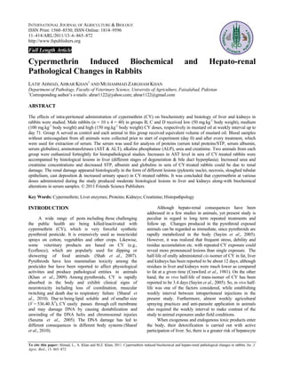
- 5. HEPATO-RENAL PATHOLOGY IN RABBITS / Int. J. Agric. Biol., Vol. 13, No. 6, 2011 Table III: Frequency and incidence of gross and Fig. 3: Photomicrograph of liver of rabbit treated with histological lesions in liver and kidneys of cypermethrin (100 mg.kg-1 body weight) at cypermethrin treated rabbits experimental day 57 showing condensed nuclei (arrow) and necrosed hepatocytes. H and E, Lens 40X Organ/Lesion Groups (Doses of CY: mg.kg-1 body weight) B (50) C (100) D (150) F* I ** F I F I Liver Paleness (grossly) +--- 30 +--- 50 ++-- 70 Fat deposits (grossly) ---- 0 +--- 50 ++-- 70 Hyperplasia of bile +--- 20 ++-- 40 ++++ 80 ducts Degeneration +--- 30 ++-- 60 +++- 90 Cytoplasmic ++-- 60 ++-- 80 +++- 100 vacuolation Kidneys Swelling (grossly) +--- 40 ++-- 60 +++- 80 Paleness (grossly) +--- 40 +--- 60 +++- 80 Hemorrhage +--- 60 ++-- 80 ++++ 90 Degeneration ++-- 40 ++-- 70 +++- 100 Increased urinary space +--- 30 ++-- 60 ++++ 90 Sloughed epithelium ++-- 40 ++-- 80 +++- 90 Cast deposition +--- 30 ++-- 60 ++++ 90 *F= Frequency; **I = Incidence (%). Various concentrations of Fig. 4: Photomicrograph of liver of rabbit treated with cypermethrin mixed in mustard oil were injected IP to male rabbits (n = 10 cypermethrin (150 mg.kg-1 body weight) on x 4 = 40) at weekly interval. experimental day 57 showing bile duct hyperplasia Fig. 2: Liver showing enhanced lobular pattern at day (arrow) and newly formed bile ducts (arrow head) 71 in the rabbit treated with high (150 mg.kg-1 body along with necrosis of hepatocytes. H and E, Lens 10X weight) cypermethrin dose of paleness of liver (Table III). The liver in group D (150 tubules, pyknosis and increased urinary space (Fig. 5) in mg.kg-1 body weight) showed enhanced lobular pattern treated animals were observed. Tubular epithelial (Fig. 2) during later stages of the study (d 57-71). detachment from the basement membrane and cast Swollen and pale kidneys of variable intensity were deposition in the renal tubules along with different stages of observed in all treated groups at various experimental days degeneration (Fig. 6) were noted. The kidney lesions (Table III). appeared earlier in group D, followed by groups C and B. Rabbits of control group (A) did not show any Variable frequency and incidence of different lesions were histological alteration in liver and kidneys. Condensation of observed in dose related pattern (Table III). hepatic nuclei and cytoplasmic vacuolation (Fig. 3) were observed initially in 50% animals and later in all treated DISCUSSION animals. In the later stages, these changes became more severe and extensive. Bile duct hyperplasia/newly formed In the present study, liver was slightly enlarged bile ducts along with various degrees of cellular with pale color and friable consistency at initial stage, degeneration in the parenchyma were observed in dose but darker/hemorrhagic at later stage with the high dose dependent manner (Fig. 4). Regenerating hepatocytes were group showing enhanced lobular pattern; the latter found in later stages of exposure. Hemorrhages in renal denoting decreased partial pressure of oxygen in blood. 869
- 6. AHMAD et al. / Int. J. Agric. Biol., Vol. 13, No. 6, 2011 Fig. 5: Photomicrograph of kidney from cypermethrin considered indicators of overall health status of an (100 mg.kg-1 body weight) treated rabbit on day 29 individual especially hepatocyte injury and related stress showing increased urinary space (blank arrows) and (Khan et al., 2009). Increases of blood aminotransferases condensation of nuclei (arrow head) along with are associated with liver injury and change in hepatic degeneration of lining epithelium in renal tubules functions (Manna et al., 2003). Increase of these enzymes (filled arrow). H and E, Lens 40X has been credited to their seepage into the blood stream (Yousef et al., 2006). Free radicals produced by pyrethroids lead to hepatocyte alterations (Manna et al., 2004). Chemical-induced cellular alterations vary from simple increase of metabolism to cell death. Since liver stores AST and synthesizes ALT (Nduka, 1999), focal, multifocal and diffused hepatic necrosis lead to variable magnitude and frequency of alteration in these enzymes (Onyesom & Anosike, 2007). Due to mitochondrial as well as cytoplasmic origin, AST activity is increased in large proportion in various types of liver damage (Theal & Scott, 1996). The increase or decrease of enzyme activity depends on the intensity of cellular damage. Rarely serum AST and ALT levels decrease in toxicology studies. Among the reasons of decreased aminotransferases could include low synthesis/release from hepatocytes, inhibited/reduced Fig. 6: Photomicrograph of kidney from cypermethrin enzyme activity, assay interference and decreased vitamin B (150 mg.kg-1 body weight) treated rabbit on day 71 (pyridoxal 5-phosphate). However, decreased amino- showing degenerated tubules (filled arrows), condensed trasferases have been documented to not have any nuclei (arrow heads) and cast deposition in the lumen toxicologically significant effects on liver, regardless of the of renal tubules (blank arrow). H and E, Lens 40X mechanism involved (Hayes, 2007). ALP is membrane bound enzyme, it is found on all cell membranes, where active transport occurs and is hydrolase and transphosphorylase in function. The highest concentrations of ALP are found in the liver, biliary tract epithelium, bone and intestinal mucosa (Ravel, 1995). Serum ALP activity increases in case of damage to hepatic cells and obstruction of bile duct through proliferation of hepatic cells (El-Demerdash et al., 2003). Its decreased activity is taken as an index of parenchymal damage (Anwar, 2003). Differences in concentrations of ALP were in-consistent, which might rule out the cholestatsis in CY pathogenesis (Wulkan & Leijnse, 1986), although moderate bile duct hyperplasia was observed histologically. The latter The greatly branched portal venous arrangement retorts might have been produced due to non-cholestatic conditions quickly to minute alterations in metabolic activity. This (Hulzebos et al., 2005). striking vascular reaction to cellular damage and the Microscopically, hemorrhages in renal tubules, intricacy of hepatic acinar blood streaming model may different stages of degeneration, cast deposition and elucidate the enhanced lobular pattern in a variety of liver increased urinary spaces were observed with dose injuries (Zimmon, 1977). Histologically, liver parenchyma dependent frequency and incidence in CY treated rabbits in in treated animals exhibited vacuolar degeneration and the present study. In kidneys, filtration barrier is made up of moderate proliferation of bile ducts in the present study. The endothelium and podocytes, which are arranged on latter is mostly obvious in cholestatic injuries (Burt & basement membrane inside and outside, respectively and all MacSween, 1993). Pathologic changes in hepatocytes due to these structures are negatively charged whereas protein is CY can be related to its inhibitory effect on total adenine tri- also negatively charged (Khan, 2008). Pyrethroids produce phosphate activity in the liver, which may disturb active oxidative stress by generating free radicals which could transport of Na+, K+ and Ca2+ ions, thus injuring hepatocytes damage the filtration barrier and negative charge on the (Khan et al., 2009). structures (endothelium, podocytes & basement membrane) The present study revealed increased serum AST with become positive or charge is removed, thereby proteins go CY treatment, whereas serum ALT activity at the end of the out freely. Due to various degenerative changes in the study was decreased significantly. Serum enzyme levels are kidneys caused by CY, epithelial cells are detached from the 870
- 7. HEPATO-RENAL PATHOLOGY IN RABBITS / Int. J. Agric. Biol., Vol. 13, No. 6, 2011 tubular basement membranes. When detached epithelial proteins by pyrethroids might occur due to oxidative stress cells are mixed with leaked proteins, the resultant mixture as discussed above in the mechanism of epithelial cast appears in the form of epithelial casts (Khan et al., 2009). formation or it may occur due to increased activities of urea The significant increase (P≤0.05) in urea and enzymes. These enzymes are mostly associated with liver creatinine levels noticed in this study is a classical sign damage, since the urea cycle is confined to liver in which that the kidney was unfavorably affected by CY proteins are broken down into urea (Woodman, 1980). exposure. Creatinine is more specific to kidneys since Decreased STP could be credited due in part to the renal damage is the only significant parameter that damaging outcome of pyrethroids on hepatocytes as increases serum creatinine in mammals (Garba et al., 2007). confirmed by the increase in the activities of liver enzymes Like many other waste products of metabolism, most (> ¾th) (Yousef et al., 2006). Decreased proteins may be of the creatinine is removed from the body through encountered in renal and intestinal protein loss, glomerular filtration and the rest (<25%) through tubular hemorrhages, malabsorption and liver failure (Khan, 2008). secretion (Ravel, 1995). The increased serum concentrations Decreased serum proteins in the present study implied that of creatinine thus might be a result of alteration in these two increase in serum urea levels could be due to increased mechanisms. Urea is also excreted by kidneys, so impaired protein breakdown, in which case the hypoproteinemia kidney function causes diminished ability to excrete urea could be due to increased breakdown of proteins and not from the blood into urine (Aslam et al., 2010). Other than lesser synthesis. renal tissue damage, causes of increased serum urea include: Decreased albumin in animals treated with pesticides (1) Rapid urea production from ammonia and proteins, (2) might be related with altered metabolic activities of proteins Hampered excretion of urea (Garba et al., 2007), (3) and free amino acids in liver (Rivarola & Balegno, 1991). Dehydration, (4) Increased activities of urea enzymes When amino acids in the liver are altered, albumin synthesis (ornithine carbomoyl transferase, arginase) (Guven et al., is hampered and decreased serum albumin results. Decreased 2006), (5) Decreased serum proteins and (6) Low blood serum globulins were recorded in the present study with CY- volume (Garba et al., 2007). Possibly dehydration with CY- treatment. The endoplasmic reticulum (ER) of plasma cells is treatment (Sharaf et al., 2010) led to increased serum principally involved with globulin synthesis, which is also proteins first, which were used for rapid urea production reported to be adversely affected during pesticide toxicity leading to high serum urea. Excessive urea production from (Reyes & More, 1979). It has been proposed that ER might proteins might have then led to hypoproteinemia and more accumulate calcium by Ca2+ pump, then either inositol 1, 4, urea production might continue after development of 5-trisphosphate or cyclic adenosine di-phosphate ribose hypoproteinemia. The erythropoeitin production is inhibited cause release of Ca2+. When ER is disturbed, there is also in renal malfunctioning leading to high urea (Garba et al., lesser globulin synthesis (He et al., 2006). Therefore, it is 2007). Definitely blood volume decreases along with apparent that decrease in serum globulin was due to decreased erythrocytes, which might be a reason for reduction in its synthesis by the plasma cells. anaemia reported in CY-treated animals (Ahmad et al., From the results of the present work, it was concluded 2009) but the types of cells involved in inflammation and that CY at various doses administered produced moderate white blood cells are increased, making the body more histological lesions in liver and kidneys along-with vulnerable to infections (Garba et al., 2007). Alterations in increased levels of various enzymes, urea and creatinine and cellular renal structure diminish the ability of the kidneys to decreased levels of proteins in serum samples. All of these filter the waste products from the blood and excrete them. changes were mostly dose and time dependent. As a result clearance values for creatinine and urea in CY- Acknowledgement: Financial assistance provided by treated animals might be lowered and blood levels of Higher Education Commission, Islamabad under Merit 200 creatinine and urea were increased (Yousef et al., 2006). Scholarship Scheme with the PIN code MLA 0543318 is Present study revealed a dose and time dependent highly acknowledged. decreasing trend in the concentration of STP, serum albumin and serum globulins with CY treatment. Decreased REFERENCES STP results either from reduced synthesis or increased breakdown/degradation. Both mechanisms have been Ahmad, L., A. Khan, M.Z. Khan and I. Hussain, 2009. Cypermethrin induced anaemia in male rabbits. Pakistan Vet. J., 29: 191–195 proposed for pyrethroid induced hypoproteinemia. Firstly Ahmad, L., A. Khan and M.Z. Khan, 2011. Pyrethroid-induced the pyrethroids have been reported to prevent adenine reproductive toxico-pathology in non-target species. Pakistan Vet. triphosphate production by inhibiting mitochondria complex J., 31: In Press I (Gassner et al., 1997) and oxygen consumption (Reddy & Aslam, F., A. Khan, M.Z. Khan, S. Sharaf, S.T. Gul and M.K. Saleemi, 2010. Toxico-pathological changes induced by cypermethrin in Philip, 1992) in the cell. In this way, Na+/K+ pumps are broiler chicks: Their attenuation with Vitamin E and selenium. disturbed (Khan et al., 2009), with sodium and water being Exp. Toxicol. Pathol., 62: 441–450 transported into the cell cytosol, resulting in cellular water Anwar, K., 2003. Cypermethrin, a pyrethroid insecticide induces overload and deranged protein synthesis (Guyton & Hall, teratological and biochemical changes in young chick embryos. Pakistan J. Biol. Sci., 6: 1698–1705 2000) leading to hypoproteinemia. Secondly, degradation of 871
- 8. AHMAD et al. / Int. J. Agric. Biol., Vol. 13, No. 6, 2011 Awaad, M.H.H., G.A. Abdel-Alim, K.S.S. Sayed, Kawkab, A. Ahmed, Manna, P.R., D.W. Eubank, E. Lalli, P. Sassone-Corsi and D.M. Stocco, A.A. Nada, A.S.Z. Metwalli and A.N. Alkhalaf, 2010. 2003. Transcriptional regulation of the mouse steroidogenic acute Immunostimulant effects of essential oils of peppermint and regulatory protein gene by the cAMP response-element binding eucalyptus in chickens. Pakistan Vet. J., 30: 61–66 protein and steroidogenic factor 1. J. Mol. Endocrinol., 30: 381–397 Burt, A.D. and R.N.M. MacSween, 1993. Bile duct proliferation: Its true Nduka, N., 1999. Clinical Biochemistry for Students of Pathology. significance? Histopathology, 23: 599–602 Longman, Nigeria Crawford, M.J., A. Croucher and D.H. Hutson, 1981. Metabolism of cis- Onyesom, I. and E.O. Anosike, 2007. Changes in rabbit liver function markers and trans-cypermethrin in rats: Balance and tissue retention study. J. after chronic exposure to ethanol. Asian J. Biochem., 2: 334–342 Agric. Food Chem., 29: 130–135 Ravel, R., 1995. Clinical Application of Laboratory Data, In: Clinical El-Demerdash, F.M., M.I. Yousef and K.S. Al-Salhen, 2003. Protective Laboratory Medicine, 6th edition, pp: 309–330. Mosby-Year Book effects of isoflavone on some biochemical parameters affected by Inc., St Louis cypermethrin in male rabbits. J. Environ. Sci. Health B., 38: 365–378 Reddy, P.M. and H. Philip, 1992. Changes in the levels of respiration and Garba, S.H., A.B. Adelaiye and L.Y. Mshelia, 2007. Histopathological and ions in the tissue of fresh water fish, Labeo rohita under fenvalerate biochemical changes in the rats’ kidney following exposure to a stress. Chemosphere, 25: 843–852 pyrethroid based mosquito coil. J Appl. Sci. Res., 3: 1788–1793 Reyes, M.D. and E.C. More, 1979. Tissue residues and ultrastructural Gassner, B., A. Wüthrich, G. Scholtysik and M. Solioz, 1997. The pyrethroids changes induced by DDT in chicken. Poult. Sci., 58: 1183–1191 permethrin and cyhalothrin are potent inhibitors of the mitochondrial Rivarola, V. and H. Balegno, 1991. 2,4-dichlorophenoxyacetic acid effects complex I. J. Pharmacol. Exp. Therapeut., 281: 855–860 on polyamine synthesis. Toxicology, 68: 109–119 Guven, A., M. Erisir, N.N. Kamiloglu, I. Kaya, A. Devecl and A.K. Saxena, P.N., L.K.S. Chauhan and S.K. Gupta, 2005. Cytogenetic effects of Deverim, 2006. Kafkas Univ Vet Fakultesi Dergisi, 12: 53–56 commercial formulation of cypermethrin in root meristem cells of Guyton, A.C. and J.E. Hall, 2000. Text Book of Medical Physiology, 10th Allium sativum: Spectroscopic basis of chromosome damage. edition, pp: 14–20. WB Saunders, Philadelphia Toxicology, 216: 244–252 Hartley, D. and H. Kidd, 1990. The Agrochemicals Handbook, 2nd edition. Sayim, F., N.U.K. Yavasolglu, Y. Uyanikgil, H. Aktug, A. Yavasolglu and Royal Society of Chemistry, Cambridge, UK M. Turgut, 2005. Neurotoxic effects of cypermethrin in Wistar rats: a Hayes, A.W., 2007. Principles and Methods of Toxicology, 5th edition, pp: hematological, biochemical and histopathological study. J. Health 1343–1345. Informa Healthcare, London, UK Sci., 51: 300–307 He, J., J.F. Chen, R. Liu, L. Song, H.C. Chang and X.R. Wang, 2006. Shah, M.K., A. Khan, F. Rizvi, M. Siddique and S.U. Rehman, 2007. Effect Fenvalerate-induced alterations in calcium homeostasis in rat ovary. of cypermethrin on clinico-haematological parameters in rabbits. Biomed. Environ. Sci., 19: 15–20 Pakistan Vet. J., 27: 171–175 Hulzebos, C.V., P.J. Voshol, H. Wolters, J.K. Kruit, R. Ottenhof, A.K. Sharaf, S., A. Khan, M.Z. Khan, F. Aslam, M.K. Saleemi and F. Mahmood, Groen, F. Stellaard, H.J. Verkade and F. Kuipers, 2005. Bile duct 2010. Clinico-hematological and micronuclear changes induced by proliferation associated with bile salt-induced hypercholeresis in cypermethrin in broiler chicks: Their attenuation with vitamin E and Mdr2 P-glycoprotein-deficient mice. Liver Int., 25: 604–612 selenium. Exp. Toxicol. Pathol., 62: 333–341 Javed, M.T., L. Ahmad, M. Irfan, I. Ali, A. Khan, M. Wasiq, F.A. Farooqi, Theal, R.M. and K. Scott, 1996. Evaluating asymptomatic patients with M.S. Latif and M. Cagiola, 2010. Haematological and serum protein abnormal liver function test results. American Family Phys., 53: values in tuberculin reactor and non-reactor water buffaloes, cattle, 2111–2119 sheep and goats. Pakistan Vet. J., 30: 100–104 Wight, D.G.D., 1982. Atlas of Liver Pathology, pp: 176–181. MTP Press Khan, A., 2008. Veterinary Clinical Pathology, University of Agriculture Limited, Lancaster, UK Press, Fasialabad, Pakistan Woodman, D.D., 1980. Assessment of hepatic function and damage in Khan, A., H.A.M. Faridi, M. Ali, M.Z. Khan, M. Siddique, I. Hussain and animal species: A review of the current approach of the academic, M. Ahmad, 2009. Effects of cypermethrin on some clinico-hemato- governmental and industrial institutions represented by the Animal biochemical and pathological parameters in male dwarf goats (Capra Clinical Chemistry Association. J. Appl. Toxicol., 8: 249–254 hircus). Exp. Toxicol. Pathol., 61: 151–160 Wulkan, R.W. and B. Leijnse, 1986. Alkaline phosphatase and cholestasis. Khan, I.A., A. Khan, A. Hussain, A. Riaz and A. Aziz, 2011. Hemato- Ann. Clin. Biochem., 4: 405–412 biochemical alterations in cross bred cattle affected with bovine Yousef, M.I., T.I. Awad and E.H. Mohamed, 2006. Deltamethrin-induced theileriosis in semi arid zone. Pakistan Vet. J., 31: 137–140 oxidative damage and biochemical alterations in rat and its Khan, W.A., M.Z. Khan, A. Khan and I. Hussain, 2010. Pathological effects attenuation by Vitamin E. Toxicology, 227: 240–247 of aflatoxin and their amelioration by vitamin E in White Leghorn Zimmon, D.S., 1977. The hepatic vasculature and its response to hepatic layers. Pakistan Vet. J., 30: 155–162 injury: A working hypothesis. Yale J. Biol. Med., 50: 497–506 Manna, S., D. Bhattacharyya, T.K. Mandal and S. Das, 2004. Repeated dose toxicity of alfa-cypermethrin in rats. J. Vet. Sci., 5: 241–245 (Received 11 July 2011; Accepted 30 September 2011) 872