Disorders of adrenal gland
•Télécharger en tant que DOCX, PDF•
20 j'aime•6,662 vues

أهداء لمن صبرت وساندتني زوجتي

Recommandé
Contenu connexe
Tendances
Tendances (20)
En vedette
En vedette (20)
Similaire à Disorders of adrenal gland
Similaire à Disorders of adrenal gland (20)
Plus de cardilogy
Plus de cardilogy (20)
Dernier
Dernier (20)
Disorders of adrenal gland
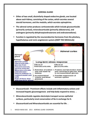
- 1. 1 MAGDI AWAD SASI 2013 ADRENAL GLAND DISORDERS ADRENAL GLAND Either of two small, dissimilarly shaped endocrine glands, one located above each kidney, consisting of the cortex, which secretes several steroid hormones, and the medulla, which secretes epinephrine. The adrenal cortex produces corticosteroids which include glucocorticoids (primarily cortisol), mineralocorticoids (primarily aldosterone), and androgens (primarily dehydroepiandrosterone and androstenedione). Function is regulated by the neuroendocrine hormones from the pituitary, hypothalamus and renin-angiotensin system.((NOT THE MEDULLA)) Glucocorticoid-- Prominent effects include anti-inflammatory actions and increased hepatic gluconeogenesis and help body respond to stress. . Mineralocorticoids regulate electrolyte transport across epithelial surfaces, particularly renal conservation of Na in exchange for K. Glucocorticoid and Mineralocorticoids are essential for life .
- 2. 2 MAGDI AWAD SASI 2013 ADRENAL GLAND DISORDERS Adrenal androgens--chief physiologic activity occurs after conversion to testosterone. The adrenal medulla is composed of chromaffin cells, which synthesize and secrete catecholamines (mainly epinephrine and lesser amounts of norepinephrine). Chromaffin cells also produce bioactive amines and peptides (eg, histamine, serotonin, chromogranins, neuropeptide hormones Epinephrine and norepinephrine(( the major effector amines of the sympathetic nervous system)), are responsible for the “flight or fight” response (ie, chronotropic and inotropic effects on the heart; bronchodilation; peripheral and splanchnic vasoconstriction with skeletal muscular vasodilation; metabolic effects including glycogenolysis, lipolysis, and renin release). Hypofunction may be primary (malfunction of the adrenal gland itself, as in Addison's disease) or secondary (due to lack of adrenal stimulation by the pituitary ((2ry)) or hypothalamus((3ry)). Hyperfunction produces distinct clinical syndromes. Hypersecretion of androgens results in adrenal virilism; of glucocorticoids, Cushing's syndrome; And of aldosterone, hyperaldosteronism (aldosteronism). These syndromes frequently have overlapping features. Hyperfunction may be compensatory, as in congenital adrenal hyperplasia, or due to acquired hyperplasia, adenomas, or adenocarcinomas. Excess quantities of epinephrine and norepinephrine are produced in pheochromocytoma .
- 3. 3 MAGDI AWAD SASI 2013 ADRENAL GLAND DISORDERS FUNCTION OF THE GLAND: Cortex -Stimulated by ACTH Hormone precursor: Cholestrol Secretes: Cortisol Aldosterone Sex hormones: Androgen ,Estrogen HORMONE FUNCTION ALDOSTERONE Renal : Na & Cl reabsorption; K excretion GI : Na absorption GLUCO-CORTICOIDS Increase serum glucose by gluconeogenesis & glycogenolysis especially during STRESS Blocks inflammation(IL6,PG,lymphokines) Counteracts effect of histamine-inflammation Required for ANG2-maintain vasculature SEX HORMONE Physiologically insignificant Becomes useful during menopause in women
- 4. 4 MAGDI AWAD SASI 2013 ADRENAL GLAND DISORDERS SYMPTOMATOLOGY: ALDOSTERONE DEFICIENCY Decrease in plasma volume leading to dehydration. Hypotension to shock. Increased K. Metabolic acidosis . CORTISOL DEFICIENCY ANOREXIA, N/V, ABDOMINAL PAIN, WT LOSS, LETHARGY HYPOGLYCEMIA HYPOTENSION HYPERKALEMIA, WEAK PULSE HYPERPIGMENTATION IMPAIRED STRESS TOLERANCE SEX HORMONE DEFICIENCY LOSS OF BODY HAIR LOSS OF LIBIDO OR IMPOTENCE MENSTRUAL & FERTILITY DISORDER ADRENAL CORTEX DISORERS ADRENAL INSUFFICIENCY ADRENAL CRISIS CUSHING’S SYNDROME ALDOSTERONISM
- 5. 5 MAGDI AWAD SASI 2013 ADRENAL GLAND DISORDERS ADRENAL INSUFFICIENCY ADDISON’S DISEASE Is a hormone deficiency caused by damage to the outer layer of the adrenal gland (adrenal cortex). Addison's disease is a disorder that results in the body producing insufficient amounts of certain hormones produced by the adrenal glands. Sometimes, Addison's disease also involves insufficient production of aldosterone, one of the mineralocorticoid hormones. Insufficiency of corticosteroids can be total when the operation of all hormones drops out, and partial fallout of activity of one adrenal hormone. Addison's disease can be life-threatening. Addison's disease can occur at any age, but is most common in people ages 30 to 50. ETIOLOGY: Autoimmune processes -80% of cases-corticosteroid, aldost,androgen-all Pituitary failure( atrophy, necrosis)-no HYPERKALEMIA/PIGMENTATION TB, Syphilis, coccid mycosis ,Histoplasmosis, CMV(AIDS) Tumor ,Metastases , Amyloidosis, Hemochromatosis Hemorrhage-warfarin, shock, major surgery, antiphospholipid. Drugs that block corticosteroid synthesis (eg, ketoconazole). All groves syndrome-achalasia, alacrima,neurologic disease. Polyglandular atrophy Addison's disease may coexist with diabetes mellitus or hypothyroidism.
- 6. 6 MAGDI AWAD SASI 2013 ADRENAL GLAND DISORDERS SIGNS AND SYMPTOMS: Signs and symptoms of Addison's disease usually develop slowly, often over several months, and may include: Mineralocorticoid deficiency ↑ in urinary excretion of Na, ↓ in urinary excretion of K Inability to concentrate urine, with electrolyte imbalance severe dehydration plasma hypertonicity, acidosis, hypotension circulatory collapse Glucocorticoid deficiency Disturbances in carbohydrate, fat and protein metabolism, decrease liver glycogen Hypoglycaemia and anorexia Destruction of adrenal gland Mineralocorticoid & glucocorticoid deficiency • Symptoms & Signs usually do not appear until ~90% of adrenal gland has been compromised. A. Manifestation, connected with the falling of mineral corticoids: 1) DEHYDRATION develops owing to loss of sodium ions (decreases rearbsortion) with the loss of water (polyuria). 2) HYPOTENSION - by decrease of circulating blood volume. 3) HEMOCONCENTRATION is connected with liquid loss, results to disorders of microcirculation and hypoxia.
- 7. 7 MAGDI AWAD SASI 2013 ADRENAL GLAND DISORDERS 4) Decreasing of kidney blood circulation is stipulated by increase of arterial pressure with disturbances of glomerular filtration and development of intoxication (nitrogenemia); 5) HYPERKALEMIA -- by decrease secretion of potassium ions and their output from the damaged cells. 6) Distal canales acidosis. It is connected with disorders of acidogenesis in distal nephron canals. 7)HYPEREMESIS- Gastro-intestinal disorders (nausea, vomiting, diarrhea). Loss of sodium (osmotic diarrhea) and intoxication have significant meaning. This disorders without appropriate correction result to death. Think of Addison’s in all those with unexplained abdominal symptoms. B. Manifestations by disorders of glucocorticoid function of adrenal: Muscle weakness and fast tiredness Decrease of ability to remove water during water load (water poisoning). Weight loss and decreased appetite (HYPOGLYCEMIA) HYPOTENSION -- (permissive reaction on catecholamine’s)90% Darkening of the skin (HYPERPIGMENTATION-- bronzed disease) Diffuse tanning over nonexposed as well as exposed areas over knuckles, elbows, knees, nail beds, palmer creases, and posterior neck. Nipples and areolar darken, new scars pigmented ,skin in pressure areas Depression , emotional changes, mental irritability. Salt craving
- 8. 8 MAGDI AWAD SASI 2013 ADRENAL GLAND DISORDERS Irritability Fever ,myalgia, arthralgia,vitiligo 10%. Sometimes, however, the signs and symptoms of Addison's disease may appear suddenly.
- 9. 9 MAGDI AWAD SASI 2013 ADRENAL GLAND DISORDERS
- 10. 10 MAGDI AWAD SASI 2013 ADRENAL GLAND DISORDERS • Examine for hyperpigmentation: Hand: palmar creases Mouth and lips Areas usually covered by clothing: nipple Areas irritated by belts, straps, collars or rings • Look for vitiligo • Look for sparse axillary hair and pubic hair • Examine the abdomen for adrenal scar • Examine blood pressure for postural hypotension • Signs of critical deterioration (Addisonian crisis): – Shock (low BP, tachycardia) – hypothermia
- 11. 11 MAGDI AWAD SASI 2013 ADRENAL GLAND DISORDERS SCREENING AND DIAGNOSIS Doctor will talk first about the medical history and the signs and symptoms. If doctor thinks that patient may have Addison's disease, He may undergo some of the following tests: Blood test. ACTH stimulation test. Insulin-induced hypoglycemia test. Imaging tests. BLOOD TEST: Measuring blood levels of sodium, potassium, cortisol and ACTH gives the doctor an initial indication of whether adrenal insufficiency may be causing the signs and symptoms. A blood test can also measure antibodies associated with autoimmune Addison's disease. BUSE • Low Na • High K • Low HCO3 • Azotemia FBC • Elevated hematocrit • Low WBC count • Relative lymphocytosis • Increased eosinophils RBS / DXT • Hypoglycemia (< 3 mmol/L)
- 12. 12 MAGDI AWAD SASI 2013 ADRENAL GLAND DISORDERS Blood chemistry • Low serum Na (< 135 mEq/L) • High serum K (> 5 mEq/L) • Ratio of serum Na:K: < 30:1 • Low fasting blood glucose (< 50 mg/dL [< 2.78 mmol/L]) • Decreased plasma HCO3 (< 20 mEq/L) • Elevated BUN (> 20 mg/dL [> 7.1 mmol/L]) ACTH stimulation test This test involves measuring the level of cortisol in the blood before and after an injection of synthetic ACTH. Plasma ACTH -INCREASED 1RY , DECREASED 2RY ACTH signals – adrenal glands to produce If adrenal glands are damaged, the ACTH stimulation test shows that the output of cortisol in response to synthetic ACTH is blunted or nonexistent. Preinjection plasma cortisol level (N= 138 – 690 nmol/L) IV Tetracosactide 250 µg Plasma cortisol level at 30 – 90 minutes post injection (≥ 500 nmol/L Failure to respond → adrenal insufficiency (? Primary / secondary)
- 13. 13 MAGDI AWAD SASI 2013 ADRENAL GLAND DISORDERS Insulin-induced hypoglycemia test Occasionally, doctors suggest this test if pituitary disease is a possible cause of adrenal insufficiency (secondary adrenal insufficiency). The test involves checking the blood sugar (blood glucose) and cortisol levels at various intervals after an injection of insulin. In healthy people, glucose levels fall and cortisol levels increase. Imaging tests Abdominal x ray- Calcifications in the adrenal areas - PTB Chest x ray----Small heart Computerized tomography (CT) scan of the abdomen to check the size of adrenal glands and look for other abnormalities that may give insight to the cause of the adrenal insufficiency. And may also suggest a CT scan or magnetic resonance imaging (MRI) scan of the pituitary gland if testing indicates the patient have secondary adrenal insufficiency. Negative Synacthen test High plasma ACTH level ADDISON’S DISEASE
- 14. 14 MAGDI AWAD SASI 2013 ADRENAL GLAND DISORDERS TREATMENT If patient receive an early diagnosis of Addison's disease, treatment may involve taking prescription corticosteroids. Because the body isn't producing sufficient steroid hormones, doctor may have recommend to take one or more hormones to replace the deficiency. Cortisol is replaced using hydrocortisone (Cortef), prednisone or cortisone. Fludrocortisone (Florinef) replaces aldosterone, which controls the body's sodium and potassium needs and keeps the blood pressure normal. In addition, doctor may recommend treating androgen deficiency with an androgen replacement called dehydroepiandrosterone. Some studies indicate that, for women with Addison's disease, androgen replacement therapy may improve overall sense of well-being, libido and sexual satisfaction. These hormones are given orally in daily doses that mimic the amount body normally would make, thereby minimizing side effects. If facing a stressful situation, such as an operation, an infection or a minor illness, doctor will suggest a temporary increase in dosage. If ill with vomiting and can't retain oral medications, may need corticosteroid injections. COPING SKILLS: Carry a medical alert card and bracelet at all times. Stay in contact with doctor. Keep extra medication handy
- 15. 15 MAGDI AWAD SASI 2013 ADRENAL GLAND DISORDERS Addisonian crisis An addisonian crisis is a life-threatening situation that results in HYPOTENSION, HYPOGLYCEMIA and HYPERKALEMIA. This situation requires immediate medical care. Treatment typically includes intravenous injections of: 1. Hydrocortisone 2. Saline solution 3. Sugar (dextrose) ADRENAL CRISIS IS : Acute episodes from stress that taxes the adrenal cortical function beyond its capabilities. Examples of acute insufficiency are: а) State after removal of adrenals b) Hemorrhage in adrenals which arises during sepsis, meningococci infection (syndrome Waterhouse-Friderixan); chronic anticoagulant therapy, DIC. c) Rapid withdrawal of steroids (from previous treatment). • Fast falling of the adrenals function causes development of collapse and the patients can die during the first day.
- 16. 16 MAGDI AWAD SASI 2013 ADRENAL GLAND DISORDERS Occur in : 1.Following stress , infection, surgery ,trauma. 2. Prolonged fasting in patient with latent insufficiency. 3. Sudden with drawl of adrenocortical hormone in pt with insufficiency 4. Following bilateral adrenlectomy or removal of functioning adrenal tumor that has suppressed other adrenal. 5. Following sudden destruction of pituitary gland( pituitary necrosis) OR when the thyroxine is given to a patient with hypoadrenalism. 6. Following injury to both adrenals by: TRAUMA ANTICOAGULANT THERAPY INFECTION HEMORRHAGE THROMBOSIS 2RY CARCINOMA ABDOMINAL DISCOMFORT EMOTIONAL UPSET HIGH TEMPERATURE C/F: CNS-headache , confusion ,lassitude , coma. GIT- nausea , vomiting , diarrhea , abdominal pain Fever with fatigubilty. SIGNS: FLUID LOSS/DEHYDRATION HYPOTENSION CYANOSIS
- 17. 17 MAGDI AWAD SASI 2013 ADRENAL GLAND DISORDERS ANY PATIENT WITH GIT SYMPTOMS AND SHOCK IN STEROID WITHDRAWL. Skin hyperpigmentation , meningococcimeia with purpura LAB: Hyponatremia ,Hyperkalemia, Hypoglycemia ,Hypercalcemia, High eosinophil. Hyperuricemia , lymphocytosis, Hyperuremia IF YOU FIND HYPERPIGMENTATION LOOK FOR OTHER (( H))= CRISIS Adrenal hormone assay : hydroxycorticoid &17ketsteroid in 24HR urine detected Cosyntropin stimulation test : Cosyntropin(ACTH) 0.25mg I/V S.cortisol 30 , 60 min Normally raises to 20 micgm/dl Stope hydrocortisone before 8hr of the test Plasma ACTH is markedly elevated in 1ry adrenal disease ((200pg/dl)) GOALS OF CARE: TO REVERSE SHOCK REPLENISH NEEDED STEROID TREATMENT: Dxtose 5% , Normal saline ADRENAL CORTICAL HORMONE REPLACEMENT: INJECTABLE HIGH SALT DIET ANTIBIOTICS.
- 18. 18 MAGDI AWAD SASI 2013 ADRENAL GLAND DISORDERS CUSHING’S SYNDROME CAUSE: Sustained over production of glucocorticoids by adrenal gland from ACTH by pituitary tumor.
- 19. 19 MAGDI AWAD SASI 2013 ADRENAL GLAND DISORDERS Excessive glucocorticoid administration. S/SX:
- 20. 20 MAGDI AWAD SASI 2013 ADRENAL GLAND DISORDERS Truncal obesity Buffalo hump Moon- face Weight gain Sodium retention with hypertension Thinning of extremities – from loss of muscle tissue due to protein catabolism Purple striae – from thinning of skin Echymosis from slight trauma poor wound healing hypertension, hyperglycaemia ANDROGENIC EFFECTS: Oligomenorrhea Hirsutism Gynaecomastia Decreased libido TREATMENT & NURSING CARE: PSYCHOLOGICAL SUPPORT PREVENT INFECTION – INFLAM & IMMUNE RESPONSE ARE SUPPRESSED PROMOTE SAFETY SURGERY – SUB/TOTAL ADRENALECTOM
- 21. 21 MAGDI AWAD SASI 2013 ADRENAL GLAND DISORDERS
- 22. 22 MAGDI AWAD SASI 2013 ADRENAL GLAND DISORDERS Aldosterone is a hormone that controls sodium and potassium levels in the blood. Hyperaldosteronism leads to retention of salt and loss of potassium, which then leads to hypertension (high blood pressure). PRIMARY – CONN’S SYNDROME(73%)unilateral adenoma -B/L CORTICAL HYPERPLASIA A.D .,defect allowing ACTH stimulation of Aldosterone production. SECONDARY Who is affected by Hyperaldosteronism? People in their 30’s-50’s (adulthood), with prevalence increasing with age Females are more likely to be affected than males African Americans have a significantly greater risk of fatality from the disease than other races. CONN’S SYNDROME PRIMARY ALDOSTERONISM CAUSE: ADRENAL ADENOMA S/SX: HYPOKALEMIA FATIGUE HYPERNATREMIA, HPN, TETANY MANAGEMENT: SURGERY ALDACTONE – ALDOSTERONE ANTAGONIST
- 23. 23 MAGDI AWAD SASI 2013 ADRENAL GLAND DISORDERS Conn’s disease Usually caused by a benign tumor of the adrenal gland or bilateral adrenal hyperplasia (enlargement of an organ caused by an increase in the reproduction rate of its cells) . Features: - Hypertension (0.7%HTN PTS) - headache Malignant HTN RARE, typically moderate, diastolic only w out S&S. - Hypokalemia metabolic alkalosis - tateny ¶sthesia - Fatigue - polyuria and polydipsia - Nocturia escape phenomenon prevents edema - Muscle weakness -Paralysis simulating periodic paralysis. Secondary Hyperaldosteronism is when the excess aldosterone is caused by something outside the adrenal gland and mimics the primary condition. The problem is outside the adrenal gland: e.g. RENIN – ANGIOTENSIN SYSTEM Causes of Secondary Hyperaldosteronism: Increased renin production, commonly caused by the ingestion of oral contraceptives and toxemia from pregnancy. Renin - An enzyme secreted by and stored in the kidney area that stimulates aldosterone and therefore, raises blood pressure
- 24. 24 MAGDI AWAD SASI 2013 ADRENAL GLAND DISORDERS Conditions like congestive heart failure, liver failure, kidney disease, cirrhosis, and dehydration Certain medicines like diuretics and fludrocortisone. SYMTOMS: Symptoms are similar to those of Primary Hyperaldosteronism Moderate hypertension (high blood pressure) Most individuals have no other specific symptoms. However, some may have: ◦ Muscle weakness ,Fatigue ◦ Temporary paralysis ,Cramping ◦ Headaches ◦ Low potassium level ◦ Tingling ◦ Muscle spasms
- 25. 25 MAGDI AWAD SASI 2013 ADRENAL GLAND DISORDERS DIAGNOSIS: High NA intake and low k Elevated aldosterone levels can be measured in the blood or urine. 24 HR urine collection is assayed for Aldosterone ,Cortisol , Creatinine. Plasma assayed for Aldosterone at 8 AM while PT is supine after overnight recumbency and again after 4 HR upright. Adrenal Adenoma ---more than 20mg/dl not raise Adrenal hyperplasia----less than 20mg/dl raise upright Low plasma rennin less than 5mg/dl with 24 hr urine Aldosterone more than 20mg/dl indicates HYPERALDOSTERONISM. Urine Ald. less 20mg /24hr seen in gonadal/adrenal enzyme defect. Once hyperaldosterone is diagnosed, plasma assayed for 18hydroxy- corticosterone—above 85mg/dl—adrenal neoplasm. ◦ In a blood test, PRA (Plasma renin activity), is used to distinguish between primary (low PRA) and secondary Hyperaldosteronism (high PRA). Abdominal CT scans can show adrenal masses Electrocardiograms (ECGs) can show abnormalities in heart rhythm that are often associated with low potassium level. It is likely that many cases of secondary Hyperaldosteronism are never detected. Treatments: Secondary Hyperaldosteronism is treated by treating the underlying cause. Typically, medication and diet (but not surgery) are used Spironolactone is the treatment of choice.
- 26. 26 MAGDI AWAD SASI 2013 ADRENAL GLAND DISORDERS When untreated, the disease can lead to uncontrolled hypertension (which can become a risk factor for stroke and heart disease). The outlook for the patient depends on the cause of the condition Adrenocortical Neoplasms • May be responsible for a variety of hyperadrenalisms a) functional adenomas most commonly associated with hyperaldosteronism and Cushing syndrome • A virilizing neoplasm high incidence to be carcinogenic • Functional and non-functional adrenocortical neoplasms cannot be differentiated solely on basis of morphology a) Hormone measurements b) Most adrenocortical adenomas do not cause hyperfunction • Adrenocortical carcinomas are rare a) two rare inherited adrenal cortical carcinomas i) Li-Fraumeni syndrome - autosomal dominant - predisposition to develop other cancers due to mutations in p53 ii) Beckwith-Wiedemann syndrome • Generally, adrenal adenomas are small (1-2cm) whereas adrenal carcinomas are large, invasive lesions a) adrenal carcinomas metastasize via lymphatics and inferior vena cava i) mean survival is ~2 yrs.
- 27. 27 MAGDI AWAD SASI 2013 ADRENAL GLAND DISORDERS Adrenal Medulla • Most important diseases of adrenal medulla are the neoplasms a) neuronal i) Neuroblastoma ii) Mature ganglionic cell tumors b) Chromaffin cells i) pheochromocytoma • Pheochromocytoma a) neoplasm composed of chromaffin cells i) synthesize and release catecholamine’s. b) familial syndromes (~10%) • i) MEN2A and MEN2B ii) type I neurofibromatosis iii) Von Hippel-Lindau disease iv) Sturge-Weber syndrome c) Extra-adrenal source (~10%) i) carotid body ii) organ of Zuckerkandl • d) are bilateral (~10%) i) may be as high as 50% in familial cases e) malignant (~10%) i) more common when arise in extra-adrenal sites HORMONES: EPINEPHRINE NOREPINEPHRINE PHEOCHROMOCYTOMA: This is a rare tumor of chromaffin tissue which secrets catecholamine’s & is responsible for less than 0.1% of causes of H.T.
- 28. 28 MAGDI AWAD SASI 2013 ADRENAL GLAND DISORDERS The tumors are usually benign /10% malignant //in over 90% of cases the tumor found in the adrenal medulla, while it may arise elsewhere in the body in sympathetic ganglia (paragangliomas). There is a useful rule of tens’ in this condition: 10% are malignant , 10% in children 10% are extraadrenal , 10%extreedrenal are extraabdominal 10% are familial , 10% bilateral adrenal tumors 10% are non hypertensive It is associated with MEN type II. • Occasionally, this tumor produces other biogenic steroids or peptides associated with Cushing’s Syndrome. • Extra-medulla tumors – 1-3% in chest and neck (usually malignant) • 20% multiple, 10% malignant SYMPTOMS AND SIGNS: Hypertension, may be paroxysmal or persistent. Due to secretion of one or more of catecholamine hormones or precursors: norepinephrine, epinephrine, dopamine or dopa. Occasionally the pt. may be hypotensive ( dopamine – secreting tumors). some pt. may present with a complication of the hypertension e.g.: stroke, MI, LVF There may be abdominal pain with vomiting, constipation, wt. loss, glucose intolerance. 90% Presented with Palpitation, Hypertension,Sweating &Anxiety.
- 29. 29 MAGDI AWAD SASI 2013 ADRENAL GLAND DISORDERS During the attack the pt. presents with: pallor (some time flushing), palpitation, sweating, headache, anxiety (feeling of death). Tachycardia, sweating, postural hypotension, tachypnea, flushing, cold and clammy skin, severe headache, angina, palpitation, dyspnoea Paroxysmal attacks may be provoked by exercise, anaesthesia, palpation of tumor, postural changes, urination, beta-blockers Hyperglycemia Cardiac arrhythmia and congestive heart failure. DIAGNOSTIC TEST : Urine Sample Blood Work Abdominal CT Scan MRI of the Abdominal Region Adrenal Gland Biopsy Screening - Two 24 h urines for catecholamines is the best screening investigation - 24 h urine for VMA (15% false negative) and metanephrine (10% false negative) - needs vanilla-free diet before collection If the diagnosis is established, or strongly suspected MIBG scan - meta-iodo-benzylguanidine labeled with 131 I - Increased uptake by pheochromocytoma
- 30. 30 MAGDI AWAD SASI 2013 ADRENAL GLAND DISORDERS CT scan of adrenals - patient should be alpha- and beta-blocked to avoid hypertensive episode after contrast administration End products of catecholamine metabolism. DRUGS & FOOD TO BE WITHHELD 24H B4 THE TEST: COFFEE & TEA BANANA VANILLA CHOCOLATES MANAGEMENT: SURGERY MEDICAL : ADRENERGIC BLOCKING AGENTS: PHENTOLAMINE NURSING CARE: MONITOR BP IN SUPINE & STANDING MONITOR URINE FOR GLUC & ACETONE
- 31. 31 MAGDI AWAD SASI 2013 ADRENAL GLAND DISORDERS This requires excision of the tumor or long term TX with α and (usually β) adrenoceptor blockade. Prior to surgery give α –antagonist drug such as phenoxybenzamine 10-20 mg orally 3-4 times / day for a minimum of 6 weeks to allow restoration of normal plasma volume if tachycardia is there then β – antagonist as propranolol (10- 20 mg ) 3 times daily should be added or combined α and β-antagonist(e.g. labetalol)can be added. β-antagonist should not be given before the α-antagonist as it may cause a paradoxical rise in blood pressure due to unopposed α-mediated vasoconstriction. During surgery Na-nitropruside & the short acting α-antagonist phentolamine are very useful in controlling hypertension episodes which may occur during anesthesia or mobilization of the tumor.post-operative hypotension may occur and require volume expansion and very occasionally noradrenalin infusion. After surgery, it is necessary to continually monitor all vital signs in an intensive care unit. When the tumor cannot be surgically removed, medication is needed to manage it. This usually requires a combination of medications to control the effects of the excessive hormones. Radiation therapy and chemotherapy have not been effective in curing this kind of tumor. a. Pre-op: Sympathetic blocking agents= Minipress (prazosin), Hytrin (terazosin), Cardura (doxazosin) to reduce BP and other symptoms of of catecholamine excess Since change in BP sudden, client may experience orthostatic hypotension Use Beta blocking agents such as Inderal to dec. heart rate, BP and force of contraction and calcium channel blocking agents also used. b. General management Diet: high in vitamin, mineral, calorie, no caffeine Sedatives; Monitor BP Eliminate attacks; If attack- complete bedrest and HOB 45 degrees c. Surgery via laparoscopic adrenalectomy or open abdominal incision; complete removal of the tumor cures hypertension in 10-30% of the cases
- 32. 32 MAGDI AWAD SASI 2013 ADRENAL GLAND DISORDERS Short Acting Preparations (t1/2 < 12 h) Drug Anti-inflam. Salt retaining Preapartions & dose Cortisol 1 1.0 • 5 mg tablet • 100 mg/vial (i.m., i.v) • Topical; enema Cortisone 0.8 0.8 • 5 mg tablet • 25 mg/vial (i.m) Intermediate Acting Preparations (t1/2 = 12 -36 h) Prednisone 4 0.8 - Prednisolone 5 0.3 • 5, 10 mg tablet • 20 mg/vial (i.m, intrarti) Methyl prednisolone 5 0 • 0.5, 1.0 gm inj. for i.m. or slow i.v. Triamcinolone 5 0 • 4 mg Tab., • 10, 40 mg/ml for i.m. & intrarticular inj. Drug Anti- inflam. Salt retaining Preapartions & dose Long Acting Preparations (t1/2 > 36 h) Dexamethasone 25 0 0.5 mg tab. 4mg/ml inj (i.m., i.v.) Betamethasone 25 0 0.5, 1 mg tab. 4mg/ml inj (i.m., i.v.) Paramethasone 10 0 2- 20 mg/day (oral) Mineralocorticoids - Preparations Drug Anti- inflammatory Salt retaining Preapartions & dose Fludrocortisone 10 150 100 mcg tab. DOCA 0 100 2.5 mg sublingual Aldosterone 0.3 3000 Not used clinically
- 33. 33 MAGDI AWAD SASI 2013 ADRENAL GLAND DISORDERS
- 34. 34 MAGDI AWAD SASI 2013 ADRENAL GLAND DISORDERS Multiple Endrocrine Neoplasia Syndromes (MEN) • Group of inherited diseases a) hyperplasias, adenomas and carcinomas of multiple endocrine organs i) Occur at younger age vs. cancers ii) Arise in multiple endocrine organs iii) Even in one organ, they are multifocal iv) Tumors preceded by hyperplasia v) More aggressive and recur more frequently vs. sporadic tumors • MEN-1(( WEMERS SYNDROME)) a) Inherited as autosomal dominant b) Is a tumor suppressor gene i) Loss of MEN-1 therefore causes tumor genesis - Parathyroid (95% involved) - Pancreas (>40%) - Pituitary (>30%) I- Parathyroid gland A) Primary hyperparathyroidism arising from hyperplasia is a common feature MEN-1 or adenoma of several parathyroid gland. B) Occurs in 90% of cases. C) Presents with hypercalcemia. II-Pancreas-pancreatic islet cell tumors a) Leading cause of death in MEN-1 b) Aggressive c) Often functional tumors i) Gastrinomas (Zollenger-Ellison syndrome)-MOST COMMON ii) Insulinomas and resultant hypoglycemia
- 35. 35 MAGDI AWAD SASI 2013 ADRENAL GLAND DISORDERS III- Pituitary adenoma A) most common in MEN-1 is prolactinoma B)May secret prolactin ,Growth hormone , ACTH. C)May cause local pressure effects and hypopituitrism. D)Usually non functional. IV- Adrenal cortical adenoma A)Occur in 70% B)Can be adenoma or hyperplasia C)Bilateral in about half D) They are generally benign and nonfunctional • MEN-2b (William syndrome) a) Involve also the thyroid b)Adrenal (medulla) c) Major differences between MEN-2a and MEN-2b is i) Do not develop primary hyperparathyroidism (MEN-2b) ii) Develop at extra endocrine sites - lips - tongue - GI tract • MEN-2 carry RET Protooncogene a) persons are advised to have prophylactic thyroidectomy to prevent carcinoma. • Adrenogenital syndrome results from the hereditary stipulated blockade of cortisole synthesis and amplified formation of androgens from general intermediate products.
- 36. 36 MAGDI AWAD SASI 2013 ADRENAL GLAND DISORDERS • Depending on the level of blockade of cortisole synthesis there are three variants of adrogenital syndrome. • І. Disorders of early stages of synthesis – deficiency of glucocorticoides, mineralcorticoides and androgens hyperproduction. Manifestations: signs of insufficiency of gluco- and mineralocorticoidal functions of adrenal cortex features of early sexual maturing in males, virilization in women (appearance of man's sexual features). • ІІ. Disorders of intermediate stages – deficiency of glucocorticoides, surplus of androgens, formation of mineralocorticoides is not infringed (classical androgenic syndrome). Manifestations are the same, as in the first case, only without signs of insufficiency of mineralocorticoidal function. • ІІІ. Disorders at final stages of cortirol synthesis – deficiency of glucocorticoides, hyperproduction of androgens and mineralocorticoide. Features of hyperaldosteronism are connected with manifestations of classical androgenital syndrome. Adrenocortical neoplasms Adrenocortical neoplasms associated with symptoms of excess of androgen are more likely to be androgen secreting adrenal carcinomas than adenomas. It is also often assoc with hypercortisolism (mixed syndrome) The tumour secretes androgen thus increasing in circulation and converted to testosterone at the peripheral tissues. May occur at any age , relatively rare. should be suspected when the onset of androgenic symptoms is sudden (i.e., generally <2 yr) and the pace of symptoms is rapid, and when they lead to virilization and masculinization.
- 37. 37 MAGDI AWAD SASI 2013 ADRENAL GLAND DISORDERS may be associated with other systemic symptoms including weight loss, anorexia, a feeling of abdominal bloating, back pain. CAH Depends on the nature and severity of the enzymytic defect. Onset of clinical symptoms can occur in the Perinatal period Later childhood Adulthood (less common) Anterior pituitary ACT H Cholesterol Pregnolone 17- hydroxypregnenolone 17- hydroxyprogesterone 21 11– deoxycortisol Cortisol Glucocorticoids Progesterone 21 Aldosterone Corticosterone 11- deoxycortisone Mineralocorticoids Testosterone Androstenedione Dehydroxypiandrosterone Sex steroids Adrenal cortex (bilateral hyperplasia) Congenital adrenal hyperplasia Autosomal recessive deficiency of an enzyme in the cortisol synthetic pathways.
- 38. 38 MAGDI AWAD SASI 2013 ADRENAL GLAND DISORDERS ◦ Cortisol secretion is reduced and feedback leads to increased ACTH secretion to maintain adequate cortisol leading to adrenal hyperplasia. ◦ Diversion of the steroid precursors into the androgenic steroid pathways occurs. Thus, 17-hydroxyprogesterone, androstenedione and testosterone levels are increased, leading to virilization. Virilism==(excessive androgens may cause masculinization). Male excessive estrogen cause feminisation—breast enlargement. Androgens have important functions in women ◦ Essential in the production of E2 (in ovary & adipose tissue) ◦ Responsible for delivery. & maint. of axillary & pubic hair ◦ Important for libido. Virilization: The development of exaggerated masculine characteristics, usually in women, often as a result of overproduction of androgens So, if hyperandrogenism becomes extreme, virilization occurs Symptoms of virilization include 1. Excess facial and body hair (hirsutism), 2. Baldness 3. Acne 4. Deepening of the voice 5. Increased muscularity
- 39. 39 MAGDI AWAD SASI 2013 ADRENAL GLAND DISORDERS 6. An increased sex drive. In women, 1. The uterus shrinks 2. The clitoris enlarges (clitoromegaly) 3. The breasts become smaller 4. normal menstruation stops (amenorrhea) Clinical Manifestation: • Cortisol deficiency – hypoglycemia, inability to withstand stress, vasomotor collapse, hyperpigmentation, apneic spells, muscle weakness & fatigue. • Aldosterone deficiency – hyponatremia, hyperkalemia, vomiting, urinary sodium wasting, salt craving, acidosis, failure to thrive, volume depletion, hypotension, dehydration, shock, diarrhea. • Androgen excess – ambiguous genitalia, virilization of external genitalia , hirsutism, early appearance of pubic hair, penile enlargement , excessive height gain and skeletal advance. *Late onset CAH – normal genitalia, have acne, hirsutism, irregular menses/amenorrhea. ANDROGEN Testosterone, dehydroepiandrosterone sulfate (DHEAS), dehydroepiandrosterone (DHEA), androstenedione, and androstenediol The ovaries produce 50% of circulating testosterone, 50% of the androstenedione and 20% of DHEA. The adrenal glands produce all the DHEAS and 80% of the DHEA. The adrenals also secrete 50% of androstenedione and 25% of testosterone.
- 40. 40 MAGDI AWAD SASI 2013 ADRENAL GLAND DISORDERS Adrenal androgens increase in response to ACTH stimulation LH stimulates theca cells of the ovaries to secrete androgens Hyperandrogenism Excess of androgens may be caused by: ◦ primary gonadal disorders ◦ primary adrenal disorders ◦ iatrogenic In practice though, the causes are restricted to a few conditions:
- 41. 41 MAGDI AWAD SASI 2013 ADRENAL GLAND DISORDERS ◦ PCOS ◦ Cushing’s syndrome ◦ CAH ◦ Tumours Lab Testosterone and Dehydroepiandrosterone sulphate (DHEAS) ◦ DHEAS hyperandrogenemia of adrenal origin Serum prolactin thyroid stimulating hormone (TSH) Serum 17 hydroxyprogesterone (17-OHP) test –if suspect CAH LH and FSH ( suggestive of PCOS if ratio >2) Lipid profile OGTT ◦ Relying on a fasting glucose level alone is inadequate as it is a poor predictor of impaired glucose tolerance or diabetes Effect of androgens Fat deposition (small breast) ◦ Androgens inhibit the ability of some fat cells to store lipids Muscle mass (heavy mascular mass) ◦ Androgens promote the enlargement of skeletal muscle cells Brain ◦ Enhanced libido.