The document outlines the anatomy and examination of the head and neck region. It describes the bones, muscles, nerves, blood vessels and structures of the eyes, ears, nose, mouth, throat and neck. Key points include identifying the cranial nerves involved in vision and hearing, describing visual field defects and causes of abnormal eye movements. Examination techniques are covered such as visual acuity tests, otoscopy, lymph node palpation and assessment of the thyroid gland. The overall goal is to teach students to obtain a relevant history and perform a complete physical exam of the head and neck.
Call Girls Hosur Just Call 9630942363 Top Class Call Girl Service Available
Head and neck
1. Head and Neck
Cognitive Objectives
Upon completion of this lesson, the student should be able to:
1. Identify the bones of the head. (see picture on pg. 204). Abnormal findings: enlarged
skull may signify hydrocephalus or Paget’s disease of bone. Tenderness or step-offs are
common after trauma
• Frontal bone
• Parietal bone
• Occipital bone
• Temporal bone
• Mastoid portion of temporal bone: behind the ears
• Maxilla bone
• Mandible bone
• Zygomatic bone
• Nasal bone
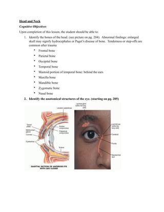
2. Identify the anatomical structures of the eye. (starting on pg. 205)
2. 3. Describe the function of each part of the eye.
- Eyelid: covers and protects the eyes
- Sclera: outer layer of the eye containing collagen and elastic fiber also known as the
white of the eye. Maintains the shape of the globe, offering resistance to internal and
external forces, and provides an attachment for the extraocular muscle insertions.
- Conjunctiva: helps lubricate the eye by producing mucus and tears. It also contributes to
immune surveillance and helps to prevent the entrance of microbes into the eye.
- Iris: responsible for controlling the diameter and size of the pupils and the amount of
light reaching the pupil.
- Pupil: allows light to enter the retina.
3. - Cornea: the transparent front part of the eye that covers the iris, pupil, and anterior
chamber. Together with the lens, the cornea refracts light, accounting for approximately
two-thirds of the eye's total optical power
- Eyelashes: protect the eye from debris; are sensitive to being touched, thus providing a
warning that an object (such as an insect or dust mite) is near the eye (which is then
closed reflexively).
- Tarsal plate: elongated plates of dense connective tissue, about 2.5 cm. in length; one is
found in each eyelid, and contributes to its form and support.
- Levator palpebrae: muscle in the orbit that elevates the superior (upper) eyelid.
- Bulbar conjunctiva: the conjunctiva covering the eyeball.
- Palpebral conjunctiva: lines the inside of the eyelids.
- Lacrimal gland: produces tears.
- Lacrimal sac: connects the lacrimal canaliculi, which drain tears from the eye's surface,
and the nasolacrimal duct, which conveys this fluid into the nasal cavity.
- Puncta: collect tears produced by the lacrimal glands
- Nasolacrimal duct: carries tears from the lacrimal sac into the nasal cavity.
- Ciliary body: has three functions: 1) accommodation (Accomommodation essentially
means that when the ciliary muscle contracts, the lens becomes more convex, generally
improving the focus for closer objects. When it relaxes,it flattens the lens, generally
improving the focus for farther objects); 2) aqueous humor production (responsible for
providing most of the nutrients for the lens and the cornea and involved in waste
management of these areas), 3) anchors lens in place.
- Lens: transmits and refracts light, converging or diverging the beam.
- Canal of Schlemm: circular channel in the eye that collects aqueous humor from the
anterior chamber and delivers it into the bloodstream via the anterior ciliary veins.
- Vitreous body: in contact with the retina and helps to keep it in place by pressing it
against the choroid, it does not adhere to the retina, except in three places: around the
anterior border of the retina; in the macula and at the optic nerve disc.
- Choroid: vascular layer containing connective tissue, of the eye lying between the retina
and the sclera. Provides oxygen and nourishment to the outer layers of the retina
- Retina: is a light-sensitive tissue lining the inner surface of the eye. The optics of the eye
create an image of the visual world on the retina, which serves much the same function as
the film in a camera. Contains rods and cones.
- Fovea: responsible for sharp central vision
- Extraocular muscle: six muscles that control the movements of the eye.
4. - Central retinal artery: supplies all the nerve fibers that form the optic nerve that carries
the visual information to the occipital lobe cerebral cortex, including those that reach
over the fovea.
- Central retinal vein: runs through the optic nerve and drains blood from the capillaries of
the retina into the larger veins outside the eye.
- Optic nerve: also called cranial nerve II, transmits visual information from the retina to
the brain.
- Optic disc: location where ganglion cell axons exit the eye to form the optic nerve.
4. Explain the causes of visual field defects. (pg. 254)
Horizontal Defect Occlusion of a branch of the central retinal artery and ischemia of the
optic nerve may cause a horizontal (altitudinal) defect.
Blind Right Eye (right optic nerve) A lesion of the optic nerve, and of course of the eye
itself, produces unilateral blindness.
Bitemporal Hemianopsia (optic chiasm) A lesion at the optic chiasm may involve only
fibers crossing over to the opposite side. Since these fibers originate in the nasal half of each
retina, visual loss involves the temporal half of each field.
Left Homonymous Hemianopsia (right optic tract) A lesion of the optic tract interrupts
fibers originating on the same side of both eyes. Visual loss in the eyes is therefore similar
(homonymous) and involves half of each field (hemianopsia).
Homonymous Left Superior Quadrantic Defect (right optic radiation, partial) A partial
lesion of the optic radiation in the temporal lobe may involve only a portion of the nerve
fibers, producing, for example, a homonymous quadrantic defect.
5. Left Homonymous Hemianopsia (right optic radiation) A complete interruption of fibers in
the optic radiation produces a visual defect similar to that produced by a lesion of the optic
tract.
5. Explain the causes of an abnormal extra ocular movement exam (EOM's).
If one of these muscles is paralyzed, the eye will deviate from its normal position in that
direction of gaze and the eyes will no longer appear conjugate, or parallel.
6. Developmental dysconjugate gaze is caused by an imbalance in ocular muscle tone. This
imbalance has many causes, may be hereditary, and usually appears in early childhood. These
gaze deviations are classified according to direction:
exotropia
esotropia
Disorders of cranial nerves: New onset of dysconjugate gaze in adult life is usually the
result of cranial nerve injuries, lesions, or abnormalities from such causes as trauma,
multiple sclerosis, syphilis, and others. Ex:
• Left cranial nerve VI paralysis:
o Looking to the right: eyes are conjugate.
o Looking straight ahead: estropia appears
o Looking to the left: esotroia is maximum
• Left cranial nerve IV paralysis:
o Left eye cannot looking down when turned inward.
• Left cranial nerve III paralysis:
o Eye is pulled outward by action of the 6th nerve. Upward, downward and
inward movements are lost. Ptosis and pulpillary dilation may be
associated.
7. 6. Discuss the difference between papillary reaction to light by direct and consensual
reaction.
Direct reaction: papillary constriction in the same eye. Consentual reaction: papillary
constriction in the opposite eye.
7. Discuss the importance of the cover / uncover test.
A cover-uncover test may reveal a slight or latent muscle imbalance not otherwise seen with
just inspection.
8. Define ptosis: is lid lag, when the lid does not overlap the iris when eyes move up and
down. In hyperthyroidism: a rim of sclera is visible above the iris w/ downward gaze
called protosis.
9. Describe and identify corneal arcus.
Corneal Arcus. A thin grayish white arc or circle not quite at the edge of the cornea.
Accompanies normal aging but also seen in younger people, especially African-Americans.
In young people, suggests possible hyperlipoproteinemia. Usually benign.
10. Describe normal findings on insufflation of the tympanic membrane with a
pneumatic otoscope.
This normal right eardrum (tympanic membrane) is pinkish gray. Note the malleus lying behind
the upper part of the drum. Above the short process lies the pars flaccida. The remainder of the
drum is the pars tensa. From the umbo, the bright cone of light fans anteriorly and downward.
Posterior to the malleus, part of the incus is visible behind the drum. The small blood vessels
along the handle of the malleus are normal.
8. 11. Identify the anatomical parts of the outer, middle, and inner ear.
12. 14. Identify lymph nodes in the head and neck. (p. 238) (Ch. 18 p.773 Child)
1. Preauricular
2. Postterior auricular
3. Occipital
4. Tonsillar
5. Submandibular
6. Submental
7. Superficial cervical
8. Posterior cervical
9. Deep cervical chain
10. Supraclavicular
13. Note: Tonsillar, Submandibular, Submental nodes: drain portions of mouth, throat, and face.
15. Discuss function of lymph nodes and sites of drainage. (p. 239)
a. Preauricular – in front of ear.
b. Posterior auricular – superficial to maistoid process
c. Occipital – base of skull posteriorly
d. Tonsillar – angle of mandible
• Small, hard, tender, high deep btwn mandible & sternomastoid is probably the
styloid process.
• If it pulsates, it is really the Carotid artery.
e. Submandibular – midway btwn angle & tip of mandible. Smaller and smoother than the
lobulated submandibular gland against which they lie.
f. Submental – midline a few cm behind tip of mandible.
g. Superficial cervical – superficial to sternomastoid.
h. Posterior cervical – along anterior edge of trapezius
i. Deep cervical chain – deep to sternomastoid. Hook thumb and fingers around either side
of sternomastoid muscle to find them.
j. Supraclavicular – deep in angle formed by clavicle and sternomastoid.
14. • Enlarged: especially on the Left: possible metastasis from a thoracic / abdominal
malignancy.
Note: Tender Nodes: suggests Inflammation.
Enlarge or Tender Nodes: If unexplained
1. Reexamine of regions they drain.
2. Assess lymph nodes elsewhere to distinguish btwn regional and generalized
lymphadenopathy. (Diffuse Lymphadenopathy raises suspicion of HIV / AIDS)
Hard/Fixed Nodes: suggests Malignancy.
16. Identify anatomical landmarks of the neck. (p. 236 - 236)
a. Anterior triangle: Mandible above, Sternomastoid laterally, & Midline of neck medially
forms the anterior angle of neck.
b. Posterior triangle: Sternomastoid, Trapezius, & Clavicle form the posterior angle.
Omohyoid muscle crosses lower portion of this triangle .
c. Great Vessels: Carotid artery & Internal jugular vein locate deep to the sternomastoids.
External jugular vein passes diagonally over the sternomastoid (helpful when trying to
identify JVP).
d. Midline Structures & Thyroid Gland:
1. Hyoid bone: below mandible
2. Thyroid cartilage: identified by notch on superior edge.
3. Cricoid cartilage
4. Tracheal rings
5. Thyroid gland
15. Clinical Objectives
1. Obtain a relevant history for complaints relating to the head, eyes, ears, nose, mouth.
throat, and neck, to include the history of present illness (HPI), relevant past medical
history (PMH) , social history (SH) and family history (FH) and review of system(s)
(ROS) as outlined in Bickley and H&P Plus Booklet.
2. The student will demonstrate a complete and systematic examination of the head and
neck by completion of the following objectives:
a. Describe and document appropriate examination of the head, including scalp and
hair.
16. b. Obtain relevant history for complaints related to the eye, ear, nose, mouth, and
neck.
c. Demonstrate and document appropriate examination of the eye, including the
cover/uncover test and screening of visual fields by confrontation.
d. Describe the steps to use an ophthalmoscope.
e. Demonstrate skill in the use of the ophthalmoscope.
f. Discuss and demonstrate the functional assessment of hearing.
g. Describe and demonstrate the Weber and Rinne tests.
h. Describe a normal tympanic membrane and common abnormalities of the
tympanic membrane (TM).
i. Demonstrate a complete ear examination using the otoscope.
j. Demonstrate ability to document a complete ear examination (objective).
k. Demonstrate an appropriate examination of the nose, frontal, and maxillary
sinuses.
l. Demonstrate a complete oral examination and document findings.
m. Identify cranial nerves that innervate the eye, ear, face, tongue, soft palate, and
neck (also covered in Neurology).
n. Demonstrate proper lymph node examination techniques.
o. Demonstrate and document an appropriate examination of the neck to include
lymph nodes and thyroid gland.
p. Accurately record history and physical exam findings related to the HEENT
systems using appropriate terminology