

Recommandé
Contenu connexe
Tendances
Tendances (19)
En vedette
En vedette (20)
Similaire à B wayang kulit
Similaire à B wayang kulit (20)
Dernier
Dernier (20)
B wayang kulit
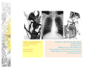
- 1. CHEST RADIOGRAPHS, NORMAL VARIANTS IN THE CHEST A WAYANG KULIT RADIOGRAPH Part Two Dr Ng Kian Seng MBBS (Singapore) MCGP (Malaysia) Second Edition Master Of Medicine (Internal Medicine, Singapore) February 2012 FAFP (Malaysia) Cert In Occupational Medicine Ph D (Theology, USA)
- 2. An Album On The Normal Variants In The Chest Radiograph You need to know the normal well enough so that you will not mistake a normal variant for some pathological condition. Practise looking at the normal Chest Radiograph…do not disdain it because it has no bizarre or frightful shadows you can be excited about! The “practice of looking” at the normal Chest radiograph is what I call a “trifle of medicine” & in Medicine “Trifles Makes Perfection & Perfection Is No Trifle”.
- 3. ACESSORY FISSURE, THE AZYGOS FISSURE The azygos lobe appears starting in a teardrop shape at around the level of T5 to the right of the midline as a pale line curving outward . and upward and then back in to meet the root of the neck, the line is the infolding of the pleura. Also described as a “curvilinear opacity, Inverted comma, tadpole.”
- 5. NIPPLE SHADOWS RIGHT NIPPLE LEFT NIPPLE Confirm these are indeed nipple shadows by using metal markers!
- 6. ASYMMETRY OF THE BREASTS Breast asymmetry is very common, even to the extent that no breast tissue is visible on one side. It should not be assumed that the patient has had a mastectomy, unless this is known from the history.
- 7. BONE ISLAND IN THE RIB
- 8. DROMEDARY HUMP IN THE DIAPHRAGM
- 9. “Ripley’s Believe It Or Not” The See Saw Diaphragm These two Chest Radiographs belong to the same Nepalese Worker who presented for a Fomema ME on 27 Dec 2011. The first “shocked” me. It showed a high right dome shaped diaphragm with a medial dromedary hump. It is 8 cm higher than the left. He was asymptomatic and a clinical examination was unremarkable, specifically there was no Hepatomegaly . I asked for a repeat CXR. The second was taken minutes later. Imagine my second “shock”. The second CXR is reproduced here on the right. Now the Left Hemidiaphragm appears to be slightly higher than the right, and it appeared to have been pushed up by the Splenic flexure of the colon. A very mobile see saw diaphragm.
- 10. Tenting In The Diaphragm Note the triangular opacity at mid part of right hemi-diaphragm (arrow). Diaphragmatic tenting is a localized Diaphragmatic tenting is due to fibrosis accentuation of the normal convexity and may not have any clinical significance. of the hemidiaphragm as if "pulled upwards by a string." This finding is minor, may be due to any inflammatory condition and not suggestive of TB. Source : Nexradiology
- 11. Diaphragmatic Hump Scalloping In The Diaphragm Note multiple arcuate elevations of the right This is due to incomplete muscularization of the hemi-diaphragm. Scalloping is seen in about diaphragm. Instead of the normal diaphragmatic 10% of normal CXR. muscle, the diaphragm is now consists of a thin membranous sheet. This is a very common abnormality. Most of the time, the abnormality is partial, involving one half to one third of the hemidiaphragm. Usually the anteromedial portion is affected. Source : Nexradiology
- 12. Normal Variants in the Rib Cage 1.Discontinuity of the first rib 2. Bridge formation posteriorly, forked rib anteriorly 3. Costal bridge 4. Bridge-shaped fusion 5. Fusion dorsally 6. Suggestion of costal bridging 7. Bifurcation suggested 8. Luschka's bifurcated rib
- 13. EXAMINE THE FIRST & SECOND RIBS ON BOTH SIDES See Next Two Slides For The Answers
- 14. FUSION OF FIRST & SECOND RIB ON THE RIGHT A bicipital rib is seen in relation to the first thoracic rib. It appears to be the result of the fusion of two ribs, either of a cervical and first thoracic or of the first two thoracic ribs. Fusion of the first two ribs is common.
- 15. PSEUDO-ARTHROSIS OF THE FIRST TWO RIBS ON THE LEFT
- 16. BIFURCATED RIB Ribs bifurcated at their sternal ends are occasionally observed, with the two extremities joined to a bifid costal cartilage.
- 17. What is the bony abnormality in this patient? Chest radiograph is showing well developed bilateral cervical ribs.
- 18. The Cervical Rib is an extra rib that arises from the 7th Cervical Vertebrae. How do you know these are Cervical Ribs and not the 1st Thoracic Ribs? Cervical Transverse Processes Points Downwards= CD Thoracic Transverse Processes Points Upwards = TU Look at the transverse processes that articulate with these ribs. Cervical transverse processes points down while thoracic transverse processes points up.
- 19. At first sight there appears to be an oval opacity the Left apical region which could be a coin lesion or something ominous…click to see! What do you think this is? Ossification at the anterior end of the first rib, which is a common finding!
- 20. Look at the ossified costal cartilages of these two individuals, a female, aged 78 on the left & a male, aged 79 on the right. What is the difference? There is a Sexual Dimorphism Of Ossified Costal Cartilage… Female, Aged 78 Male, Aged 79
- 21. Male, the peace sign The first is the “Peripheral Ossification Pattern”, the male pattern, in which there is subperichondral deposits which contour the upper and lower margin of cartilage. Some radiologists described this appearance as that of 2 fingers making a “peace sign”. Male, Aged 79 Another Image of The peace sign
- 22. Female, A solitary Finger The second is the “Central Lingual Ossification Pattern”, the female pattern which is characterized by the pyramidal (lingual) shape of ossifications with a peak towards the sternum. The ossification involves the central portion of the cartilage and is described by Radiologists as a solitary finger. Female, Aged 78
- 23. What is the abnormality In this Indonesian man ? A “Charm Needle” inserted into the chest wall, a common practice among Indonesian men
- 24. Fat Tissue Soft tissue fat This close-up demonstrates a normal fat plane between layers of muscle. Fat is less dense than muscle and so appears blacker. Note that the edge of fat is smooth. Irregular areas of black within the soft tissues may represent air tracking in the subcutaneous layers. This is known as surgical emphesyma
- 25. Pectus Excavatum, Funnel Chest Pectus excavatum is usually an isolated anomaly but can be associated with Marfan’s Syndrome, Noonan’s Syndrome, Fetal Alcohol syndrome and Homocystinuria
- 26. Pectus Excavatum, Funnel Chest (1)Indistinct R heart border, sometimes mimic R Middle Lobe Pathology (2)Decreased Heart density (3)Displacement of heart to Left (4)Anterior ribs have an accentuated downward slope so that the ribs appear heart shaped
- 27. Dextrocardia with Situs Inversus If you did not look at the side marker you would have missed the diagnosis of Dextrocardia
- 28. Collage, Shanghai Girl Series, By Ng Kian Seng Copyright : Please Do Not Post This PowerPoint On The Net
Notes de l'éditeur
- Ce
- Pectus excavatum (funnel chest) is a congenital chest wall deformity characterised by concave depression of the sternum. Compression of the heart causes characteristic findings on frontal CXR of an indistinct right heart border, decreased heart density and displacement of the heart to the left. The anterior ribs have an accentuated downward slope so that the ribs appear heart-shaped. The indistinct right heart border can mimic right middle lobe pathology but a lateral CXR confirms the sternal deformity. Surgical repair is performed in severe cases. Pectus excavatum is usually an isolated anomaly but can be associated with Marfan’s syndrome, Noonan’s syndrome, fetal alcohol syndrome and homocystinuria