Contenu connexe
Similaire à Acs0409 Chest Wall Procedures
Similaire à Acs0409 Chest Wall Procedures (20)
Plus de medbookonline (20)
Acs0409 Chest Wall Procedures
- 1. © 2005 WebMD, Inc. All rights reserved. ACS Surgery: Principles and Practice
4 THORAX 9 CHEST WALL PROCEDURES — 1
9 CHEST WALL PROCEDURES
Seth D. Force, M.D.
Chest wall procedures are an important component of any tho- maximum voluntary ventilation.3 In a study that compared 37
racic surgeon’s practice. The approach to these procedures is patients who had undergone surgical repair of pectus excavatum
somewhat different from the approach to esophageal or pul- both with normal persons and with persons who had uncorrected
monary resections and requires specific knowledge of thoracic deformities, no differences in physical working capacity among the
musculoskeletal anatomy, as well as of the different types of autol- three groups were noted.4 Other studies have reported improve-
ogous and artificial grafts available for chest wall reconstruction. ments in exercise tolerance and regional ventilation and perfusion
Broadly, chest wall procedures may be divided into those per- after surgical repair of pectus excavatum.5,6 On the other hand,
formed to treat congenital chest wall disease and those done to some investigators have reported decreases in pulmonary function
treat acquired disease. In what follows, I describe the major surgi- in symptomatic patients after corrective surgery. One group attrib-
cal techniques in both categories and review the pitfalls that may uted this result to overly aggressive resection in very young pa-
accompany them. tients that led to growth restriction of the chest wall; accordingly,
they recommended delaying surgical repair until 6 to 8 years of
age.7
Procedures for Congenital Chest Wall Disease Severe pectus excavatum has also been reported to cause car-
Congenital chest wall defects arise from abnormal development diac dysfunction secondary to sternal compression of the right
of the sternum, the costal cartilages, and the ribs. Such defects ventricle. Several early studies found stroke volume and cardiac
include pectus excavatum (funnel chest), pectus carinatum (pigeon output to be lower in exercising upright patients than in supine
chest), cleft sternum, and Poland syndrome (absence of the breast patients.8,9 However, improvement in cardiac function after pectus
and the underlying pectoralis muscle and ribs). Of these, pectus excavatum repair has not been universally documented. In one
excavatum is by far the most common, accounting for more than study, first-pass radionuclide angiocardiography failed to show
90% of all congenital chest wall procedures; accordingly, the ensu- any improvements in left ventricular function after repair of pec-
ing discussion focuses on the surgical aspects of pectus excavatum tus excavatum.10 At present, there is no consensus on the car-
repair. diopulmonary benefits of pectus excavatum repair, and the major
reasons for surgical treatment are still patient discomfort and dis-
REPAIR OF PECTUS EXCAVATUM
satisfaction with appearance.
Preoperative Evaluation Operative Technique
Because pectus excavatum occurs in varying degrees of severi- A number of different procedures have been employed to treat
ty, patients may seek surgical treatment for any of a number of dif- pectus excavatum, but for present purposes, I focus on (1) the
ferent reasons, such as shortness of breath, early fatigue with exer- Ravitch procedure (and variations thereof) and (2) the Nuss pro-
cise, or simple dissatisfaction with their appearance. Thus, one of cedure. For historical reasons, the turnover technique, originally
the most important tasks for surgeons treating pectus excavatum described by Judet and Judet11 and later employed by Wada,12
is determining which patients are candidates for operative man- warrants a brief mention. Wada’s series included 199 patients
agement. In an attempt to facilitate this determination, the Con- whose deformities were corrected with a version of this technique;
genital Heart Surgery Nomenclature and Database Project has good results were achieved in 63% of patients, and there were only
developed a classification system for pectus excavatum, in which a three instances of partial sternal necrosis. Today, however, the
deformity less than 2 cm in depth is classified as mild, a deformi- turnover technique is rarely used because of the good results that
ty 2 to 3 cm in depth is classified as moderate, and a deformity can be achieved with techniques that do not carry a risk of sternal
greater than 3 cm in depth is classified as severe.1 A computed necrosis. It is usually reserved for extreme cases of pectus excava-
tomography–based index has also been devised, in which the tum, which often include deformities of the sternum in addition
transverse chest diameter is divided by the anteroposterior diam- to abnormalities of the costal cartilages.
eter; an index greater than 3.2 is considered indicative of severe
disease.2 Ravitch procedure Repair of pectus excavatum is based on
These classification attempts notwithstanding, the precise indi- the principle that the deformity is secondary to abnormal growth
cations for surgery remain unclear. Many studies have attempted of the costal cartilages. Accordingly, correction involves (1) resec-
to show that the depressed sternum leads to pulmonary compro- tion of the abnormal cartilages, (2) a transverse anterior sternal
mise, but for the most part, these studies have had small sample osteotomy to allow anterior displacement of the sternum, and (3)
sizes and have employed differing measures of lung function, both sternal fixation to prevent posterior displacement after the repair.
of which have made accurate comparisons difficult. In one study Most of the variations in the Ravitch procedure have to do with
that included 25 United States Air Force personnel with symp- the use of different sternal fixation techniques.
tomatic pectus excavatum, lung volumes were comparable to
those in normal persons, but there was a significant difference in Step 1: initial incision and exposure. Either a midline incision or a
- 2. © 2005 WebMD, Inc. All rights reserved. ACS Surgery: Principles and Practice
4 THORAX 9 CHEST WALL PROCEDURES — 2
a
Costal
c Cartilages
Edge of Reflected
Pectoralis Major
b Sternum
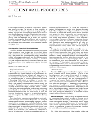
Figure 1 Repair of pectus excavatum: Ravitch procedure. The procedure begins
with a midline incision (a) or a bilateral inframammary incision (b). The pectoralis
muscles are then dissected off the chest wall (c).
bilateral inframammary incision is made [see Figure 1a, b]; the latter The posterior plane between the cartilage and the perichondrium is
incision yields superior cosmetic results, especially in female pa- then developed in one area, and the cartilage is divided with a scalpel
tients, but necessitates the elevation of large subcutaneous skin flaps between the jaws of a right-angle clamp [see Figure 2b]. The cut end
to the level of the angle of Louis or the sternal notch superiorly and of the cartilage is grasped with a clamp, and the rest of the cartilage
to the xiphoid process inferiorly. The pectoralis muscles are then is dissected from the perichondrium. Once the correct plane is
mobilized from the chest wall, beginning medially and proceeding established, the dissection can be facilitated by gently pushing the
laterally until the costal cartilages are exposed [see Figure 1c]. perichondrium off the cartilage with a finger. The entire cartilage
should be removed from the sternum to the rib, with every attempt
Step 2: resection of abnormal cartilages. For each abnormal costal made to maintain the integrity of the perichondrium. During this
cartilage, the anterior perichondrium is scored with the electro- part of the procedure, the xiphoid process is also detached from the
cautery along the length of the cartilage, and the cartilage is dissect- sternum.The extent of cartilage removal depends on the individual
ed from the perichondrium with a periosteal elevator [see Figure 2a]. defect present but usually includes the third rib.
a b
Figure 2 Repair of pectus excavatum: Ravitch procedure. (a) The anterior perichondrium is opened,
and the abnormal cartilage is dissected free with a periosteal elevator. (b) The cartilage is divided.
- 3. © 2005 WebMD, Inc. All rights reserved. ACS Surgery: Principles and Practice
4 THORAX 9 CHEST WALL PROCEDURES — 3
a
c
b 1 1
2 2
3 3
4
4
5
6 5
7
30°–35° 6
Figure 3 Repair of pectus 7
excavatum: Ravitch procedure. After
Before
re
(a) An osteotomy is made in
the upper sternum. (b) The
sternum is angled anteriorly;
when the desired angle is
reached, the osteotomy is
closed. (c) Shown is a lateral
view of the sternal angle before
and after correction. Osteotomy Closure
with Thumb Pressure
Step 3: sternal osteotomy. An osteotomy is made in the upper the defect. The bar that will be used for the repair is shortened to
anterior table of the sternum with either a periosteal elevator or a a length equivalent to the measured distance between the two
small reticulating bone saw [see Figure 3a], and the posterior table midaxillary lines minus 1 cm. A complex series of bends are then
of the sternum is fractured.The sternum can then be angled ante- placed in the bar to match its contours to those of the patient’s
riorly. When the desired angle is reached, the osteotomy is closed deformity.
with three interrupted nonabsorbable sutures or with microplates
and screws [see Figure 3b, c]. At this point, rotational sternal de- Step 2: initial incisions and creation of intrathoracic tunnel. In-
fects can be corrected by making anterior and posterior lateral cisions are made in the right and left midaxillary lines at the level
osteotomies on either side of the sternum and then closing the of the marks, and a subcutaneous flap is raised from each inci-
osteotomies with sutures or microplates. sion and extended to the defect. A Crawford vascular clamp or a
Step 4: sternal fixation. Sternal fixation can be accomplished by
any of several means. Posterior sternal support can be achieved by
placing a Kirschner wire or retrosternal bar that is secured to the
periosteum of the rib and left in place for approximately 3 months
after operation [see Figure 4]. Alternatively, the sternum can be
supported with a piece of polypropylene mesh or with two poly-
propylene sutures sutured to the xiphoid process and then brought
around the right and left second ribs.13
Step 5: closure and drainage. The pectoralis muscles are reap-
proximated in the midline, closed suction drains are placed in the
subcutaneous flaps, and the subcutaneous layer and the skin are
closed. To prevent seroma formation, one closed suction drain
may be placed posterior to the pectoralis muscles and another
between the pectoralis muscles and the subcutaneous layer; the
right pleural space may then be opened anteriorly and a right
pleural tube placed through a separate incision.14
Nuss procedure Minimally invasive repair of pectus excava-
tum, also referred to as the Nuss procedure, has gained populari-
ty over the past decade.
Step 1: configuration of bar. The patient is placed in the supine Figure 4 Repair of pectus excavatum: Ravitch pro-
position with the arms abducted, and marks are made on either cedure. Sternal fixation is accomplished through
side of the chest at spots that correspond to the deepest point of placement of a retrosternal bar.
- 4. © 2005 WebMD, Inc. All rights reserved. ACS Surgery: Principles and Practice
4 THORAX 9 CHEST WALL PROCEDURES — 4
Outcome Evaluation
In general, the results of pectus excavatum repair are good, and
the overall complication rate is low. In one study, 90% of 76 pa-
tients operated on over a 30-year period experienced excellent
outcomes, and only one patient required reoperation for a recur-
rent defect.14 The incidence of complications (pleural effusions,
pneumonia, and wound seromas) was 14%.14 In another study, no
operative deaths occurred in more than 800 repairs, and only a few
cases of serious infections and bleeding were reported.16 Other
investigators have reported rare complications arising from the
migration of sternal support bars and wires.17
REPAIR OF PECTUS CARINATUM
Surgical repair of pectus carinatum resembles surgical repair of
pectus excavatum in several respects. The same skin incision is
employed, and the pectoralis major muscles are elevated in a sim-
ilar manner. Subperichondrial resection of the abnormal cartilages
Figure 5 Repair of pectus excavatum: Nuss procedure. Incisions is then carried out, usually extending to the second costal carti-
are made on either side of the chest. A Crawford vascular clamp is lage. Next, a generous V-shaped osteotomy is made in the upper
inserted through the right intercostal space and advanced along portion of the sternum at the point of maximal protrusion, which
the sternum and out the left intercostal space.
is usually near the insertion of the second cartilage. Occasionally,
a second osteotomy is required near the caudal end of the sternum
Lorenz pectus introducer is then placed through the right inter- to facilitate elevation of the manubrium and depression of the ster-
costal space under thoracoscopic visualization and advanced along num. Finally, the osteotomy is closed with nonabsorbable mono-
the posterior sternum and out the corresponding left intercostal filament sutures, drains are placed, and soft tissue is closed as in a
space [see Figure 5]. pectus excavatum repair.
The results of pectus carinatum repair are generally compara-
Step 3: placement and fixation of bar. An umbilical tape is pulled ble to those of pectus excavatum repair. Most patients experience
through the anterior mediastinum and attached to the bar, which good outcomes, and operative morbidity is low.
is then gently pulled, with the concave side up, through the inter-
costal space. A Lorenz pectus bar rotator is employed to flip the
bar over, and the ends of the bar are positioned in the subcuta- Procedures for Acquired Chest Wall Disease
neous space [see Figure 6]. Occasionally, for proper alignment, the
TRANSAXILLARY FIRST RIB RESECTION FOR THORACIC OUTLET
bar may have to be removed and rebent, or stabilizers may have to
SYNDROME
be placed alongside it. When the bar is correctly positioned, it is
sutured to the chest wall musculature with an absorbable suture
on one end and a permanent suture on the other. Preoperative Evaluation
The bar is usually left in place for 2 years. Excellent results have TOS results from compression of the subclavian blood vessels
been reported.15 Significant complications include bar displace- or the brachial plexus as these structures exit the bony thorax.
ment necessitating reoperation (9.2% of procedures), pneumo- Symptoms may be primarily vascular (e.g., arm swelling or loss of
thorax (4.8%), infection (2%), and pleural effusion (2%). Rare pulse) or neurogenic (e.g., pain and paresthesias).The workup for
complications include cardiac injury, thoracic outlet syndrome TOS includes a detailed physical examination, as well as imaging
(TOS), pericarditis, and sternal erosion caused by the bar. and nerve conduction studies.
a b
Figure 6 Repair of pectus excavatum: Nuss procedure. (a) The pectus bar is pulled into the tunnel
opened by the vascular retractor, then flipped to provide the desired chest contour. (b)The ends of the
bar are then sutured to the chest wall musculature.
- 5. © 2005 WebMD, Inc. All rights reserved. ACS Surgery: Principles and Practice
4 THORAX 9 CHEST WALL PROCEDURES — 5
Subclavian via several different approaches, including posterior, supraclavicu-
Artery lar, infraclavicular, transthoracic, and transaxillary. I focus here on
the transaxillary approach, which provides good exposure of the
first rib and allows the surgeon to avoid the subclavian blood ves-
sels and the brachial plexus. Regardless of the specific surgical
approach followed, any surgeon embarking on a first rib resection
Subclavian
Vein
must have a detailed knowledge of the thoracic outlet to keep from
injuring the neurovascular structures in the area.
Middle
Scalene
Operative Technique
Muscle The patient is placed in the lateral decubitus position, and the
affected arm is kept at a 90° angle either by an arm holder or, alter-
natively, by an assistant. Care must be taken not to hyperabduct or
hyperextend the shoulder. The arm, the axilla, and the chest are
Brachial
prepared and draped into the sterile field.
Posterior Plexus
Scalene Step 1: initial incision and exposure An incision is made
Muscle
just below the axillary hair line and extended from the pectoralis
Apex of major to the latissimus dorsi [see Figure 7]. The subcutaneous tis-
Pleura
sue is incised down to the chest wall with the electrocautery, with
care taken to stay perpendicular to the axis of the chest. Dissection
1st Rib is then begun along the chest wall and carried toward the first rib.
Anterior The intercostal brachial nerve is identified where it exits between
Scalene
Muscle the first and second ribs. This nerve should be spared: dividing it
leads to numbness of the upper inner biceps region.
Latissimus
Dorsi Border Step 2: dissection and division of anterior portion of first
Pectoralis Major rib When the first rib is encountered, it is dissected from the
Skin Incision Border periosteum with a periosteal elevator. Dissection is continued
anteriorly along the rib until just past the subclavian vein, at which
Figure 7 Transaxillary first rib resection. Shown are the point a right-angle clamp can be passed around the rib in the sub-
transaxillary incision and the thoracic outlet anatomy. periosteal plane. A Gigli saw or a first rib cutter is then used to
divide the anterior portion of the rib [see Figure 8a].
Next, the first rib is retracted inferiorly to permit visualization
Operative Planning of the anterior scalene muscle, which is then divided at its attach-
Surgical treatment of TOS typically involves resection of the ment to the rib. To prevent thermal injury to the phrenic nerve, a
first rib, which widens the thoracic outlet and relieves the neu- scalpel rather than an electrocautery is used to divide the muscle
rovascular impingement. First rib resection can be accomplished [see Figure 8b]. Care should also be taken not to injure the subcla-
a b
Posterior Anterior Posterior Anterior
Figure 8 Transaxillary first rib resection. (a) The anterior portion of the first rib is cut. (b) The anterior
scalene muscle is then divided.
- 6. © 2005 WebMD, Inc. All rights reserved. ACS Surgery: Principles and Practice
4 THORAX 9 CHEST WALL PROCEDURES — 6
[see Figure 9], and magnetic resonance imaging if vertebral involve-
ment is suspected. The other preoperative tests ordered are much
the same as those required for any other large thoracic procedure,
including pulmonary function testing, nutritional assessment, and
cardiac stress testing in patients who are older or have a history of
cardiac disease.
Operative Planning
Operative planning for chest wall resection should include
establishing the extent of the resection, weighing options for chest
wall stabilization, and deciding on the method of tissue coverage
to be employed. A multidisciplinary approach, involving the
participation of a neurosurgeon and a plastic surgeon, may be
required.
The technique of chest wall resection is essentially the same for
benign conditions as for malignant ones and is mainly dependent
on the location of the lesion. For malignant tumors of the chest
Figure 9 Chest CT reveals a large pulmonary and chest wall mass.
wall, a 5 cm margin, or at least resection of one uninvolved rib
above and below the tumor, is required. Additionally, any involved
vian vein and artery, which lie anterior and posterior to the ante- skin and any biopsy site must be resected along with the chest wall
rior scalene muscle, respectively. As an alternative, the anterior specimen. For infection or osteoradionecrosis, the resection must
scalene muscle may be divided before the anterior portion of the include all nonviable skin and underlying bone; if it does not, skin
rib is cut. and muscle flaps may not heal properly. Any destroyed lung tissue
may also have to be resected along with the chest wall specimen.
Step 3: dissection and division of posterior portion of In addition, recurrent cancer must be ruled out before the opera-
first rib The subperiosteal dissection is continued posteriorly, tion can proceed. A particular challenge is posed by breast cancer
freeing the first rib from the pleura, the subclavian vessels, and the patients who have already had muscle flaps for breast reconstruc-
brachial plexus. The posterior portion of the rib is then divided tion; in these patients, tissues other than muscle (e.g., omentum)
with a first rib cutter as close as possible to the articulation of the may be required for tissue coverage after chest wall resection.
rib with the transverse process. Every effort should be made to
keep from injuring the C8 and T1 nerve roots. Standard Chest Wall Resection
In cases in which a concomitant lung resection is required, the
Step 4: closure The incision is closed without drainage. If
the pleura was inadvertently entered, air may be aspirated from the
chest with a red rubber tube, which is removed before the subcu-
taneous tissue is closed. One authority recommends further neu- Anterior
Resection Margin
rolysis of the C7 to T1 nerve roots and the middle and lower
trunks of the brachial plexus, as well as resection of the anterior
and middle scalene muscles up into the neck.18
Complications
Surgical complications include injuries to the subclavian vein
and artery (leading to massive blood loss), the brachial plexus, the
phrenic nerve, the long thoracic nerve, and the thoracic duct.
Outcome Evaluation
The long-term results of first rib resection appear to be inde-
pendent of the exposure technique employed. Good results, de-
fined as relief of major symptoms, have been reported in as many
of 90% of patients in the first year and in as many as 70% of
patients 5 to 10 years after operation. There continues to be con-
siderable debate over the preferred surgical approach, but to date,
no studies have shown any one approach to have significant advan-
tages over any of the others. Posterior
Resection
CHEST WALL RESECTION Margin
Chest wall resection has become a critical component of the
thoracic surgeon’s armamentarium. It may be performed to treat
either benign conditions (e.g., osteoradionecrosis, osteomyelitis,
and benign neoplasms) or malignant disease.
Figure 10 Chest wall resection. The anterior and posterior mar-
Preoperative Evaluation
gins of the required resection are determined. The anterior mar-
Preoperative imaging studies may include chest x-ray, chest CT gin is completed first.
- 7. © 2005 WebMD, Inc. All rights reserved. ACS Surgery: Principles and Practice
4 THORAX 9 CHEST WALL PROCEDURES — 7
Osteotome the rib. Alternatively, the intercostal bundle can be doubly ligated
and divided at the anterior resection margin. Once the intercostal
vessels are cleared from the lowest rib, the electrocautery is used to
divide the pleura below the rib toward the anterior boundary of the
resection.The rib is then divided with a rib cutter, with care taken
to ensure a margin of at least 5 cm from the tumor [see Figure
10]. Next, the intercostal bundle of the next higher rib is li-
gated and divided, the intercostal muscle is divided with the elec-
trocautery, and the rib is cut with a rib cutter in the same manner
as the previous rib. This process is repeated until the anterior
boundary of resection is completed. A subperiosteal plane is then
developed over the highest rib to be resected, the adjacent inter-
costal bundle is separated from the rib, and the parietal pleura is
divided with the electrocautery.
Transverse Step 4: completion of posterior boundary of resection. If the tumor
Process margin does not involve the vertebrae, the posterior portion of the
chest wall resection is identical to the anterior portion [see Step 3,
above]. If, however, the tumor appears to encroach on the head of
Divided Erector the rib or the transverse process, it will be necessary to disarticu-
Spinae Muscle late the rib from the transverse process or, in the latter situation,
remove the transverse process entirely.
Figure 11 Chest wall resection. Depicted is disarticulation of the Disarticulation of the rib from the transverse process is per-
rib from the transverse process. formed by dissecting the paraspinal ligament and erector spinae
muscles away from the spine with the electrocautery, thereby
chest wall resection is usually performed first; this measure renders exposing the joint between the head of the rib and the transverse
the lung more mobile and facilitates the pulmonary resection.The process. The ligaments attaching the rib to the transverse process
lateral decubitus position is the best choice for most combined are then incised with the electrocautery, and an osteotome is
lung–chest wall procedures, whereas the supine position is prefer- inserted into the joint, which is then levered anteriorly and poste-
able for isolated anterior chest wall procedures. If a larger chest riorly to disarticulate the rib from the transverse process [see Figure
wall resection is expected, every attempt should be made to spare 11]. The intercostal neurovascular bundle must be ligated and
major muscle groups so that these muscles can be used later to divided at this point: failure to do so will result in bleeding and
cover any prosthetic material used in reconstruction. possibly in leakage of cerebrospinal fluid. If bleeding occurs, it can
be controlled with bipolar electrocauterization and temporary pack-
Operative technique Step 1: initial incision and exposure. The ing with a hemostatic agent.The hemostatic agent must not be left
usual incision is a standard posterolateral thoracotomy incision in place permanently, because it may expand or result in a neural
through the fifth interspace. foramen hematoma, and either of these events can lead to spinal
cord compression and significant neurologic injury. If at any time
Step 2: determination of extent of required chest wall resection. As the surgeon feels uncomfortable about ongoing intercostal bleed-
soon as the pleura is opened, the surgeon should palpate the tumor ing or a possible CSF leak, intraoperative neurosurgical consulta-
to evaluate the extent of chest wall involvement, which determines tion should be obtained. In cases in which the tumor involves the
the extent of the resection. Removal of uninvolved ribs may make transverse process, this structure must be removed from the verte-
reconstruction of the chest wall more complicated. For example, bral body with an osteotome and a mallet or with a first rib cutter.
posterior resections that do not require removal of the fifth rib are If the tumor has invaded the vertebral body and resection is still
protected by the scapula, so that reconstruction is unnecessary. If being considered, neurosurgical consultation should be obtained.
the fifth rib is removed, however, the tip of the scapula will tend to Generally, if the tumor involves more than one quarter of the ver-
become stuck under the sixth rib with shoulder movement; this is tebral body or extends into multiple vertebral levels, it is consid-
very uncomfortable for the patient, and chest wall reconstruction ered unresectable.
will therefore be required at the time of resection.
At this point, the surgeon should also rule out diffuse pleural Step 5: lung resection (if required). Once the posterior chest wall
disease before proceeding with resection. In some cases, the tumor margin has been completed, the lung resection (if required) is per-
can be removed by means of extrapleural dissection, without any formed.The entire lung–chest wall specimen is then be submitted
need for chest wall resection. If there is any suspicion of chest wall for pathologic examination, and histopathologic margins are ob-
involvement, however, chest wall resection is mandatory because tained both on the lung and on the chest wall. If the chest wall
leaving any tumor behind guarantees a recurrence. margins are positive, the involved area must be trimmed back and
The extent of the chest wall resection is marked with the elec- a new margin submitted.
trocautery on the outside of the thoracic cavity. At least one gross-
ly uninvolved rib should be included both above and below the Step 6: chest wall reconstruction. Chest wall reconstruction is
tumor. required for all anterior defects and for posterior defects that
involve any rib lower than the fourth rib. Reconstruction can be
Step 3: completion of anterior boundary of resection. Initially, the performed either with polypropylene or Gore-Tex (W. L. Gore and
periosteum over the lowest rib to be resected is scored, and a Associates, Flagstaff,Arizona) mesh or with a polypropylene-methyl-
periosteal elevator is used to separate the intercostal bundle from methacrylate sandwich. The latter is employed when rigid recon-
- 8. © 2005 WebMD, Inc. All rights reserved. ACS Surgery: Principles and Practice
4 THORAX 9 CHEST WALL PROCEDURES — 8
a Polypropylene Methylmethacrylate
Mesh Layers Layer between
Polypropylene Layers b
Figure 12 Chest wall resection. (a) A polypropylene-methyl-
methacrylate sandwich is created by spreading a layer of
methylmethacrylate cement between two pieces of polypropy-
lene mesh. When sufficiently hardened, the sandwich is sutured
to the ribs. (b) Photograph shows a polypropylene-methyl-
methacrylate sandwich sutured in place.
struction is warranted (as in anterior reconstruction); it not only tion resulting from the use of synthetic material. In particular, radi-
provides added protection of pleural and mediastinal structures ation injury may involve all layers of the chest wall, necessitating
but also creates a better cosmetic effect by recreating the shape of very large resections [see Figure 13a]. Muscle or omental flaps with
the chest wall. split-thickness skin grafts may be required for coverage; thus, pre-
To create the polypropylene-methylmethacrylate sandwich, two operative consultation with an experienced plastic surgeon is
pieces of polypropylene mesh are cut to the size of the defect. A advisable. A particular concern is what to use to reconstruct the
thin layer of methylmethacrylate cement is spread on one of the chest wall. Various tissues (e.g., fascia lata and ribs) have been
mesh pieces, and the other piece is then applied over the methyl- employed, but an easier substitute that works quite well is an
methacrylate layer. As this sandwich begins to harden, it is mold- absorbable synthetic mesh (e.g., Vicryl). The mesh is sewn to the
ed to the contours of the chest wall, with care taken to protect the ribs as previously described [see Step 6, above], and the tissue flap
patient’s skin against injury from the heat given off by the harden- is placed on top of the mesh, followed by a skin graft [see Figure
ing cement.When the sandwich is sufficiently hardened, it is sewn 13b, c]. Alternatively, some authors recommend the use of muscle
to the ribs with 0 polypropylene sutures [see Figure 12a]. The or myocutaneous flaps without rigid chest wall reconstruction
sutures may be passed around the uppermost and lowermost ribs after resection, particularly in infected fields.19
and may be placed directly through the anterior and posterior
margins [see Figure 12b]. If rib disarticulation was required to com- Outcome evaluation The results achieved after major chest
plete the posterior margin, holes may be drilled in the transverse wall resection have generally been excellent. One study reviewed
processes and the sutures passed through these holes; alternative- 200 patients who underwent resection and reconstruction over a
ly, the sandwich may be sutured to the paraspinal ligament. 25-year period.20 The reconstructions ranged from relatively
If polypropylene or Gore-Tex mesh is used without cement, it straightforward two-rib resections to more complex forequarter
should be cut to a size smaller than that of the defect. Thus, the amputations. The indications for resection were lung cancer
mesh will effectively be stretched when it is sutured to the chest (38%), osteoradionecrosis (29%), chest wall tumor (27%), and
wall, and any laxity in the reconstruction will thereby be alleviated. osteomyelitis (16%). Immediate reconstruction was performed in
98% of patients. The major muscle flaps utilized were latissimus
Step 7: closure and drainage. The serratus anterior and the latis- dorsi (20%), rectus abdominis (17%), pectoralis major (16%), and
simus dorsi are closed in the standard fashion, as are the subcuta- serratus anterior (9%). Free flaps were utilized in only 9% of cases,
neous and skin layers. With the exception of pleural tubes, drains and split-thickness skin grafts were required in 12% of patients.
are not routinely used. Special attention should be paid to postop- Reconstruction was performed with Prolene mesh (25%), Marlex
erative analgesia: patients who have undergone extensive resec- mesh (11%),Vicryl mesh (6%), or a polypropylene-methylmetha-
tions often experience considerable pain and are therefore prone crylate sandwich (6%). Operative mortality was 7%, and major
to atelectasis and pneumonia. Epidural analgesia should be em- morbidity occurred in 24% of patients. Most of the morbidity was
ployed routinely in such cases. accounted for by pneumonia (14%) and acute respiratory distress
syndrome (6%).
Troubleshooting If chest wall infection is a possibility (as
with osteoradionecrosis or osteomyelitis), alternative reconstruc- Manubrial and Clavicular Resection
tive techniques are required to obviate concerns about superinfec- Resection of the manubrium or the clavicle may be necessary if
- 9. © 2005 WebMD, Inc. All rights reserved. ACS Surgery: Principles and Practice
4 THORAX 9 CHEST WALL PROCEDURES — 9
a b
c
Figure 13 Chest wall resection. The presence of
osteoradionecrosis may necessitate very large
resections and resulting defects (a). Such defects
may be covered with absorbable mesh (b), followed
by an omental flap (c) or a muscle flap.
Resection of sternoclavicular joint for infection Clavic-
ular resections are rarely performed but may be required to treat
tumors, vascular compression from healed fractures, or infection.
Occasionally, infections involve the sternoclavicular joint (SCJ).
Patients with osteomyelitis of this joint are often immunosup-
pressed and may have had an indwelling subclavian vein catheter
that became infected. In a study of seven patients who underwent
SCJ resection for infection, five of six patients initially treated with
these structures become infected or involved with tumors. Clavic- antibiotics and simple drainage experienced recurrences, whereas
ular and manubrial resections follow the same operative approach six of six patients treated with resection of the joint and pectoralis
as other chest wall resections. Specifically, attention must be paid muscle advancement flaps were cured. None of the patients expe-
to how much bone to resect, how to reconstruct the defect, and rienced problems with arm mobility in the course of long-term
how to provide tissue coverage. follow-up.21
a b
Figure 14 Manubrial resection and reconstruction.
(a) The clavicles and ribs are divided as in clavicular
and other chest wall resections. (b) A polypropylene-
methylmethacrylate sandwich may be used to recon-
struct the chest wall.
- 10. © 2005 WebMD, Inc. All rights reserved. ACS Surgery: Principles and Practice
4 THORAX 9 CHEST WALL PROCEDURES — 10
Skin Resection of manubrium for cancer Manubrial resections
may be required for rare cases of primary or metastatic cancers.
Rib
Operative technique. Because of the relative paucity of tissue
overlying the manubrium, cancers in this area may involve the der-
mis. In such cases, it may be necessary to resect skin along with
the specimen. Alternatively, if the skin is not involved, an upper
midline incision may be employed. The incision is carried down
circumferentially to the chest wall, with care taken to maintain a 2
to 3 cm margin from the tumor.The clavicles and ribs are divided
in the same fashion as for chest wall and clavicular resections [see
Pleura Figure 14a]. Associated structures (e.g., the thymus) can be resect-
ed along with the manubrium; these tumors rarely involve the
innominate vein.
A polypropylene-methylmethacrylate sandwich is useful for
reconstruction of this area of the chest wall [see Figure 14b]. The
patch is secured to the remaining ribs and clavicles with 0 poly-
propylene sutures. Coverage is then provided with a pectoralis major
a
Figure 15 Open chest drainage (Eloesser flap). Once the ribs
have been resected, the skin overlying the thoracostomy is mar-
supialized to the parietal pleura to permit packing and open
pleural drainage.
Operative technique. An incision is made that extends along the
distal clavicle and curves down onto the manubrium.The soft tis-
sue is divided with the electrocautery down to the clavicle and the
manubrium. The muscular attachments of the pectoralis major
and the sternocleidomastoid muscle are dissected off the clavicle
and the manubrium with a periosteal elevator. Dissection in the
subperiosteal plane is then continued circumferentially around the
distal clavicle, with special care taken to keep from injuring the
subclavian vessels that lie deep to the clavicle. A Gigli saw is passed b
around the clavicle with a right-angle clamp and used to divide the
distal clavicle. The distal cut end of the clavicle is grasped with a
penetrating towel clamp and bluntly dissected away from the deep
tissue toward the manubrium. Any pockets of infection encoun-
tered should be cultured, drained, and debrided.
At this point, a large separation in the SCJ, caused by the infec-
tion, should be apparent. Resection of a small portion of the ma-
nubrium is usually required to remove all of the infected bone.
Once the tissue deep to the manubrium has been dissected, a
small band retractor is placed beneath the manubrium, and an
oscillating sternal saw is used to resect the lateral portion of the
manubrium, adjacent to the SCJ. Alternatively, a rongeur may be
used to debride infected bone from the manubrium. All tissue
should be sent for culture.
Severe infections may necessitate more extensive resection of
bone or soft tissue, but if the infection is caught early, simple resec-
tion of the SCJ is generally curative. In more extensive resections,
muscle flap coverage may be required, but in simple SCJ resec-
tions, good results can be obtained by using only deep closed suc- Figure 16 Open chest drainage (Eloesser flap).
tion drainage, followed by multilayer closure of the wound.To pre- (a) Photograph shows a right Eloesser flap 8 months
vent any recurrent osteomyelitis, antibiotics should be continued after creation. (b) Photograph shows an Eloesser flap
for several weeks after resection. that was closed with a muscle flap.
- 11. © 2005 WebMD, Inc. All rights reserved. ACS Surgery: Principles and Practice
4 THORAX 9 CHEST WALL PROCEDURES — 11
advancement flap or, if skin was excised, a pedicled pectoralis space is opened with the electrocautery, any pus present is
myocutaneous flap. A pleural drain may be placed if either pleural drained, and the chest cavity is manually and visually explored.
space was entered, but this measure is not routinely employed. Next, 6 to 8 cm segments of two or three adjacent ribs are resect-
ed according to the same principles employed for other chest wall
Open Chest Drainage (Eloesser Flap) resections. The resulting thoracostomy is large enough to permit
Open drainage procedures are usually included in discussions drainage and packing.The skin overlying this thoracostomy is then
of treatment of empyema, but they really represent a type of chest marsupialized to the thickened parietal pleura with absorbable
wall resection. Open drainage techniques for empyema were first sutures [see Figure 15]. If the pleura does not possess sufficient
described in the late 1800s by Poulet and subsequently by Schede. integrity to hold the sutures, they can be placed through the
Graham, who headed the Army Empyema Commission during periosteum of the ribs.
World War I, is credited with the observation that ensuring pleu-
ral-pleural symphysis was the key to preventing the often fatal Step 3: packing and drainage. The wound is irrigated with nor-
complication of pneumothorax.22 Indications for open chest drain- mal saline and packed with saline-moistened gauze. Postopera-
age include postpneumonectomy empyema or bronchopleural tively, a chest x-ray should be obtained to rule out pneumothorax,
fistula, long-standing empyema in a patient who cannot undergo and twice- to thrice-daily packing is initiated. Packing is continued
decortication, and chronic bronchopleural fistula in a high-risk on an outpatient basis, and the wound is monitored. The wound
patient. will begin to close over the next several weeks. If the empyema or
bronchopleural fistula has not healed by the time the wound starts
Operative technique The technique currently employed by closing, the thoracostomy will have to be revised. In some cases,
most thoracic surgeons follows Symbas’s modification of Eloesser’s this can be accomplished merely by manually dilating the opening
open drainage technique.23 This procedure has come to be known in the operating room; in others, the entire thoracostomy must be
as the Eloesser flap. Preoperative chest CT is essential for identi- revised. In either case, the goal is to maintain a large enough open-
fying the exact location of the empyema, which determines the ing to allow adequate packing.
placement of the incision.
Step 4: closure of thoracostomy. Once the lung and the pleural
Step 1: initial incision and exposure. The patient is placed in the space have healed, the thoracostomy is closed. The procedure for
decubitus position, and a 6 to 8 cm incision is made over the area closing the thoracostomy depends on the size and nature of the
corresponding to the most dependent area of the infected cavity. remaining defect [see Figure 16a]. For small defects, simple closure
Symbas employed a U-shaped incision; however, a simple linear of the skin will suffice. For larger defects or residual spaces in the
incision can also be used with good results.The subcutaneous tis- pleura, however, muscle flap closure will be required [see Figure
sue and muscle are then divided down to the chest wall with the 16b]. Improvements in radiographic techniques and greater
electrocautery. emphasis on early intervention for empyemas have significantly
reduced the need for open chest drainage; however, this technique
Step 2: resection of ribs and creation of thoracostomy. The pleural can still be valuable in the appropriate clinical situation.
References
1. Backer CL, Mavroudis C: Congenital heart 8. Bevegard S: Postural circulatory changes at rest tum. Chest Surg Clin N Am 10:277, 2000
surgery nomenclature and database project: vascu- and during exercise in patients with funnel chest, 17. Stefani A, Morandi U, Lodi R: Migration of pec-
lar rings, tracheal stenosis, pectus excavatum. Ann with special reference to the influence on the tus excavatum correction metal support into the
Thorac Surg 69(4 suppl):S308, 2000 stroke volume. Acta Physiol Scand 49:279, 1960 abdomen. Eur J Cardiothorac Surg 14:434, 1998
2. Haller JA, Kramer Ss, Lietman SA: Use of CT 9. Gattiker H, Buhlmann A: Cardiopulmonary func- 18. Urschel HC: The transaxillary approach for
scans in selection of patients for pectus excava- tion and exercise tolerance in supine and sitting treatment of thoracic outlet syndrome. Chest
tum surgery: a preliminary report. J Pediatr Surg position in patients with pectus excavatum. Helv Surg Clin N Am 9:771, 1999
22:904, 1987 Med Acta 33:122, 1967
19. Arnold PG, Pairolero PC: Use of pectoralis
3. Weg JG, Krumholz RA, Harkleroad LE: Pul- 10. Peterson RJ, Young WG Jr, Godwin JD, et al: major muscle flaps to repair defects of anterior
monary dysfunction in pectus excavatum. Am Rev Noninvasive assessment of exercise cardiac func- chest wall. Plast Reconstruct Surg 63:105, 1979
Respir Dis 96:936, 1967 tion before and after pectus excavatum repair. J
20. Mansour KA, Thourani VH, Losken A, et al:
Thorac Cardiovasc Surg 90:251, 1985
4. Gyllensward A, Irnell L, Michaelsson M, et al: Chest wall resections and reconstruction: a 25-
Pectus excavatum: a clinical study with long 11. Judet J, Judet R: Sternum en entonnoir par resec- year experience. Ann Thorac Surg 73:1720, 2002
term postoperative follow-up. Acta Paediatr tion et retournement. Mem Acad Chir 82:250,
21. Song HK, Guy TS, Kaiser LR, et al: Current
255(suppl): 2, 1975 1956
presentation and optimal surgical management
5. Cahill JL, Lees GM, Robertson HT: A summary 12. Wada J, Ikeda K, Ishida T, et al: Results of 271 of sternoclavicular joint infections. Ann Thorac
funnel chest operations. Ann Thorac Surg 10: Surg 73:427, 2002
of preoperative and postoperative cardiorespira-
526, 1970 22. Somers J, Faber LP: Historical developments in
tory performance in patients undergoing pectus
excavatum and carinatum repair. J Pediatr Surg 13. Robicsek F, Cook JW, Daugherty HK, et al: the management of empyema. Chest Surg Clin
19:430, 1984. Pectus carinatum. J Thorac Cardiovasc Surg 78: N Am 6:404, 1996
52, 1979 23. Symbas PN, Nugent JT, Abbott OA, et al: Nontu-
6. Blickman JG, Rosen PR, Welch KJ, et al: Pectus
excavatum in children: pulmonary scintigraphy 14. Mansour KA, Thourani VH, Odessey EA, et al: berculous pleural empyema in adults. Ann Thorac
before and after corrective surgery. Radiology Thirty-year experience with repair of pectus Surg 12:69, 1971
156:781, 1985 deformities in adults. Ann Thorac Surg 76:391,
2003
7. Haller JA, Colombani PM, Humphries CT, et al: Acknowledgment
Chest wall constriction after too extensive and 15. Hebra A: Minimally invasive pectus surgery.
too early operations for pectus excavatum. Ann Chest Surg Clin N Am 10:329, 2000 Figures 1 through 8, 10 through 12, 14, and 15 Alice
Thorac Surg 61:1618, 1996 16. Robicsek F: Surgical treatment of pectus excava- Y. Chen.
![© 2005 WebMD, Inc. All rights reserved. ACS Surgery: Principles and Practice
4 THORAX 9 CHEST WALL PROCEDURES — 2
a
Costal
c Cartilages
Edge of Reflected
Pectoralis Major
b Sternum
Figure 1 Repair of pectus excavatum: Ravitch procedure. The procedure begins
with a midline incision (a) or a bilateral inframammary incision (b). The pectoralis
muscles are then dissected off the chest wall (c).
bilateral inframammary incision is made [see Figure 1a, b]; the latter The posterior plane between the cartilage and the perichondrium is
incision yields superior cosmetic results, especially in female pa- then developed in one area, and the cartilage is divided with a scalpel
tients, but necessitates the elevation of large subcutaneous skin flaps between the jaws of a right-angle clamp [see Figure 2b]. The cut end
to the level of the angle of Louis or the sternal notch superiorly and of the cartilage is grasped with a clamp, and the rest of the cartilage
to the xiphoid process inferiorly. The pectoralis muscles are then is dissected from the perichondrium. Once the correct plane is
mobilized from the chest wall, beginning medially and proceeding established, the dissection can be facilitated by gently pushing the
laterally until the costal cartilages are exposed [see Figure 1c]. perichondrium off the cartilage with a finger. The entire cartilage
should be removed from the sternum to the rib, with every attempt
Step 2: resection of abnormal cartilages. For each abnormal costal made to maintain the integrity of the perichondrium. During this
cartilage, the anterior perichondrium is scored with the electro- part of the procedure, the xiphoid process is also detached from the
cautery along the length of the cartilage, and the cartilage is dissect- sternum.The extent of cartilage removal depends on the individual
ed from the perichondrium with a periosteal elevator [see Figure 2a]. defect present but usually includes the third rib.
a b
Figure 2 Repair of pectus excavatum: Ravitch procedure. (a) The anterior perichondrium is opened,
and the abnormal cartilage is dissected free with a periosteal elevator. (b) The cartilage is divided.](data:image/gif;base64,R0lGODlhAQABAIAAAAAAAP///yH5BAEAAAAALAAAAAABAAEAAAIBRAA7)