SlideShare une entreprise Scribd logo
1  sur  35
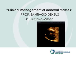
―Clinical management of adnexal masses”
          PROF. SANTIAGO DEXEUS
            Dr. Gustavo Missón
Introduction

   Adnexal masses are the fourth most

    common gynecological cause for

    hospitalization and 90% have benign

    characteristics.
Adnexal Mases USA
ANNUAL HOSPITALIZATION: 289000 PATIENTS

RISK OF
MALIGNANCY

13% in pre menopause
45% in post menopause
L Van Lie (2000)
48 Meeting of the ACOG
ADNEXAL MASSES


N= 4.359
October 1991 – October 1999
average: 37.22 years (14-85)


Rate of malignancy: 2.1%
IUDEXEUS-1999
ADNEXAL MASSES
             Color Doppler
       Absence of pathological flow


3.0% Malignant tumor
                                Kurjak et al,1993


4.2% Malignant tumor
                                MªA Pascual y col.,1996
PRIORITY


 Differential diagnosis
 Diagnostic studies and interpretation
 Management
Anatomy

   ―Adnexa‖
    › Area next to the
      uterus containing
      ligaments, vessels,
      tubes, ovaries
Background
       Prevalence of adnexal masses is 2
        to 8%
    › Random TVUS of 335 asymptomatic
         premenopausal women, 7.8% with
         adnexal masses 2.5 cm or larger (6.6%
         were ovarian cysts.

    ›    Transvaginal sonographic ovarian findings in a random sample of women 25-40 years old.
         Ultrasound Obstet Gynecol 1999 May;13(5):345-50.
Background
       Prevalence of adnexal masses is 2
        to 8%

    › TVUS in 8794 asymptomatic
         postmenopausal women, 2.5% were
         found to have adnexal cysts

    ›    Alcazar JL; Jurado M. Natural history of sonographically detected simple unilocular adnexal
         cysts in asymptomatic postmenopausal women. Gynecol Oncol 2004 Mar;92(3):965-9.
Differential Diagnosis

   Physiologic cysts
    › Follicle develops but never ruptures, continues to
      grow
    › Simple, smooth-walled
   Functional cysts
    › Corpus luteum does not involute or continues to
      grow
 Most are small (<2.5 cm), but can be larger
 Usually no symptoms, unless rupture or torsion
Differential Diagnosis

   Ectopic pregnancy
   PID
   Hydrosalpinx
   Benign neoplasms
    ›   Serous or mucinous cystadenoma
    ›   Endometrioma
    ›    Cy.Dermoid
    ›   Fibroids (exophytic, broad ligament)
   Malignancy
    › Primary vs. mts
Non-Gyn Etiology
   Abdominal
    › Appendicitis
    › Diverticulitis
    › Inflammatory bowel
      disease
   Inclusion cysts
    › Peritoneal or omental
   Retroperitoneal
    masses
    › Pelvic kidney
Diagnosis: History
   History
    › Pain
       Midcycle physiologic or functional cyst
       Dysmenorrhea/dyspareunia endometriosis
       Sudden onset, severetorsion, rupture, hemorrhage
       Chronic aching, bloatingneoplasm
    › Nonspecific GI symptoms
       May suggest ovarian cancer in postmenopausal female
       May suggest appendicitis or GI etiology in younger
        women
    › FH
       Breast, colon, or ovarian cancer
Diagnosis: Physical Exam

   Physical exam—should include bimanual
    and rectovaginal exam

    › Fever PID, appy, diverticulitis
    › Shouldn’t be able to palpate a
      postmenopausal ovary
    › Cul de sac nodularity, tender ligaments
      endometriosis
    › Cervical motion tendernessPID
    › Fixed, irregular, solid may suggest neoplasia
Diagnosis: Physical Exam

       Will probably need more than an H&P
        to make a diagnosis
    ›    84 women underwent pelvic examination
         prior to surgery, blinded to surgical
         indication
    ›    Attending, resident, student examined
         patient
    ›    Padilla L, Radosevich D, Milad M. Limitations of the pelvic examination for evaluation of the
         female pelvic organs . Int J of Gyn 2005; 88 (1): 84 – 88.
Diagnosis: Physical Exam


› Exam is a ―limited screening tool‖ for
 detection of adnexal masses


› Sensitivity at detecting adnexal masses: p
 >0.04
Diagnosis: Labs

   Labs
    › β-HCG to exclude ectopic
    › RPC if infection suspected
    › Tumor makers
       CA-125 (more to come)
       Others useful in adolescents/premenopaual
        women with adnexal masses and high
        suspicion
         LDHDysgerminoma
         HCGchoriocarcinoma
         AFPEndodermal sinus tumor
Malignancy
   Postmenopausal
    › Roughly 50 per 100,000 women, relative risk of ~3.5
    › 80% of ovarian cancers occur in women over 50
   Family history
   Symptoms
    › Vague, chronic aching, bloating, +/- GI symptoms
   Physical examination
    › Remember. . . Not really useful
   Ultrasound findings
   CA-125
Family History

       Lifetime risk of ovarian cancer in
        general population 1.5%
    ›     In BRCA 1 carrier 45-55%
    ›     In BRCA 2 carrier 15-25%
       Not all mutations have been identified
    ›     Two to three relatives with ovarian cancer
          increases lifetime risk to 5% (15% if first
          degree relatives)
    ›     Carlson KJ; Skates SJ; Singer DE. Screening for ovarian cancer. Ann Intern Med 1994 Jul 15;121(2):124-32.
CA-125
   Not specific to ovarian cancer, also elevated
    in:
       Other cancers (endometrial, fallopian tube, germ
        cell, cervical, pancreatic, breast, colon)
       Benign conditions
        (endometriosis, fibroids, PID, adenomyosis, functional
        ovarian cysts, pregnancy)
       Other diseases (renal, heart, liver, and many others)
       Also abnormal in 1% of normal females

         Bast R; Klug T; St John E; Jenison E; Niloff J; Lazarus H; Berkowitz R; Leavitt T; Griffiths C; Parker L;
          Zurawski V; Knapp R. A radioimmunoassay using a monoclonal antibody to monitor th
         course of epithelial ovarian cancer. N Engl J Med 1983 Oct 13;309(15):883
CA-125
   Normal value <35
    › Rarely >100-200 in benign conditions
CA-125
   Utility as screening tool for ovarian cancer
    ›   CA-125 increased in roughly 80% of ovarian cancers
    ›   About 50% sensitivity for Stage I, 90% for Stage II

   Study of 5550 healthy Swedish women
    ›   Followed women with elevated and normal CA-125
        levels
    ›   Serial pelvic exams, U/S, serial CA-125 levels
    ›   Of 175 women with elevated CA-125, 6 with ovarian
        cancer
    ›   Of the remaining women with normal CA-125
        levels, 3 had ovarian cancer
    ›   Einhorn N; Sjovall K; Knapp RC; Hall P; Scully RE; Bast RC Jr; Zurawski VR Jr. Prospective evaluation of serum CA 125
        levels for early detection of ovarian cancer. Obstet Gynecol 1992 Jul;80(1):14-8.
CA-125 (follow)
BIOMARKERS
› Ca 125
› Ca 19.9
› Ca 15.3
› BCGH
› Alpfa-phetoprotein


› HE-4
Ultrasound

                         Simple cyst
                          › Less than 2.5 cm
                          › Unlikely malignant
                          › Probably a follicle


                         Homogeneous
                         appearance may
                         suggest
                         endometrioma


www.uptodate.com
Ultrasound

   Features suggestive
    of malignancy:
    › Solid component
    › Doppler flow
    › Thick septations
    › Size
    › Presence of ascites or
      other peritoneal
      masses
Ultrasound: The DePriest Score
  De Priest PD, Shenson D, Fried A, Hunter JE, Andrew SJ, Gallion HH, et al A morphology index based on sonographic
  findings in ovarian cancer. Gynecol Oncol. 1993 Oct;51(1):7-11


                                                               Morphology index
                                                               U/S on 121 patients who
                                                                underwent exlap
                                                               Morphology score <5
                                                                (80)all benign, 100% NPV
                                                               Morphology score >10 (5)
                                                                all malignant, 100% PPV
                                                               Morphology score ≥ 5, 45%
                                                                PPV for malignancy
                                                                (but, PPV only 14% for
                                                                premenopausal women)
                                                               There are other
                                                                morphology indices—this is
                                                                not the only one
So now what?
                 Management
   Premenopausal females
    › If size <10 cm, mobile, cystic,
      unilateralfollow, place patient on
      monophasic OC, repeat U/S in 2-3 months
       70% of these will resolve8
    › If size >10 cm, fixed, solid, or other
      concerning featurestake it out
    › If mass persists or enlarges at repeat
      scantake it out
What about the Postmenopausal
 Female?
   Modesitt study9
    ›   15,106 asymptomatic women over 50 who underwent TVUS
    ›   If no abnormalitiesannual screening
    ›   If abnormalrepeat U/S in 4-6 weeks with Doppler and CA-125
    ›   18% with unilocular ovarian cysts <10 cm in diameter
           69.4% resolved
           5.8% developed solid component
           16.5% developed septum
           6.8% persisted as unilocular
    › 10 patients with unilocular lesion who developed ovarian cancer, all
        of whom either:
         developed a septum or solid component on U/S,
         underwent complete resolution of the cyst,
         or developed cancer in the contralateral ovary
    › Thus. . . The risk of developing ovarian cancer in a woman with a
        unilocular, small cyst is VERY low (0.1%)
Management
   Postmenopausal
     › If asymptomatic, normal exam, simple cyst on U/S, normal
       CA-125,unilateral, ≤ 5 cm
        follow with serial U/S and CA-125 q 3-6 months until 12 months,
         then annually thereafter
    › If above except complex appearance and ≤ 5 cm
        Repeat U/S and CA-125 in 4 weeks
          Resolution
          Persistence or decreasing complexityfollow q 3-6 months with U/S
           and CA-125
          Increasing CA-125 or increasing complexitysurgery
    › If complex, ≤ 5 cm, and elevated CA-125
        Take it out
    › If symptomatic, ≥ 5 cm, clinically apparent, non-simple in
      appearance, or elevated CA-125take it out.
Management Algorithm (there are many of these)


Van Nagell, JR, et al. Am J of Obstet & Gynecol 2005:193,30-35
ADNEXAL MASSES

Anatomical Pathology in surgery


Biopsy of peritoneal implants

Biopsy of growths ovarian / tubal

Cystectomy / oophorectomy


               Concordance with definitive biopsy > 95%
When should I refer to an
                       oncologist?
       ACOG Guidelines:

          Premenopausal (< 50 Years Old)
            › CA-125 > 200 U/mL
            › Ascites
            › Evidence of abdominal or distant metastasis (by exam or imaging
              study)
            › Family history of breast or ovarian cancer (in a first-degree relative)

          Postmenopausal (>= 50 Years Old)
            › CA-125 > 35 U/mL
            › Ascites
            › Nodular or fixed pelvic mass
            › Evidence of abdominal or distant metastasis (by exam or imaging
              study)
            › Family history of breast or ovarian cancer (in a first-degree relative)
 ACOG Committee Opinion: number 280, December 2002. The role of the generalist
obstetrician-gynecologist in the early detection of ovarian cancer. Obstet Gynecol 2002;100:1413–6
Thank you by you attention
 www.santiagodexeus.com

Contenu connexe

Tendances

Vaginal Hysterectomy
Vaginal HysterectomyVaginal Hysterectomy
Vaginal HysterectomyVijay Balaji
 
Management of ovarian cysts in postmenopausal women
Management of ovarian cysts in postmenopausal womenManagement of ovarian cysts in postmenopausal women
Management of ovarian cysts in postmenopausal womenHesham Gaber
 
PLACENTA ACCRETA
PLACENTA ACCRETAPLACENTA ACCRETA
PLACENTA ACCRETApaviarun
 
Post menopausal bleeding seminar
Post menopausal bleeding seminarPost menopausal bleeding seminar
Post menopausal bleeding seminarmohammed abdulbast
 
OPERATIONS FOR NULLIPAROUS PROLAPSE AND VAGINAL VAULT PROLAPSE, Mob: 7289915...
OPERATIONS FOR NULLIPAROUS PROLAPSE AND VAGINAL VAULT PROLAPSE,  Mob: 7289915...OPERATIONS FOR NULLIPAROUS PROLAPSE AND VAGINAL VAULT PROLAPSE,  Mob: 7289915...
OPERATIONS FOR NULLIPAROUS PROLAPSE AND VAGINAL VAULT PROLAPSE, Mob: 7289915...Pradeep Garg
 
gynaecology.Sec amenorrhea.(dr.hana)
gynaecology.Sec amenorrhea.(dr.hana)gynaecology.Sec amenorrhea.(dr.hana)
gynaecology.Sec amenorrhea.(dr.hana)student
 
MANAGEMENT OF VAULT PROLAPSE BY DR SHASHWAT JANI
MANAGEMENT OF VAULT PROLAPSE BY DR SHASHWAT JANIMANAGEMENT OF VAULT PROLAPSE BY DR SHASHWAT JANI
MANAGEMENT OF VAULT PROLAPSE BY DR SHASHWAT JANIDR SHASHWAT JANI
 
Antenatal fetal surveillance
Antenatal fetal surveillanceAntenatal fetal surveillance
Antenatal fetal surveillanceguptasarika79
 
Post menopausal bleeding
Post menopausal bleedingPost menopausal bleeding
Post menopausal bleedingdr.hafsa asim
 
Vaginal Hysterectomy
Vaginal HysterectomyVaginal Hysterectomy
Vaginal HysterectomyRafi Rozan
 
Uterine fibroids
Uterine fibroidsUterine fibroids
Uterine fibroidsdrmcbansal
 

Tendances (20)

Vaginal Hysterectomy
Vaginal HysterectomyVaginal Hysterectomy
Vaginal Hysterectomy
 
Postmenopausal bleeding for undergraduate
Postmenopausal bleeding for undergraduatePostmenopausal bleeding for undergraduate
Postmenopausal bleeding for undergraduate
 
Management of ovarian cysts in postmenopausal women
Management of ovarian cysts in postmenopausal womenManagement of ovarian cysts in postmenopausal women
Management of ovarian cysts in postmenopausal women
 
Endometrial Carcinoma
Endometrial CarcinomaEndometrial Carcinoma
Endometrial Carcinoma
 
PLACENTA ACCRETA
PLACENTA ACCRETAPLACENTA ACCRETA
PLACENTA ACCRETA
 
Post menopausal bleeding seminar
Post menopausal bleeding seminarPost menopausal bleeding seminar
Post menopausal bleeding seminar
 
OPERATIONS FOR NULLIPAROUS PROLAPSE AND VAGINAL VAULT PROLAPSE, Mob: 7289915...
OPERATIONS FOR NULLIPAROUS PROLAPSE AND VAGINAL VAULT PROLAPSE,  Mob: 7289915...OPERATIONS FOR NULLIPAROUS PROLAPSE AND VAGINAL VAULT PROLAPSE,  Mob: 7289915...
OPERATIONS FOR NULLIPAROUS PROLAPSE AND VAGINAL VAULT PROLAPSE, Mob: 7289915...
 
gynaecology.Sec amenorrhea.(dr.hana)
gynaecology.Sec amenorrhea.(dr.hana)gynaecology.Sec amenorrhea.(dr.hana)
gynaecology.Sec amenorrhea.(dr.hana)
 
Endometriosis
EndometriosisEndometriosis
Endometriosis
 
Adenomyosis
AdenomyosisAdenomyosis
Adenomyosis
 
Abnormal uterine bleeding
Abnormal  uterine bleedingAbnormal  uterine bleeding
Abnormal uterine bleeding
 
Colposcopy examination
Colposcopy examinationColposcopy examination
Colposcopy examination
 
Tubal patency tests
Tubal patency testsTubal patency tests
Tubal patency tests
 
MANAGEMENT OF VAULT PROLAPSE BY DR SHASHWAT JANI
MANAGEMENT OF VAULT PROLAPSE BY DR SHASHWAT JANIMANAGEMENT OF VAULT PROLAPSE BY DR SHASHWAT JANI
MANAGEMENT OF VAULT PROLAPSE BY DR SHASHWAT JANI
 
Antenatal fetal surveillance
Antenatal fetal surveillanceAntenatal fetal surveillance
Antenatal fetal surveillance
 
Post menopausal bleeding
Post menopausal bleedingPost menopausal bleeding
Post menopausal bleeding
 
Pelvic organ prolapse
Pelvic organ prolapsePelvic organ prolapse
Pelvic organ prolapse
 
Vaginal Hysterectomy
Vaginal HysterectomyVaginal Hysterectomy
Vaginal Hysterectomy
 
Uterine fibroids
Uterine fibroidsUterine fibroids
Uterine fibroids
 
OVARIAN TUMOURS
OVARIAN TUMOURSOVARIAN TUMOURS
OVARIAN TUMOURS
 

En vedette (7)

Pelvic mass of ovarian/adenexal origin
Pelvic mass of ovarian/adenexal originPelvic mass of ovarian/adenexal origin
Pelvic mass of ovarian/adenexal origin
 
Adnexal mass kauh
Adnexal mass kauhAdnexal mass kauh
Adnexal mass kauh
 
Ovarian cyst
Ovarian cystOvarian cyst
Ovarian cyst
 
Clasificación iota
Clasificación iotaClasificación iota
Clasificación iota
 
Adnexal Masses
Adnexal MassesAdnexal Masses
Adnexal Masses
 
Adnexal USG
Adnexal USGAdnexal USG
Adnexal USG
 
Classification of ovarian tumors
Classification of ovarian tumorsClassification of ovarian tumors
Classification of ovarian tumors
 

Similaire à Adnexal Masses

Similaire à Adnexal Masses (20)

Ovarian classification and Management
Ovarian classification and ManagementOvarian classification and Management
Ovarian classification and Management
 
FMIC PRESENTATION1 (OVARIAN CANCER).pptx
FMIC PRESENTATION1 (OVARIAN CANCER).pptxFMIC PRESENTATION1 (OVARIAN CANCER).pptx
FMIC PRESENTATION1 (OVARIAN CANCER).pptx
 
mati
matimati
mati
 
Ovarian ca screening
Ovarian ca screeningOvarian ca screening
Ovarian ca screening
 
121211
121211121211
121211
 
98ca screening
98ca screening98ca screening
98ca screening
 
Lets Talk Menopausal Health in Women.pptx
Lets Talk Menopausal Health in Women.pptxLets Talk Menopausal Health in Women.pptx
Lets Talk Menopausal Health in Women.pptx
 
Cervical cancer
Cervical cancer Cervical cancer
Cervical cancer
 
An apporach to ovarian pathology
An apporach to ovarian pathologyAn apporach to ovarian pathology
An apporach to ovarian pathology
 
Ovarian cancer
Ovarian cancerOvarian cancer
Ovarian cancer
 
Endo ca
Endo caEndo ca
Endo ca
 
Ca endometrium
Ca endometriumCa endometrium
Ca endometrium
 
CERVICAL CANCER & ITS PREVENTION
CERVICAL CANCER & ITS PREVENTIONCERVICAL CANCER & ITS PREVENTION
CERVICAL CANCER & ITS PREVENTION
 
РREMALIGNANT AND MALIGNANT DISORDERS OF THE CERVIX.pdf
РREMALIGNANT AND MALIGNANT DISORDERS OF THE CERVIX.pdfРREMALIGNANT AND MALIGNANT DISORDERS OF THE CERVIX.pdf
РREMALIGNANT AND MALIGNANT DISORDERS OF THE CERVIX.pdf
 
Manegement of adenexal masses in pregnancy
Manegement of adenexal masses in pregnancyManegement of adenexal masses in pregnancy
Manegement of adenexal masses in pregnancy
 
Hereditary Cancer Predisposition
Hereditary Cancer PredispositionHereditary Cancer Predisposition
Hereditary Cancer Predisposition
 
Ganz 2 13-13
Ganz 2 13-13Ganz 2 13-13
Ganz 2 13-13
 
16290 (10) benign and malignant disease of the breast
16290 (10) benign and malignant disease of the breast16290 (10) benign and malignant disease of the breast
16290 (10) benign and malignant disease of the breast
 
Kanker ginekologo prof fs
Kanker ginekologo prof fsKanker ginekologo prof fs
Kanker ginekologo prof fs
 
Shoulder to Shoulder: Cervical Cancer Screening
Shoulder to Shoulder: Cervical Cancer ScreeningShoulder to Shoulder: Cervical Cancer Screening
Shoulder to Shoulder: Cervical Cancer Screening
 

Adnexal Masses

  • 1. ―Clinical management of adnexal masses” PROF. SANTIAGO DEXEUS Dr. Gustavo Missón
  • 2. Introduction  Adnexal masses are the fourth most common gynecological cause for hospitalization and 90% have benign characteristics.
  • 3. Adnexal Mases USA ANNUAL HOSPITALIZATION: 289000 PATIENTS RISK OF MALIGNANCY 13% in pre menopause 45% in post menopause L Van Lie (2000) 48 Meeting of the ACOG
  • 4. ADNEXAL MASSES N= 4.359 October 1991 – October 1999 average: 37.22 years (14-85) Rate of malignancy: 2.1% IUDEXEUS-1999
  • 5. ADNEXAL MASSES Color Doppler Absence of pathological flow 3.0% Malignant tumor Kurjak et al,1993 4.2% Malignant tumor MªA Pascual y col.,1996
  • 6. PRIORITY  Differential diagnosis  Diagnostic studies and interpretation  Management
  • 7. Anatomy  ―Adnexa‖ › Area next to the uterus containing ligaments, vessels, tubes, ovaries
  • 8. Background  Prevalence of adnexal masses is 2 to 8% › Random TVUS of 335 asymptomatic premenopausal women, 7.8% with adnexal masses 2.5 cm or larger (6.6% were ovarian cysts. › Transvaginal sonographic ovarian findings in a random sample of women 25-40 years old. Ultrasound Obstet Gynecol 1999 May;13(5):345-50.
  • 9. Background  Prevalence of adnexal masses is 2 to 8% › TVUS in 8794 asymptomatic postmenopausal women, 2.5% were found to have adnexal cysts › Alcazar JL; Jurado M. Natural history of sonographically detected simple unilocular adnexal cysts in asymptomatic postmenopausal women. Gynecol Oncol 2004 Mar;92(3):965-9.
  • 10. Differential Diagnosis  Physiologic cysts › Follicle develops but never ruptures, continues to grow › Simple, smooth-walled  Functional cysts › Corpus luteum does not involute or continues to grow  Most are small (<2.5 cm), but can be larger  Usually no symptoms, unless rupture or torsion
  • 11. Differential Diagnosis  Ectopic pregnancy  PID  Hydrosalpinx  Benign neoplasms › Serous or mucinous cystadenoma › Endometrioma › Cy.Dermoid › Fibroids (exophytic, broad ligament)  Malignancy › Primary vs. mts
  • 12. Non-Gyn Etiology  Abdominal › Appendicitis › Diverticulitis › Inflammatory bowel disease  Inclusion cysts › Peritoneal or omental  Retroperitoneal masses › Pelvic kidney
  • 13. Diagnosis: History  History › Pain  Midcycle physiologic or functional cyst  Dysmenorrhea/dyspareunia endometriosis  Sudden onset, severetorsion, rupture, hemorrhage  Chronic aching, bloatingneoplasm › Nonspecific GI symptoms  May suggest ovarian cancer in postmenopausal female  May suggest appendicitis or GI etiology in younger women › FH  Breast, colon, or ovarian cancer
  • 14. Diagnosis: Physical Exam  Physical exam—should include bimanual and rectovaginal exam › Fever PID, appy, diverticulitis › Shouldn’t be able to palpate a postmenopausal ovary › Cul de sac nodularity, tender ligaments endometriosis › Cervical motion tendernessPID › Fixed, irregular, solid may suggest neoplasia
  • 15. Diagnosis: Physical Exam  Will probably need more than an H&P to make a diagnosis › 84 women underwent pelvic examination prior to surgery, blinded to surgical indication › Attending, resident, student examined patient › Padilla L, Radosevich D, Milad M. Limitations of the pelvic examination for evaluation of the female pelvic organs . Int J of Gyn 2005; 88 (1): 84 – 88.
  • 16. Diagnosis: Physical Exam › Exam is a ―limited screening tool‖ for detection of adnexal masses › Sensitivity at detecting adnexal masses: p >0.04
  • 17. Diagnosis: Labs  Labs › β-HCG to exclude ectopic › RPC if infection suspected › Tumor makers  CA-125 (more to come)  Others useful in adolescents/premenopaual women with adnexal masses and high suspicion  LDHDysgerminoma  HCGchoriocarcinoma  AFPEndodermal sinus tumor
  • 18. Malignancy  Postmenopausal › Roughly 50 per 100,000 women, relative risk of ~3.5 › 80% of ovarian cancers occur in women over 50  Family history  Symptoms › Vague, chronic aching, bloating, +/- GI symptoms  Physical examination › Remember. . . Not really useful  Ultrasound findings  CA-125
  • 19. Family History  Lifetime risk of ovarian cancer in general population 1.5% › In BRCA 1 carrier 45-55% › In BRCA 2 carrier 15-25%  Not all mutations have been identified › Two to three relatives with ovarian cancer increases lifetime risk to 5% (15% if first degree relatives) › Carlson KJ; Skates SJ; Singer DE. Screening for ovarian cancer. Ann Intern Med 1994 Jul 15;121(2):124-32.
  • 20. CA-125  Not specific to ovarian cancer, also elevated in:  Other cancers (endometrial, fallopian tube, germ cell, cervical, pancreatic, breast, colon)  Benign conditions (endometriosis, fibroids, PID, adenomyosis, functional ovarian cysts, pregnancy)  Other diseases (renal, heart, liver, and many others)  Also abnormal in 1% of normal females  Bast R; Klug T; St John E; Jenison E; Niloff J; Lazarus H; Berkowitz R; Leavitt T; Griffiths C; Parker L; Zurawski V; Knapp R. A radioimmunoassay using a monoclonal antibody to monitor th  course of epithelial ovarian cancer. N Engl J Med 1983 Oct 13;309(15):883
  • 21. CA-125  Normal value <35 › Rarely >100-200 in benign conditions
  • 22. CA-125  Utility as screening tool for ovarian cancer › CA-125 increased in roughly 80% of ovarian cancers › About 50% sensitivity for Stage I, 90% for Stage II  Study of 5550 healthy Swedish women › Followed women with elevated and normal CA-125 levels › Serial pelvic exams, U/S, serial CA-125 levels › Of 175 women with elevated CA-125, 6 with ovarian cancer › Of the remaining women with normal CA-125 levels, 3 had ovarian cancer › Einhorn N; Sjovall K; Knapp RC; Hall P; Scully RE; Bast RC Jr; Zurawski VR Jr. Prospective evaluation of serum CA 125 levels for early detection of ovarian cancer. Obstet Gynecol 1992 Jul;80(1):14-8.
  • 24. BIOMARKERS › Ca 125 › Ca 19.9 › Ca 15.3 › BCGH › Alpfa-phetoprotein › HE-4
  • 25. Ultrasound Simple cyst › Less than 2.5 cm › Unlikely malignant › Probably a follicle Homogeneous appearance may suggest endometrioma www.uptodate.com
  • 26. Ultrasound  Features suggestive of malignancy: › Solid component › Doppler flow › Thick septations › Size › Presence of ascites or other peritoneal masses
  • 27. Ultrasound: The DePriest Score De Priest PD, Shenson D, Fried A, Hunter JE, Andrew SJ, Gallion HH, et al A morphology index based on sonographic findings in ovarian cancer. Gynecol Oncol. 1993 Oct;51(1):7-11  Morphology index  U/S on 121 patients who underwent exlap  Morphology score <5 (80)all benign, 100% NPV  Morphology score >10 (5) all malignant, 100% PPV  Morphology score ≥ 5, 45% PPV for malignancy (but, PPV only 14% for premenopausal women)  There are other morphology indices—this is not the only one
  • 28. So now what? Management  Premenopausal females › If size <10 cm, mobile, cystic, unilateralfollow, place patient on monophasic OC, repeat U/S in 2-3 months  70% of these will resolve8 › If size >10 cm, fixed, solid, or other concerning featurestake it out › If mass persists or enlarges at repeat scantake it out
  • 29. What about the Postmenopausal Female?  Modesitt study9 › 15,106 asymptomatic women over 50 who underwent TVUS › If no abnormalitiesannual screening › If abnormalrepeat U/S in 4-6 weeks with Doppler and CA-125 › 18% with unilocular ovarian cysts <10 cm in diameter  69.4% resolved  5.8% developed solid component  16.5% developed septum  6.8% persisted as unilocular › 10 patients with unilocular lesion who developed ovarian cancer, all of whom either:  developed a septum or solid component on U/S,  underwent complete resolution of the cyst,  or developed cancer in the contralateral ovary › Thus. . . The risk of developing ovarian cancer in a woman with a unilocular, small cyst is VERY low (0.1%)
  • 30. Management  Postmenopausal › If asymptomatic, normal exam, simple cyst on U/S, normal CA-125,unilateral, ≤ 5 cm  follow with serial U/S and CA-125 q 3-6 months until 12 months, then annually thereafter › If above except complex appearance and ≤ 5 cm  Repeat U/S and CA-125 in 4 weeks  Resolution  Persistence or decreasing complexityfollow q 3-6 months with U/S and CA-125  Increasing CA-125 or increasing complexitysurgery › If complex, ≤ 5 cm, and elevated CA-125  Take it out › If symptomatic, ≥ 5 cm, clinically apparent, non-simple in appearance, or elevated CA-125take it out.
  • 31. Management Algorithm (there are many of these) Van Nagell, JR, et al. Am J of Obstet & Gynecol 2005:193,30-35
  • 32. ADNEXAL MASSES Anatomical Pathology in surgery Biopsy of peritoneal implants Biopsy of growths ovarian / tubal Cystectomy / oophorectomy Concordance with definitive biopsy > 95%
  • 33. When should I refer to an oncologist? ACOG Guidelines:  Premenopausal (< 50 Years Old) › CA-125 > 200 U/mL › Ascites › Evidence of abdominal or distant metastasis (by exam or imaging study) › Family history of breast or ovarian cancer (in a first-degree relative)  Postmenopausal (>= 50 Years Old) › CA-125 > 35 U/mL › Ascites › Nodular or fixed pelvic mass › Evidence of abdominal or distant metastasis (by exam or imaging study) › Family history of breast or ovarian cancer (in a first-degree relative) ACOG Committee Opinion: number 280, December 2002. The role of the generalist obstetrician-gynecologist in the early detection of ovarian cancer. Obstet Gynecol 2002;100:1413–6
  • 34.
  • 35. Thank you by you attention www.santiagodexeus.com