Call Girls Madurai Just Call 9630942363 Top Class Call Girl Service Available
1
1. Vascular anomalies are soft tissue lesions of congeni-
tally aberrant blood vessel growth that affect up to 10%
of newborns.1
Vascular anomalies are divided into two
main categories: vascular tumors and vascular malfor-
mations.1,2
The vascular tumors mainly include hemangiomas. They
are made up of rapidly proliferating endothelial cells. The
blood vessel architecture is incomplete and surrounded by
hyperplastic cells in these tumors. Clinically, hemangio-
mas usually appear in early infancy, grow rapidly during
the first 18-24 months of life and slowly involute over the
next 5 or 6 years. In contrast, vascular malformations
consist of progressively enlarging aberrant and ectatic ves-
sels of particular vascular architecture such as arteries,
veins, lymphatic vessels.
The vascular malformations are further subdivided into
low-(slow) and high-(fast) flow lesions based upon the
velocity of fluid motion through their system. Capillary,
venous, and lymphatic malformations are considered low-
flow malformations while arteriovenous malformations
(AVMs) have fast-flow characteristics. Unlike hemangio-
mas, vascular malformations are uncommon, rarely regress,
continue to expand, and have high rates of recurrence fol-
lowing intervention.3-5
Approximately 51% of vascular malformations occur in
the head and neck region, and the male-to-female ratio is
1 : 1.5.3
Extracranial AVMs of the head and neck (extra-
cranial) are high-flow lesions and among the most serious
of the vascular malformations because they are difficult
to diagnose, treat, and cure. They grow throughout life
with frequent, dramatic, and aggressive growth spurts under
various environmental influences. AVMs are very destruc-
tive, infiltrative and often life-threatening secondary to
massive bleeding. Most common areas of occurrence are
the cheek, lips, neck, scalp, neck, ear, tongue, and mandi-
ble.1
Signs and symptoms reported are commonly soft tis-
sue swelling, pain of variable intensity, teeth mobility and
migration, discoloration of overlying skin and intraoral mu-
cosal surface, paresthesia, facial asymmetry, local pulsa-
tions, bruits, erythematous gingivae and bleeding around
the teeth, and bone resorption with palpable thrill as well
as resorption of the roots in the affected area with no
evidence of related cause or periapical pathoses.6
─ 123 ─
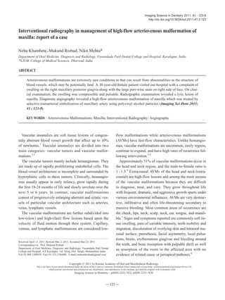
Interventional radiography in management of high-flow arteriovenous malformation of
maxilla: report of a case
Neha Khambete, Mukund Risbud, Nikit Mehta*
Department of Oral Medicine, Diagnosis and Radiology, Vasantdada Patil Dental College and Hospital, Kavalapur, India
*S.D.M. College of Medical Sciences, Dharwad, India
ABSTRACT
Arteriovenous malformations are extremely rare conditions in that can result from abnormalities in the structure of
blood vessels, which may be potentially fatal. A 30-year-old female patient visited our hospital with a complaint of
swelling on the right maxillary posterior gingiva along with the large port-wine stain on right side of face. On clini-
cal examination, the swelling was compressible and pulsatile. Radiographic examination revealed a lytic lesion of
maxilla. Diagnostic angiography revealed a high-flow arteriovenous malformation of maxilla which was treated by
selective transarterial embolization of maxillary artery using polyvinyl alcohol particles. (Imaging Sci Dent 2011;
41 : 123-8)
KEY WORDS : Arteriovenous Malformations; Maxilla; Interventional Radiography; Angiography
Received April 13, 2011; Revised May 2, 2011; Accepted May 23, 2011
Correspondence to : Prof. Mukund Risbud
Department of Oral Medicine, Diagnosis and Radiology, Vasantdada Patil Dental
College and Hospital, A/P Kavalapur, Tal: Miraj, Dist: Sangli, Maharashtra, India
Tel) 91-988-1260939, Fax) 91-233-2364400, E-mail) mukundrisbud@gmail.com
Imaging Science in Dentistry 2011; 41 : 123-8
http://dx.doi.org/10.5624/isd.2011.41.3.123
Copyright ⓒ 2011 by Korean Academy of Oral and Maxillofacial Radiology
This is an Open Access article distributed under the terms of the Creative Commons Attribution Non-Commercial License (http://creativecommons.org/licenses/by-nc/3.0)
which permits unrestricted non-commercial use, distribution, and reproduction in any medium, provided the original work is properly cited.
Imaging Science in Dentistry∙pISSN 2233-7822 eISSN 2233-7830
2. Various treatment modalities have been discussed for
the management of AVM.7,8
Many studies have reported
the use of selective transarterial embolization of AVM
with various substances.6,9,10
This may be followed by sur-
gical resection or sclerotherapy. Here we report a case in
which high flow AVM of maxilla was imaged using angio-
graphy. The angiogram revealed the feeder vessel which
was branch of internal maxillary artery. Selective trans-
arterial embolization of the feeder artery was performed
using polyvinyl alcohol particles.
Case Report
A 30-year-old female patient reported to the outpatient
department of Vasantdada Patil Dental College and Hos-
pital, Sangli with a chief complaint of gingival swelling on
the right maxillary posterior region. The patient noticed a
painless swelling 6 months ago, which was small in size
initially, started growing gradually and reached the present
size. The patient revealed a history of similar swelling in
the same region 2 years ago. Surgical removal of the
mass had been attempted, however it had been unsuccess-
ful due to heavy bleeding from the site. The bleeding had
been controlled by cauterization.
On general examination, the patient showed unilateral
port-wine stain on the right side of her face extending
from the superior border of the upper lip to the bridge of
the nose superoinferiorly and from the midline to the
preauricular region anteroposteriorly since her birth. On
clinical examination, the patient revealed about 3×3 cm
sized solitary extraoral swelling on the right maxilla ex-
tending from the midline to the midpupilary line antero-
posteriorly and from the superior border of the upper lip to
the base of the nose superoinferiorly. The swelling was
roughly ovoid in shape with diffuse borders. The overly-
ing skin was erythematous. There was a local increase in
temperature of the overlying skin. The swelling was soft
in consistency and non-tender on palpation. Intraorally,
about 3×6 cm sized solitary swelling, reddish pink in
color was found extending from the right central incisor
to the third molar on the buccal and palatal gingivae of
the maxilla. The swelling involved marginal, papillary,
and attached gingivae, and had smooth surface. It was
soft in consistency, non-tender on palpation, compressi-
ble, pulsatile and blanched on pressure. No bleeding on
palpation was noted. The right maxillary first and second
premolars showed grade II mobility. The right maxillary
lateral incisor was displaced laterally (Fig. 1). A provi-
sional diagnosis of vascular lesion was given and the pat-
ient was subjected to radiographic examination. Intraoral
periapical radiographs showed coarse bony trabeculae
─ 124 ─
Interventional radiography in management of high-flow arteriovenous malformation of maxilla: report of a case
Fig. 1. A. Extraoral photograph shows port- wine stain and ex-
traoral swelling. B. Intraoral photograph shows the extent of the
lesion from buccal aspect. C. Intraoral photograph shows the pal-
atal extent of lesion.
A
B
C
3. and enlarged marrow spaces on the right maxillary ante-
rior and posterior regions. Alveolar bone loss was reveal-
ed on the maxillary central and lateral incisor regions.
Widening of periodontal ligament space was seen with
lateral incisor, first and second premolar and the first
molar was supraerupted (Figs. 2A and B). Panoramic
radiograph showed ill-defined, irregular area of bone
destruction extending from the mesial surface of the left
maxillary central incisor upto the mesial surface of the
right maxillary canine. The maxillary central and lateral
incisors showed marked displacement (Fig. 2C).
The patient was referred to The Wanless Hospital, Miraj.
The ultrasound examination showed a high-flow arterio-
venous malformation. Admission laboratory tests showed
hemoglobin (Hb) of 7.3 g/dL, a platelet count of 63,000 per
mm3
, prothrombin time of 17 seconds, activated partial th-
romboplastin time of 35 sec, and international normalized
ratio of 1.3. The patient was then given blood and platelet
transfusions after which the blood investigations showed
hemoglobin of 13.3 g/dL, platelet count of 1,96,000, proth-
rombin time 15 seconds, activated partial thromboplastin
time of 28 seconds and international normalized ratio of
1.1. Diagnostic angiography was performed after gaining
access from the right femoral artery. Selective catheteriza-
tion of the right external carotid artery was performed un-
der fluoroscopic guidance. The diagnostic angiogram show-
ed a high-flow vascular malformation supplied princi-
pally by the alveolar branch of the internal maxillary arte-
ry. Surgical treatment of such lesion required extensive
resection of the maxilla and might result in the dysfunction
and disfigurement. Ligation of the external carotid artery
was not advisable, since many anastomoses promoted the
rapid appearance of a collateral circulation. Therefore,
embolization which consisted of occlusion of the vessels
which contributed to the lesion was considered. Further
selective catheterization of the maxillary artery was per-
formed. Embolization was performed using polyvinyl alco-
hol (PVA) particles (Fig. 3). The patient tolerated the pro-
─ 125 ─
Neha Khambete et al
Fig. 2. Intraoral periapical radio-
graphs show coarse bony trabecu-
lae and enlarged marrow spaces. A.
Right maxillary posterior portion.
B. Right maxillary anterior portion.
C: Panoramic radiograph shows an
ill-defined irregular area of bone de-
struction in right maxillary anterior
region.
A B
C
4. cedure well and had an unremarkable postoperative course.
One week postoperatively the patient showed complete
regression of the palatal lesion (Fig. 4). One year follow-
up revealed a significant reduction of clinical symptoms
and signs of the lesion without any further complications.
Discussion
AVMs are extremely rare conditions that can be fatal if
left untreated.6
They are caused by disturbances in the late
stages of angiogenesis, mainly abnormal differentiation
of vascular system.10
Vascular malformations can be sub-
divided further into high-flow and low-flow lesions.1,2
In
the present case, angiography was performed which de-
monstrated a high-flow AVM.
The diagnosis of AVMs can be made clinically in con-
junction with imaging studies. Most AVMs have a history
that includes the presence of a vascular blush in the overly-
ing skin in the childhood, which begins to expand more
rapidly as the patient enters puberty or undergoes other
hormonal changes. They may also reveal a history of trau-
ma to the involved area prior to the notification of the le-
sion. Bleeding, pain, and tissue destruction are often sub-
sequent signs in AVM.1,6
On physical examination, early
AVMs may have an overlying vascular blush in the skin
similar to an early port-wine stain. The mucosa is usually
thickened and vascular, and pulsation is usually present.
─ 126 ─
Interventional radiography in management of high-flow arteriovenous malformation of maxilla: report of a case
Fig. 3. A. Pre-embolization angio-
gram shows the abnormal blush in
central incisor region. B and C. An-
giogram during embolization shows
passage of polyvinyl alcohol parti-
cles. D. Post-embolization angio-
gram shows occlusion of feeder ves-
sel.
A B
C D
Fig. 4. Post-operative intraoral photograph shows regression of
palatal lesion after selective embolization.
5. The teeth may be loose or may exhibit pumping move-
ment when pressure is applied and released.1,3,4,6
AVMs
can invade the skin where ulcerations and bleeding are
common.1,11
Some “high-flow” lesions may result in con-
sumption coagulopathy, requiring transfusion therapy.12,13
Multiple imaging modalities should be used to evaluate
characteristics of AVMs such as size, flow velocity, flow
direction, relation to the surrounding structures and lesion
contents.2
There are no pathognomonic radiographic fea-
tures to distinguish AVMs on plain radiographs. They may
appear as bone erosion, sclerotic change, periosteal reac-
tion or a cyst like radiolucent lesion. A sunburst effect,
created by spicules radiating from the center, is often pre-
sent.2,14
The lesions most often have a multiloculated
appearance due to residual bone trapped around vessels.
Small radiolucent locules may resemble enlarged marrow
spaces surrounded by coarse, dense, and well defined trabe-
culae. They may have a honey comb or soap-bubble pattern
that is well demarcated from adjacent bone. The roots of
the teeth in proximity of the lesion may show displacement
or resorption. High-flow lesions tend to result in more
destructive skeletal changes, appearing as moth-eaten and
poorly defined areas of radiolucency.12
The radiographic
differential diagnosis of these lesions include amelobla-
stoma, ameloblastic fiboma, odontogenic myxoma, cent-
ral giant cell granuloma and metastatic malignant tumors.6
Color Doppler ultrasound examination can provide infor-
mation about the flow velocity. Contrast enhanced CT
can be useful in assessing the AVMs. The drawbacks of
CT are considerable exposure to ionizing radiation and
limited information about blood flow.2
Angiography and
MR imaging are the preferred imaging modalities.2,6,10,15
MRI depicts the anatomic relation of the vascular lesion
with adjacent organs and the flow velocity of lesions. It is
useful for evaluating the lesions postoperatively.2
Angio-
graphy is currently the gold standard for the determina-
tion of location and flow characteristics of vascular le-
sions. Angiography is useful to determine blood vessels
supplying blood to the lesion, and the relative venous out-
flow characteristics, and the presence or absence of arte-
riovenous shunts.3
According to Orbach, the angiographic
features of AVMs include dilatation and lengthening of
afferent arteries, early and preferential filling of shunts,
delayed filling of associated normal arteries, early opaci-
fication of draining veins and rapid flow to collateral ves-
sels.16
Intentional transarterial embolization was originally des-
cribed in 1969 by Lalli and coworkers.17
Treatment of vas-
cular malformation with selective embolization procedure
is currently highly recommended and often used. The pur-
pose of the selective embolization is to abruptly cut off the
blood supply of the lesion, reducing the risk of potentially
massive and lethal blood loss after its rupture, enabling a
more focused and selective surgical procedure with less
morbidity and with maximal preservation of important
structures.18
Various materials can be used for emboli-
zation. They can be classified as either temporary or per-
manent. Temporary materials include autologous clot or
muscle, Gelfoam19,20
and microfibrillar collagen. Perma-
nent agents include silicone spheres, lyophilized dura,
PVA, isobutyl cyanoacrylate and stainless steel or plati-
num coils. The most commonly used materials are Gel-
foam and PVA. PVA was used in present case as it is nona-
bsorbable and denser than Gelfoam.9
To conclude, vascular malformations represent some of
the most difficult lesions to diagnose and treat. Interven-
tional radiography plays an essential role in diagnosis and
management of such lesions. With the use of this techni-
que, extensive surgical procedures can be avoided so as to
avoid facial disfigurement and functional compromise.
Acknowledgements
The authors are thankful to Dr. Rahul Kumar for his
great help during preparation of manuscript. The authors
would like to thank Dr. Avinash Kshar, Dr. Nivedita Ba-
jantri, Dr, Alka Hazari, Dr. Praveen Kumar, Dr. Amit Ma-
thur and Dr. Arati Oka for their co-operation.
References
1. Buckmiller LM, Richter GT, Suen JY. Diagnosis and manage-
ment of hemangiomas and vascular malformations of the head
and neck. Oral Dis 2010; 16 : 405-18.
2. Hyodoh H, Hori M, Akiba H, Tamakawa M, Hyodoh K, Hare-
yama M. Peripheral vascular malformations: imaging, treat-
ment approaches, and therapeutic issues. Radiographics 2005;
25 (Suppl 1) : S159-71.
3. Nekooei S, Hosseini M, Nazemi S, Talaei-Khoei M. Embolisa-
tion of arteriovenous malformation of the maxilla. Dentomaxil-
lofac Radiol 2006; 35 : 451-5.
4. Lei ZM, Huang XX, Sun ZJ, Zhang WF, Zhao YF. Surgery of
lymphatic malformations in oral and cervicofacial regions in
children. Oral Surg Oral Med Oral Pathol Oral Radiol Endod
2007; 104 : 338-44.
5. Bai Y, Jia J, Huang XX, Alsharif MJ, Zhao JH, Zhao YF.
Sclerotherapy of microcystic lymphatic malformations in oral
and facial regions. J Oral Maxillofac Surg 2009; 67 : 251-6.
6. Mohammadi H, Said-A1-Naief NA, Heffez LB. Arterioven-
ous malformation of the mandible: report of a case with a note
on the differential diagnosis. Oral Surg Oral Med Oral Pathol
─ 127 ─
Neha Khambete et al
6. Oral Radiol Endod 1997; 84 : 286-9.
7. Larsen PE, Peterson LJ. A systematic approach to manage-
ment of high-flow vascular malformations of the mandible. J
Oral Maxillofac Surg 1993; 51 : 62-9.
8. Noreau N, Landry P, Morais D. Arteriovenous malformation of
the mandible: review of literature and case history. J Can Dent
Assoc 2001; 67 : 646-51.
9. Caiazzo A, Mehra P, Papageorge MB. The use of preopera-
tive percutaneous transcatheter vascular occlusive therapy in
the management of Sturge-Weber syndrome: report of a case.
J Oral Maxillofac Surg 1998; 56 : 775-8.
10. Lai QG, Li GJ, Yuan KF, Xu X, Wang YZ, Li YL, et al. Tran-
sarterial embolization of a high-flow maxillary arteriovenous
malformation fed by multiple arteries. Oral Surg Oral Med Oral
Pathol Oral Radiol Endod 2009; 108 : e17-21.
11. Donnelly LF, Adams DM, Bisset GS 3rd. Vascular malforma-
tions and hemangiomas: a practical approach in a multidisci-
plinary clinic. AJR Am J Roentgenol 2000; 174 : 597-608.
12. Kademani D, Costello BJ, Ditty D, Quinn P. An alternative
approach to maxillofacial arteriovenous malformations with
transosseous direct puncture embolization. Oral Surg Oral
Med Oral Pathol Oral Radiol Endod 2004; 97 : 701-6.
13. Speer AL, Panossian A, Arkader A. Stanley P, Anselmo DM.
Vascular surgery for arteriovenous malformations. eMedicine
from WebMD. Updated Aug 9, 2010. Available at http://eme
dicine. medscape.com/article/459927-diagnosis.
14. Johnson LM, Cook H, Friedlander A. Central arteriovenous
malformations of the maxillofacial skeleton: case report. J Oral
Maxillofac Surg 1991; 49 : 759-63.
15. Woods NK, Goaz PW. Differential diagnosis of oral lesions.
4th ed. St. Louis: CV Mosby Co; 1991. p. 285.
16. Orbach S. Congenital arteriovenous malformations of the
face. Report of a case. Oral Surg Oral Med Oral Pathol 1976;
42 : 2-13.
17. Lalli AF, Peterson N, Bookstein JJ. Roentgen-guided infarc-
tions of kidneys and lungs. A potential therapeutic technic.
Radiology 1969; 93 : 434-5.
18. Nagler RM, Braun J, Daitzchman M, Peled M, Laufer D.
Selective embolization for head and neck vasculopathies. Oral
Surg Oral Med Oral Pathol Oral Radiol Endod 2002; 93 :
127-31.
19. Anderson JH, Grisius RJ, McKean TW. Arteriovenous malfor-
mation of the mandible. Oral Surg Oral Med Oral Pathol
1981; 52 : 118-25.
20. McKenna SJ, Roddy SC Jr. Delayed management of a mandi-
bular vascular malformation. J Oral Maxillofac Surg 1989; 47 :
517-22.
─ 128 ─
Interventional radiography in management of high-flow arteriovenous malformation of maxilla: report of a case