SlideShare une entreprise Scribd logo
1  sur  61
Télécharger pour lire hors ligne
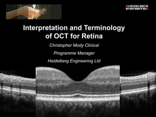
Interpretation and Terminology
of OCT for Retina
Christopher Mody Clinical
Programme Manager
Heidelberg Engineering Ltd
 Make valued judgements whilst scanning
 Utilise the fundus reference image
 Understand the significance OCT image
 Identify pathology & link to visual
symptoms
30° high resolution line scan
Fovea
Inf
Dark regions :
• Intra retinal fluid
• Sub retinal fluid
• Sub RPE fluid
• Retinal elevation
IR fundus reference image
Green areas :
• Intra retinal fluid
• Sub retinal fluid
• Sub RPE fluid
• Retinal elevation
MultiColor fundus reference image
Blue laser FAF reference image
Dark regions:
• Retinal atrophy
• RPE atrophy
Bright regions:
• Active disease
Normal OCT
 What does the SD-OCT image actually
represent?
N T
Fovea
Internal Limiting Membrane
Retinal Blood Vessels
RNFL
Ganglion Cell Layer
Inner Plexiform Layer
Inner Nuclear Layer
Outer Plexiform Layer
Outer Nuclear Layer
External Limiting Membrane
Henle fibre layer
Spectralis oct normal anatomy & systematic interpretation.
Choroid
Inner photoreceptor segments
Inner/outer photoreceptor junction
Rod/cone outer segments
RPE interdigitation
Bruch’s/RPE Complex
Pre-Euretina Normal OCT Classification
Formed Vitreous
Posterior Cortical Vitreous
Preretinal Space
Nerve Fiber Layer
Ganglion Cell Layer
Inner Plexiform Layer
Inner Nuclear Layer
Outer Plexiform Layer (dendritic)
Henle’s-ONL
junction (subtle)
Henle Fiber Layer (axonal OPL)
Outer Nuclear Layer
Sattler’s Layer (inner Choroid)
Haller’s Layer (Outer Choroid)
External Limiting Membrane
Ellipsoid Zone
Outer Segments
Interdigitation Zone
Choriocapillaris
RPE/ Bruch’s
Complex
Myoid Zone
Choroid
Sclera
Junction
Rods & Cones
OS
EZ
MZ
ELM
ONL
OPL
IZ
RPE
Inner segment
 Ellipsoid zone
 Mitochondria
 ATP production – chemical energy
 Myoid zone
 Golgi apparatus
 Protein synthesis
Ellipsoid zone
Myoid zone
Evaluating OCT images
1. Determine scan quality
2. Rate overall scan profile
3. Evaluate foveal profile
4. Identify foveal cut
5. Carry out structured assessment
 Observe alteration of layers
 Identify additional structures
 Pre retinal
 Epiretinal
 Intraretinal
 Subretinal
 Sub RPE
STEP 1
Determine overall scan quality
 Scan quality
Qualitative assessment
Step 1
 Scan quality
 Identify inner and outer retinal band
 Good signal to noise ratio
 Truncated
 Shadowing
Vitreous opacities
Vessel shadowing
STEP 2
Rate the overall scan profile
Qualitative assessment
Step 2. Rate the over-all retinal scan profile
 The normal over-all retinal profile has a slightly
concave curvature.
 Abnormal profiles would include exaggerated
concavity and convexity or retinal folds.
The over-all retinal profile
 RPE detachment
 Fibrotic/Serous
 Haemorrhagic
 Retinal detachment
 Rhegmatogenous
 Serous
 Retinal thickening
 CSMO/CMO/CNV
STEP 3
Evaluate the foveal profile
Qualitative assessment
 Step 3. Evaluate the foveal profile
 The normal foveal profile is a slight
depression in the surface of the retina.
Foveal profile
 Deformations in the foveal profile include the following:
1. Macular pucker
2. Macular pseudo-hole
3. Macular lamellar hole
4. Macular cyst
5. Macular hole, stage 1 (no depression, cyst present)
6. Macular hole, stage 2 (partial rupture of retina, increased thickness)
7. Macular hole, stage 3 (hole extends to RPE, increased thickness, some
fluid)
8. Macular hole, stage 4 (complete hole, oedema at margins, complete
PVD)
STEP 4
Identify foveal cut
Fovea
Do you have a foveal cut?
STEP 5
Carry out a structural assessment
Qualitative Assessment
Step 5. Carry out a structural assessment
a) Observe alteration of layers
b) Identify additional structures
• Pre-retinal
• Epiretinal
• Intra-retinal
• Sub-retinal
• Sub RPE
Qualitative assessment
Pre-retinal
Epiretintal
Intra-retinal
Sub-retinal
• Sub neurosensory retina
• Sub RPE
Qualitative assessment
 Pre retinal – vitreous cavity:
1. pre-retinal membrane
2. epi-retinal membrane
3. vitreo-retinal strands
4. vitreo-retinal traction
5. syneresis
6. pre-retinal neovascular membrane NVE
7. pre-papillary neovascular membrane NVD
Qualitative assessment
 Pre-retinal
 A normal pre-retinal profile is displayed as
a black or white space.
 Prepapilla/prefoveal lacunae (premacula
bursa) may be visible
Spectralis oct normal anatomy & systematic interpretation.
Qualitative assessment
 Intra-retinal changes:
1. Choroidal neovascularization
2. Diffuse intra-retinal oedema
3. Cystoid macular oedema
4. Hard exudates
5. Scar tissue
6. Atrophic degeneration
Qualitative assessment
 Sub-retinal/RPE:
1. choroidal neovascularization
2. RPE detachment
3. Drusen
4. sub-retinal fibrosis
5. scar tissue
6. RPE atrophy
What to look for…
1. Determine scan quality
2. Rate overall scan profile
3. Evaluate foveal profile
4. Identify foveal cut
5. Carry out structured assessment
 Observe alteration of layers
 Identify additional structures
 Pre retinal
 Epiretinal
 Intraretinal
 Subretinal
 Sub RPE
TERMINOLOGY
Irregularity
Fragmentation
Rupture
Interruption
Depression
Elevation
Thinning
Thickening
Alteration of Layers
Terminology
Irregularity
Fragmentation
Rupture
Interruption
Depression
Elevation
Thinning
Thickening
RPE
Terminology
Irregularity
Fragmentation
Rupture
Interruption
Depression
Elevation
Thinning
Thickening
RPE
Terminology
Irregularity
Fragmentation
Rupture
Interruption
Depression
Elevation
Thinning
Thickening
RPE
Terminology
Irregularity
Fragmentation
Rupture
Interruption
Depression
Elevation
Thinning
Thickening
RPE
Terminology
Irregularity
Fragmentation
Rupture
Interruption
Depression
Elevation
Thinning
Thickening
RPE
Terminology
Irregularity
Fragmentation
Rupture
Interruption
Depression
Elevation
Thinning
Thickening
RPE
Terminology
Additional
Structures
Macular Hole
Epiretinal Membrane (ERM)
Drusen
Blood Component
Fluid /Edema
Non Exudative Spaces
Neovascularisation
Fibrosis
Lipid Precipitates
Hyperreflective Spots & Dense
Areas
Tubulations of Outer Retina
Tumours
Identify RPE
Examine
RPE
Examine
posterior to
RPE
Examine
anterior to
RPE
Systematic Procedure
RPE
• Irregularity
• Fragmentation
• Rupture
• Interruption
• Depression
• Elevation
• Thinning
• Thickening
Posterior to RPE
• PED
• Bruch’s Membrane
• Hyperreflectivity
(atrophy of RPE
vs. fibrosis)
• Hyporeflectivity
(screen effect)
Anterior to RPE
• Vitreous
• Retinal Thickness
• Foveal Depression
• Subretinal Fluid
• ELM
• Elipsoid Zone
• Hyperreflective
Spots
• Dense Areas
• Outer Nuclear
Layer
• Intraretinal Cysts
• Inner Retinal
Layers
Identify RPESystematic Procedure
Examine
RPE
One single elevation of RPE (PED)
Undulating (wavy) PED
No interruption
No thickening or thinning of RPE
Identify EPR
Identify RPE
Examine
RPE
Examine
posterior to
RPE
Sub-RPE Reflectivity moderate
Shadow at RPE / sub - RPE
Hyperreflective Zone in inner layers
Intraretinal Cysts (in 2 layers)
Increased retinal thickness
Large Hyperreflective Precipitates
Hyperreflective Punctiform Precipitates
Dense Areas anterior to RPE
Interruption of ELM & Elipsoid Zone
Identify RPE
Examine
RPE
Retinal Angiomatous Profliferation
(RAP)
Posterio
rto RPE
Examine
anterior to
RPE
Intra & subretinal fluid
Final thoughts…
 Know your chorio retinal anatomy
 Familiarise yourself with normal variation in OCT
recordings
 Adopt a systematic approach to evaluating OCT images
 Familiarise yourself with the aetiology of macular
disease
 Don’t forget vision, signs/symptoms, history and fundus
appearance
Questions…
Acknowledgements:
Evangelos Tsiroukis MD
Institut Català de Retina
Barcelona
Professor Yit Yang
Wolverhampton Eye
Infirmary

Contenu connexe

Tendances

OCT – Introduction and Macular disorders
OCT – Introduction and Macular disordersOCT – Introduction and Macular disorders
OCT – Introduction and Macular disordersSajjan Sangai
 
22 optic-disc-evaluation-in-glaucoma
22 optic-disc-evaluation-in-glaucoma22 optic-disc-evaluation-in-glaucoma
22 optic-disc-evaluation-in-glaucomaUsamaIqbal18
 
Looking deep into retina : indirect ophthalmoscopy and fundus drawing
Looking deep into retina : indirect ophthalmoscopy and fundus drawingLooking deep into retina : indirect ophthalmoscopy and fundus drawing
Looking deep into retina : indirect ophthalmoscopy and fundus drawingPrachir Agarwal
 
Pentacam analysis
Pentacam analysisPentacam analysis
Pentacam analysisNikhil Rp
 
Newer modalities in diagnosis of glaucoma
Newer modalities in diagnosis of glaucomaNewer modalities in diagnosis of glaucoma
Newer modalities in diagnosis of glaucomaBipin Bista
 
Choroidal neovascular membranes (CNVM)
Choroidal neovascular membranes (CNVM)Choroidal neovascular membranes (CNVM)
Choroidal neovascular membranes (CNVM)Md Riyaj Ali
 
Visual Field in Glaucoma
Visual Field in GlaucomaVisual Field in Glaucoma
Visual Field in Glaucomadocsarsi
 
Polypoidal choroidal vasculopathy
Polypoidal choroidal vasculopathyPolypoidal choroidal vasculopathy
Polypoidal choroidal vasculopathySujay Chauhan
 
Iol power calculation in pediatric patients
Iol power calculation in pediatric patientsIol power calculation in pediatric patients
Iol power calculation in pediatric patientsAnisha Rathod
 
Diagnosis of pre perimetric glaucoma
Diagnosis of pre perimetric glaucomaDiagnosis of pre perimetric glaucoma
Diagnosis of pre perimetric glaucomaSadhwini Harish
 
Anterior segment OCT & UBM
Anterior segment OCT & UBMAnterior segment OCT & UBM
Anterior segment OCT & UBMDinesh Madduri
 
Pentacam and Corneal topography
Pentacam and Corneal topographyPentacam and Corneal topography
Pentacam and Corneal topographyPriyanka Raj
 
Corneal degenerations
Corneal degenerationsCorneal degenerations
Corneal degenerationsdrkvasantha
 

Tendances (20)

OCT – Introduction and Macular disorders
OCT – Introduction and Macular disordersOCT – Introduction and Macular disorders
OCT – Introduction and Macular disorders
 
OCT in Ophthalmology
OCT in OphthalmologyOCT in Ophthalmology
OCT in Ophthalmology
 
22 optic-disc-evaluation-in-glaucoma
22 optic-disc-evaluation-in-glaucoma22 optic-disc-evaluation-in-glaucoma
22 optic-disc-evaluation-in-glaucoma
 
OCT
OCTOCT
OCT
 
Looking deep into retina : indirect ophthalmoscopy and fundus drawing
Looking deep into retina : indirect ophthalmoscopy and fundus drawingLooking deep into retina : indirect ophthalmoscopy and fundus drawing
Looking deep into retina : indirect ophthalmoscopy and fundus drawing
 
Pentacam analysis
Pentacam analysisPentacam analysis
Pentacam analysis
 
Newer modalities in diagnosis of glaucoma
Newer modalities in diagnosis of glaucomaNewer modalities in diagnosis of glaucoma
Newer modalities in diagnosis of glaucoma
 
Choroidal neovascular membranes (CNVM)
Choroidal neovascular membranes (CNVM)Choroidal neovascular membranes (CNVM)
Choroidal neovascular membranes (CNVM)
 
Visual Field in Glaucoma
Visual Field in GlaucomaVisual Field in Glaucoma
Visual Field in Glaucoma
 
Polypoidal choroidal vasculopathy
Polypoidal choroidal vasculopathyPolypoidal choroidal vasculopathy
Polypoidal choroidal vasculopathy
 
Perimetry
PerimetryPerimetry
Perimetry
 
Iol power calculation in pediatric patients
Iol power calculation in pediatric patientsIol power calculation in pediatric patients
Iol power calculation in pediatric patients
 
Diagnosis of pre perimetric glaucoma
Diagnosis of pre perimetric glaucomaDiagnosis of pre perimetric glaucoma
Diagnosis of pre perimetric glaucoma
 
Anterior segment OCT & UBM
Anterior segment OCT & UBMAnterior segment OCT & UBM
Anterior segment OCT & UBM
 
Pentacam
Pentacam Pentacam
Pentacam
 
B scan
B scanB scan
B scan
 
Pentacam and Corneal topography
Pentacam and Corneal topographyPentacam and Corneal topography
Pentacam and Corneal topography
 
Aberrometry
AberrometryAberrometry
Aberrometry
 
Corneal degenerations
Corneal degenerationsCorneal degenerations
Corneal degenerations
 
IMAGING TECHNIQUES IN GLAUCOMA
IMAGING TECHNIQUES IN GLAUCOMAIMAGING TECHNIQUES IN GLAUCOMA
IMAGING TECHNIQUES IN GLAUCOMA
 

En vedette

İnterpretation of optic coherence tomography images
İnterpretation of optic coherence tomography imagesİnterpretation of optic coherence tomography images
İnterpretation of optic coherence tomography imagesSinan çalışkan
 
The basics of retinal oct ophso.net
The basics of retinal oct ophso.netThe basics of retinal oct ophso.net
The basics of retinal oct ophso.netkebaplik
 
A macular pathology and oct update for optometrists
A macular pathology and oct update for optometristsA macular pathology and oct update for optometrists
A macular pathology and oct update for optometristsTheEyeExpert
 
OCT - few basic facts
OCT - few basic factsOCT - few basic facts
OCT - few basic factsArindam Pande
 
Optical Coherence Tomography
Optical Coherence TomographyOptical Coherence Tomography
Optical Coherence TomographyManoj Aryal
 
Optical coherence tomography
Optical coherence tomographyOptical coherence tomography
Optical coherence tomographySinan çalışkan
 
Optical Coherence Tomography - principle and uses in ophthalmology
Optical Coherence Tomography - principle and uses in ophthalmologyOptical Coherence Tomography - principle and uses in ophthalmology
Optical Coherence Tomography - principle and uses in ophthalmologytapan_jakkal
 

En vedette (11)

İnterpretation of optic coherence tomography images
İnterpretation of optic coherence tomography imagesİnterpretation of optic coherence tomography images
İnterpretation of optic coherence tomography images
 
The basics of retinal oct ophso.net
The basics of retinal oct ophso.netThe basics of retinal oct ophso.net
The basics of retinal oct ophso.net
 
CURSO OCT SPECTRAL DOMAIN
CURSO OCT SPECTRAL DOMAINCURSO OCT SPECTRAL DOMAIN
CURSO OCT SPECTRAL DOMAIN
 
A macular pathology and oct update for optometrists
A macular pathology and oct update for optometristsA macular pathology and oct update for optometrists
A macular pathology and oct update for optometrists
 
oct-ujjval solanki
oct-ujjval solankioct-ujjval solanki
oct-ujjval solanki
 
TOMOGRAFIA DE COHERENCIA OPTICA
TOMOGRAFIA DE COHERENCIA OPTICATOMOGRAFIA DE COHERENCIA OPTICA
TOMOGRAFIA DE COHERENCIA OPTICA
 
OCT - few basic facts
OCT - few basic factsOCT - few basic facts
OCT - few basic facts
 
Anatomy of retina
Anatomy of retinaAnatomy of retina
Anatomy of retina
 
Optical Coherence Tomography
Optical Coherence TomographyOptical Coherence Tomography
Optical Coherence Tomography
 
Optical coherence tomography
Optical coherence tomographyOptical coherence tomography
Optical coherence tomography
 
Optical Coherence Tomography - principle and uses in ophthalmology
Optical Coherence Tomography - principle and uses in ophthalmologyOptical Coherence Tomography - principle and uses in ophthalmology
Optical Coherence Tomography - principle and uses in ophthalmology
 

Similaire à Spectralis oct normal anatomy & systematic interpretation.

MACULAR DISORDERS.pptx
MACULAR DISORDERS.pptxMACULAR DISORDERS.pptx
MACULAR DISORDERS.pptxvignapallavi
 
Optic nerve head evaluation in glaucoma
Optic nerve head evaluation in glaucomaOptic nerve head evaluation in glaucoma
Optic nerve head evaluation in glaucomaDr Laltanpuia Chhangte
 
18. Retinal detachment.pptx
18. Retinal detachment.pptx18. Retinal detachment.pptx
18. Retinal detachment.pptxannieamjad1
 
18. Retinal detachment.pptx
18. Retinal detachment.pptx18. Retinal detachment.pptx
18. Retinal detachment.pptxAnnie Amjad
 
Retina and layers
Retina and layersRetina and layers
Retina and layersLhacha
 
Retinal Detachment for medical students
Retinal Detachment for medical studentsRetinal Detachment for medical students
Retinal Detachment for medical studentsSohag University
 
Funduscopy evaluation in glaucomatous optic
Funduscopy evaluation in glaucomatous opticFunduscopy evaluation in glaucomatous optic
Funduscopy evaluation in glaucomatous opticrikhaerina
 
Hereditary macular dystrophies
Hereditary macular  dystrophiesHereditary macular  dystrophies
Hereditary macular dystrophiesShruti Laddha
 
Operating an OCT & evaluating normal scan (Optom.Saptarshi Mehta).....
Operating an OCT & evaluating normal scan (Optom.Saptarshi Mehta).....Operating an OCT & evaluating normal scan (Optom.Saptarshi Mehta).....
Operating an OCT & evaluating normal scan (Optom.Saptarshi Mehta).....Saptarshivision
 
Optical/ocular coherence tomography OCT All in one Presentation
Optical/ocular coherence tomography OCT All in one PresentationOptical/ocular coherence tomography OCT All in one Presentation
Optical/ocular coherence tomography OCT All in one PresentationKhushminaKhan
 
Optical coherence-tomography-120421562843883-4
Optical coherence-tomography-120421562843883-4Optical coherence-tomography-120421562843883-4
Optical coherence-tomography-120421562843883-4kebaplik
 
Optical coherence-tomography-120421562843883-4
Optical coherence-tomography-120421562843883-4Optical coherence-tomography-120421562843883-4
Optical coherence-tomography-120421562843883-4kebaplik
 
Anatomy and Physiology of retina
Anatomy and Physiology of retinaAnatomy and Physiology of retina
Anatomy and Physiology of retinaVivek Adwe
 
RETINAL DETACHMENT
RETINAL DETACHMENTRETINAL DETACHMENT
RETINAL DETACHMENTp K
 

Similaire à Spectralis oct normal anatomy & systematic interpretation. (20)

OCT.pptx
OCT.pptxOCT.pptx
OCT.pptx
 
OCT IN WET AMD
OCT IN WET AMDOCT IN WET AMD
OCT IN WET AMD
 
MACULAR DISORDERS.pptx
MACULAR DISORDERS.pptxMACULAR DISORDERS.pptx
MACULAR DISORDERS.pptx
 
Optic nerve head evaluation in glaucoma
Optic nerve head evaluation in glaucomaOptic nerve head evaluation in glaucoma
Optic nerve head evaluation in glaucoma
 
18. Retinal detachment.pptx
18. Retinal detachment.pptx18. Retinal detachment.pptx
18. Retinal detachment.pptx
 
18. Retinal detachment.pptx
18. Retinal detachment.pptx18. Retinal detachment.pptx
18. Retinal detachment.pptx
 
Retina and layers
Retina and layersRetina and layers
Retina and layers
 
Retinal Detachment for medical students
Retinal Detachment for medical studentsRetinal Detachment for medical students
Retinal Detachment for medical students
 
Funduscopy evaluation in glaucomatous optic
Funduscopy evaluation in glaucomatous opticFunduscopy evaluation in glaucomatous optic
Funduscopy evaluation in glaucomatous optic
 
Hereditary macular dystrophies
Hereditary macular  dystrophiesHereditary macular  dystrophies
Hereditary macular dystrophies
 
Rrd
RrdRrd
Rrd
 
MACULAR DYSTROPHY
MACULAR DYSTROPHYMACULAR DYSTROPHY
MACULAR DYSTROPHY
 
Operating an OCT & evaluating normal scan (Optom.Saptarshi Mehta).....
Operating an OCT & evaluating normal scan (Optom.Saptarshi Mehta).....Operating an OCT & evaluating normal scan (Optom.Saptarshi Mehta).....
Operating an OCT & evaluating normal scan (Optom.Saptarshi Mehta).....
 
Nw2015 rcopt oct_retina22
Nw2015 rcopt oct_retina22Nw2015 rcopt oct_retina22
Nw2015 rcopt oct_retina22
 
Optical/ocular coherence tomography OCT All in one Presentation
Optical/ocular coherence tomography OCT All in one PresentationOptical/ocular coherence tomography OCT All in one Presentation
Optical/ocular coherence tomography OCT All in one Presentation
 
RHEGMATOGENOUS Retinal detachment
 RHEGMATOGENOUS Retinal detachment RHEGMATOGENOUS Retinal detachment
RHEGMATOGENOUS Retinal detachment
 
Optical coherence-tomography-120421562843883-4
Optical coherence-tomography-120421562843883-4Optical coherence-tomography-120421562843883-4
Optical coherence-tomography-120421562843883-4
 
Optical coherence-tomography-120421562843883-4
Optical coherence-tomography-120421562843883-4Optical coherence-tomography-120421562843883-4
Optical coherence-tomography-120421562843883-4
 
Anatomy and Physiology of retina
Anatomy and Physiology of retinaAnatomy and Physiology of retina
Anatomy and Physiology of retina
 
RETINAL DETACHMENT
RETINAL DETACHMENTRETINAL DETACHMENT
RETINAL DETACHMENT
 

Dernier

HERPES SIMPLEX VIRUS 12032019 TUESDAY pptx
HERPES SIMPLEX VIRUS 12032019 TUESDAY  pptxHERPES SIMPLEX VIRUS 12032019 TUESDAY  pptx
HERPES SIMPLEX VIRUS 12032019 TUESDAY pptxPulkitMittal54
 
EMBRYOLOGY AND FOETAL DEVELOPMENT-mayu.pdf
EMBRYOLOGY AND FOETAL DEVELOPMENT-mayu.pdfEMBRYOLOGY AND FOETAL DEVELOPMENT-mayu.pdf
EMBRYOLOGY AND FOETAL DEVELOPMENT-mayu.pdfMayuriGamit2
 
CELL BLOCK PREPARATION AND ITS IMPORTANCE
CELL BLOCK PREPARATION AND ITS IMPORTANCECELL BLOCK PREPARATION AND ITS IMPORTANCE
CELL BLOCK PREPARATION AND ITS IMPORTANCEShubhadip Ghosh
 
The Art and the Science of UK General Practice A historical review and timeli...
The Art and the Science of UK General Practice A historical review and timeli...The Art and the Science of UK General Practice A historical review and timeli...
The Art and the Science of UK General Practice A historical review and timeli...JakeMatthews12
 
Brief introduction to information ecosystem x public health.pptx
Brief introduction to information ecosystem x public health.pptxBrief introduction to information ecosystem x public health.pptx
Brief introduction to information ecosystem x public health.pptxTina Purnat
 
SGK PHÙ PHỔI CẤP ĐHYHN .pdf
SGK PHÙ PHỔI CẤP ĐHYHN                   .pdfSGK PHÙ PHỔI CẤP ĐHYHN                   .pdf
SGK PHÙ PHỔI CẤP ĐHYHN .pdfHongBiThi1
 
Spinal cord Gross anatomy with Clinical Anatomy.pptx
Spinal cord Gross anatomy with Clinical Anatomy.pptxSpinal cord Gross anatomy with Clinical Anatomy.pptx
Spinal cord Gross anatomy with Clinical Anatomy.pptxsiddharthroy26587
 
airway management recorded for S2.pptx
airway management  recorded  for S2.pptxairway management  recorded  for S2.pptx
airway management recorded for S2.pptxnakera38
 
Problems associated with the production of recombinant protein.pdf
Problems associated with the production of recombinant protein.pdfProblems associated with the production of recombinant protein.pdf
Problems associated with the production of recombinant protein.pdfNetHelix
 
Laryngeal papillomatosis .pdf ENT BY QUICKMEDTALK
Laryngeal papillomatosis .pdf ENT BY QUICKMEDTALKLaryngeal papillomatosis .pdf ENT BY QUICKMEDTALK
Laryngeal papillomatosis .pdf ENT BY QUICKMEDTALKQuick MedTalk
 
Methicillin-resistant Staphylococcus Aureus (MRSA)
Methicillin-resistant Staphylococcus Aureus (MRSA)Methicillin-resistant Staphylococcus Aureus (MRSA)
Methicillin-resistant Staphylococcus Aureus (MRSA)Ahmad Thanin
 
Neutraceuticals, Herb-drug & Herb-food inetractions
Neutraceuticals, Herb-drug & Herb-food inetractionsNeutraceuticals, Herb-drug & Herb-food inetractions
Neutraceuticals, Herb-drug & Herb-food inetractionsPradnya Wadekar
 
Cholesterol Biosynthesis and catabolism for MBBS, Lab. MEd. BDS.pptx
Cholesterol Biosynthesis  and catabolism for MBBS, Lab. MEd. BDS.pptxCholesterol Biosynthesis  and catabolism for MBBS, Lab. MEd. BDS.pptx
Cholesterol Biosynthesis and catabolism for MBBS, Lab. MEd. BDS.pptxRajendra Dev Bhatt
 
TARGET DELINEATION IN RECTUM CANCER BY DR KANHU
TARGET DELINEATION IN RECTUM  CANCER BY DR KANHUTARGET DELINEATION IN RECTUM  CANCER BY DR KANHU
TARGET DELINEATION IN RECTUM CANCER BY DR KANHUKanhu Charan
 
(IDE)and(IVD),QMS,21 CFR part820 , 801)
(IDE)and(IVD),QMS,21 CFR part820  , 801)(IDE)and(IVD),QMS,21 CFR part820  , 801)
(IDE)and(IVD),QMS,21 CFR part820 , 801)chahattyagi200
 
SGK VIÊM KHỚP DẠNG THẤP YHN .pdf
SGK VIÊM KHỚP DẠNG THẤP YHN              .pdfSGK VIÊM KHỚP DẠNG THẤP YHN              .pdf
SGK VIÊM KHỚP DẠNG THẤP YHN .pdfHongBiThi1
 
INTRODUCTION TO THE FORENSIC SCIENCE.ppt
INTRODUCTION TO THE FORENSIC SCIENCE.pptINTRODUCTION TO THE FORENSIC SCIENCE.ppt
INTRODUCTION TO THE FORENSIC SCIENCE.pptKavitha Krishnan
 
NECROSIS FOR MBBS FIRST YEAR STUDENTS MADE EASY.pptx
NECROSIS FOR MBBS FIRST YEAR STUDENTS MADE EASY.pptxNECROSIS FOR MBBS FIRST YEAR STUDENTS MADE EASY.pptx
NECROSIS FOR MBBS FIRST YEAR STUDENTS MADE EASY.pptxSizan Thapa
 
Routine Medicine Laboratory Testing _ CÁC XÉT NGHIỆM THƯỜNG QUY ÁP DỤNG TRON...
Routine Medicine Laboratory Testing  _ CÁC XÉT NGHIỆM THƯỜNG QUY ÁP DỤNG TRON...Routine Medicine Laboratory Testing  _ CÁC XÉT NGHIỆM THƯỜNG QUY ÁP DỤNG TRON...
Routine Medicine Laboratory Testing _ CÁC XÉT NGHIỆM THƯỜNG QUY ÁP DỤNG TRON...DUY NGUYEN PHUC
 

Dernier (20)

HERPES SIMPLEX VIRUS 12032019 TUESDAY pptx
HERPES SIMPLEX VIRUS 12032019 TUESDAY  pptxHERPES SIMPLEX VIRUS 12032019 TUESDAY  pptx
HERPES SIMPLEX VIRUS 12032019 TUESDAY pptx
 
EMBRYOLOGY AND FOETAL DEVELOPMENT-mayu.pdf
EMBRYOLOGY AND FOETAL DEVELOPMENT-mayu.pdfEMBRYOLOGY AND FOETAL DEVELOPMENT-mayu.pdf
EMBRYOLOGY AND FOETAL DEVELOPMENT-mayu.pdf
 
Catalysts for Change in SCLC: Optimizing the Use of Standard Therapies and Ac...
Catalysts for Change in SCLC: Optimizing the Use of Standard Therapies and Ac...Catalysts for Change in SCLC: Optimizing the Use of Standard Therapies and Ac...
Catalysts for Change in SCLC: Optimizing the Use of Standard Therapies and Ac...
 
CELL BLOCK PREPARATION AND ITS IMPORTANCE
CELL BLOCK PREPARATION AND ITS IMPORTANCECELL BLOCK PREPARATION AND ITS IMPORTANCE
CELL BLOCK PREPARATION AND ITS IMPORTANCE
 
The Art and the Science of UK General Practice A historical review and timeli...
The Art and the Science of UK General Practice A historical review and timeli...The Art and the Science of UK General Practice A historical review and timeli...
The Art and the Science of UK General Practice A historical review and timeli...
 
Brief introduction to information ecosystem x public health.pptx
Brief introduction to information ecosystem x public health.pptxBrief introduction to information ecosystem x public health.pptx
Brief introduction to information ecosystem x public health.pptx
 
SGK PHÙ PHỔI CẤP ĐHYHN .pdf
SGK PHÙ PHỔI CẤP ĐHYHN                   .pdfSGK PHÙ PHỔI CẤP ĐHYHN                   .pdf
SGK PHÙ PHỔI CẤP ĐHYHN .pdf
 
Spinal cord Gross anatomy with Clinical Anatomy.pptx
Spinal cord Gross anatomy with Clinical Anatomy.pptxSpinal cord Gross anatomy with Clinical Anatomy.pptx
Spinal cord Gross anatomy with Clinical Anatomy.pptx
 
airway management recorded for S2.pptx
airway management  recorded  for S2.pptxairway management  recorded  for S2.pptx
airway management recorded for S2.pptx
 
Problems associated with the production of recombinant protein.pdf
Problems associated with the production of recombinant protein.pdfProblems associated with the production of recombinant protein.pdf
Problems associated with the production of recombinant protein.pdf
 
Laryngeal papillomatosis .pdf ENT BY QUICKMEDTALK
Laryngeal papillomatosis .pdf ENT BY QUICKMEDTALKLaryngeal papillomatosis .pdf ENT BY QUICKMEDTALK
Laryngeal papillomatosis .pdf ENT BY QUICKMEDTALK
 
Methicillin-resistant Staphylococcus Aureus (MRSA)
Methicillin-resistant Staphylococcus Aureus (MRSA)Methicillin-resistant Staphylococcus Aureus (MRSA)
Methicillin-resistant Staphylococcus Aureus (MRSA)
 
Neutraceuticals, Herb-drug & Herb-food inetractions
Neutraceuticals, Herb-drug & Herb-food inetractionsNeutraceuticals, Herb-drug & Herb-food inetractions
Neutraceuticals, Herb-drug & Herb-food inetractions
 
Cholesterol Biosynthesis and catabolism for MBBS, Lab. MEd. BDS.pptx
Cholesterol Biosynthesis  and catabolism for MBBS, Lab. MEd. BDS.pptxCholesterol Biosynthesis  and catabolism for MBBS, Lab. MEd. BDS.pptx
Cholesterol Biosynthesis and catabolism for MBBS, Lab. MEd. BDS.pptx
 
TARGET DELINEATION IN RECTUM CANCER BY DR KANHU
TARGET DELINEATION IN RECTUM  CANCER BY DR KANHUTARGET DELINEATION IN RECTUM  CANCER BY DR KANHU
TARGET DELINEATION IN RECTUM CANCER BY DR KANHU
 
(IDE)and(IVD),QMS,21 CFR part820 , 801)
(IDE)and(IVD),QMS,21 CFR part820  , 801)(IDE)and(IVD),QMS,21 CFR part820  , 801)
(IDE)and(IVD),QMS,21 CFR part820 , 801)
 
SGK VIÊM KHỚP DẠNG THẤP YHN .pdf
SGK VIÊM KHỚP DẠNG THẤP YHN              .pdfSGK VIÊM KHỚP DẠNG THẤP YHN              .pdf
SGK VIÊM KHỚP DẠNG THẤP YHN .pdf
 
INTRODUCTION TO THE FORENSIC SCIENCE.ppt
INTRODUCTION TO THE FORENSIC SCIENCE.pptINTRODUCTION TO THE FORENSIC SCIENCE.ppt
INTRODUCTION TO THE FORENSIC SCIENCE.ppt
 
NECROSIS FOR MBBS FIRST YEAR STUDENTS MADE EASY.pptx
NECROSIS FOR MBBS FIRST YEAR STUDENTS MADE EASY.pptxNECROSIS FOR MBBS FIRST YEAR STUDENTS MADE EASY.pptx
NECROSIS FOR MBBS FIRST YEAR STUDENTS MADE EASY.pptx
 
Routine Medicine Laboratory Testing _ CÁC XÉT NGHIỆM THƯỜNG QUY ÁP DỤNG TRON...
Routine Medicine Laboratory Testing  _ CÁC XÉT NGHIỆM THƯỜNG QUY ÁP DỤNG TRON...Routine Medicine Laboratory Testing  _ CÁC XÉT NGHIỆM THƯỜNG QUY ÁP DỤNG TRON...
Routine Medicine Laboratory Testing _ CÁC XÉT NGHIỆM THƯỜNG QUY ÁP DỤNG TRON...
 

Spectralis oct normal anatomy & systematic interpretation.