1. MANAGEMENT OF DUODENAL INJURIES
IN BRIEF
With most of the duodenum protected deep within the ana-
tomic confines of the retroperitoneum, injuries to this organ are
uncommon but not rare. These injuries represent approximately
4% of all abdominal injuries. However, because of difficulties
with initial assessment, establishment of the diagnosis, and, oc-
casionally, management, the morbidity and mortality rates associ-
ated with injuries of the duodenum approach 65% and 20%,
respectively.
The first successful repair of a duodenal injury after blunt trauma
was reported by Herczel in 1896. It was 1901 before Moynihan re-
paired a penetrating duodenal injury; he performed a gastrojejunos-
tomy in a patient who lived for 104 days. With subsequent improve-
ments in anesthesia, antibiotic therapy, and surgical techniques, sig-
nificant decreases in operative morbidity and mortality rates have
been reported.
The experiences of American military surgeons from the American
Civil War through the Korean and Vietnam conflicts have contributed
to our understanding of duodenal injuries. World Wars I and II, in
particular, provided surgeons the opportunity to improve the care of
many battlefield casualties.
The incidence of duodenal injuries is related to the geographic set-
ting of the traumatic incident (i.e., urban or rural). Penetrating trauma
accounts for 78% of all duodenal injuries, whereas blunt trauma ac-
counts for 22%. Retroperitoneal duodenal ruptures caused by blunt
trauma occur only rarely.
The morbidity and mortality associated with duodenal injuries are
increased with associated injuries of the liver, pancreas, small bowel,
and colon. The most commonly injured vascular structures are the
inferior vena cava and the abdominal aorta. These associated inju-
ries result in particularly high mortality from the resulting exsangui-
nating hemorrhage.
The second portion of the duodenum is injured more often than
any other portion and poses greater technical difficulties for surgical
management. Injuries affecting multiple portions of the duodenum
1026 Curr Probl Surg, November 1993
2. occur with a frequency of 14%, resulting in greater technical chal-
lenges to the surgeon.
Successful diagnosis of a duodenal injury requires a high index of
suspicion. The mechanism of injury represents important informa-
tion that can be obtained from close communication with personnel
from emergency medical services (EMS). Information such as the
presence of a bent steering wheel and related data on the velocity,
direction, and impact of the motor vehicular accident often yield im-
portant clues that alert the surgeon to the possibility of duodenal in-
jury.
Duodenal injury is often overlooked because of seemingly more
dramatic and life-threatening injuries, particularly those causing life-
threatening hemorrhage. The history and physical examination ini-
tiates the diagnostic process. The use of laboratory studies, in gen-
eral, are not helpful. Radiographic studies such as plain abdominal
films are helpful but only if positive. Important abnormal findings
such as unexplained fluid collections surrounding the duodenum
and retroperitoneal free air, particularly that outlining the upper pole
of the right kidney, strongly suggest a duodenal injury.
No studies, either retrospective or prospective, have compared the
use of upper gastrointestinal contrast study with the computed to-
mographic scan. The upper gastrointestinal contrast study is per-
formed initially by the ingestion or administration of a water-soluble
medium and should confirm or exclude the presence of a leak. If
negative, this initial study should be followed by a thin solution of
barium to provide better definition of the duodenal anatomy. Posi-
tive computed tomography scan findings include extravasation from
the lumen, perimural and intramural duodenal hematomas, and free
retroperitoneal air. The computed tomography scan also provides in-
formation that helps to diagnose other associated injuries. Magnetic
resonance imaging is not yet a useful diagnostic tool in this setting.
Diagnostic peritoneal lavage is not useful in detecting retroperito-
neal injuries. It is positive in approximately 50% of all cases resulting
from the multiple associated intraabdominal injuries. The definitive
diagnostic tool remains a meticulous exploratory laparotomy and ret-+
roperitoneal exploration.
Surgical management of duodenal injuries begins with the basic
principles of initial assessment and resuscitation recommended by
the Advanced Trauma Life Support course of the American College
of Surgeons, including early control of the airway and adequate vol-
ume resuscitation. When a decision has been made to operate, ap-
propriate broad-spectrum antibiotics are administered. The abdo-
men is entered through a generous midline incision extending from
xiphoid to pubis. A meticulous exploratory laparotomy and retroperi-
toneal exploration should avoid the severe consequences of over-
looked injuries.
Curr Probl SW,, November 1993 1027
3. When a duodenal injury is detected intraoperatively, the surgeon
must be aware of factors that increase the morbidity and mortality of
the injury, including the presence of associated biliary and pancre-
atic injuries. We recommend intraoperative grading of all duodenal
injuries by the Penetrating Abdominal Trauma Index. Injuries of
lesser grade should be treated by simpler surgical techniques, and
injuries of greater severity should be treated by more complex tech-
niques. The American Association for the Surgery of Trauma has also
devised a scoring system to grade these injuries and establish a uni-
form reporting standard.
Surgeons who treat patients with traumatic injuries to the duode-
num must be able to use an armamentarium of surgical procedures
to repair these injuries. Approximately 75% to 80% of all duodenal
injuries can be repaired safely by simple surgical techniques such as
debridement to viable tissue, primary repair by double-layer duode-
norrhaphy, and drainage using a closed system. The role of tube duo-
denostomy as an adjunct to management and as a means of decom-
pression and protection of the suture line is controversial. Complex
surgical procedures such as the jejunal serosal patch, duodenal re-
section with Roux-en-Y duodenojejunostomy, duodenal resection
with end-to-end duodenoduodenostomy, pedicled grafts, duodenal
diverticularization, pyloric exclusion, and pancreatoduodenectomy
are each useful in selected patients.
Duodenal injuries are uncommon in the trauma patient, and thus
many general surgeons do not develop the expertise necessary to
manage patients with this unique and challenging clinical problem.
The potential for morbidity and mortality is ample and is related to
the accuracy and timing of diagnosis as well as to the skill of the sur-
geon.
1028 Cur-r Probl Surg, November 1993
4. Juan A. Asensio, MD, graduated with a BS degree from the
University of Illinois. He received his MD degree from Rush
Medical College and completed his surgical residency at
Northwestern University in Chicago and the Medical CoZ-
lege of Ohio at Toledo and then completed fellowships in
trauma surgery and surgical critical care at the University
of Tezas Health Sciences Center, Dallas/Southwestern
Medical School, and Parkland Memorial Hospital. He is CUF-
rently Associate Professor of Surgery and Chief of the Di-
vision of Trauma Surgery and Surgical Critical Care at Hah-
nemann University, where he also serves in the capacity as
medical director of the Air Evacuation Service/MEDEVAC
and medical director of the Trauma Center. Dr. Asensio has
been instrumental in organizing trauma centers in both
Central and South America. ilr. Asensio’s interests include
exsanguination; penetrating neck injuries, cardiovascular
system injuries, pancreas, and duodenum; and surgical
critical care.
David V. Feliciano, MD, received his BS and MD degrees
from Georgetown University, He completed internship and
residency training in general surgery at the Mayo Clinic af-
ter active duty in the U.S. Navy. He furthered his training
in trauma surgery at Detroit General Hospital during resi-
dency and a fellowship in vascular surgery at the Baylor,
College of Medicine. Dr. Feliciano is currently Chief of Sur-
gery at Grady Memorial Hospital, attending surgeon at
Crawford Long Hospital, Professor of Surgery at Emory Uni-
versity, and Clinical Professor of Surgery at the Uniformed
Services University of Health Sciences. He is immediate
Past President of the Southwestern Surgical Corigress, the
President of the Western Trauma Association and the
Priestley Society (Mayo surgeons), and a member of the Ex-
ecutive Committee of the Committee on Trauma of the
American College of Surgeons. His interests include ab-
dominal and vascular trauma, endocrine and general SUF-
gery, and surgical critical care.
Curr Probl Surg, November 1993 1029
5. L. Delano Britt, MD, received his BA degree from the Uni-
versity of Virginia, his MD degree from the Harvard Medi-
cal School, and his MPH degree from the Harvard School
of Public Health. Dr. Britt is currently Chief of the Division
of Trauma and Critical Care at the Eastern Virginia Medi-
cal School and medical director of the Shock Trauma Cen-
ter at Sentara Norjiolk General Hospital.
Morris D. Kerstein, MD, completed his surgical training on
the First (Tufts) Surgical Service of the Boston City Hospi-
tal in 1971 afrer serving a research fellowship at Sahlgren-
ska Hospital in Goteborg, Sweden, from 1968 to 1969. After
serving on the faculty of the Yale University School of Medi-
cine, the University of Chicago, and the Tulane University
School of Medicine, Dr. Kerstein was appointed as the
Edgar J. Deissler Professor and Chairman of the Depart-
ment of Surgery at Hahnemann University School of Medi-
cine. His interest in trauma began with an active-duty tour
from 2965 to 1967 with the U.S. Navy during the Vietnam
conflict. His continued interest in trauma and the Navy con-
tributed to his appointments as Rear Admiral, U.S. Navy, at
the Bureau of Medicine and Surgery and Assistant to the
Deputy Surgeon General of the Navy for Reserve Matters.
Dr. Kerstein’s research interests have focused on vascular
surgery problems, prostaglandin metabolism in the vascu-
lature, and trauma.
Curr Probl Surg, November 1993
6. MANAGEMENT OF DUODENAL INJURIES
INTRODUCTION
The duodenum is the epitome of an organ poorly designed to with-
stand the ravages of trauma. Located in the inaccessible and dark
reaches of the retroperitoneum, injuries to the duodenum usually are
not suspected or are diagnosed rather late while more apparent in-
juries to other organs are addressed. The small, thin-walled duode-
num possesses a marginal blood supply shared with the pancreas.
Therefore this organ is not amenable to sound technical closure, and
parts of it are very difficult to resect. Lying against the vertebral col-
umn, the duodenum is highly susceptible to severe crushing inju-
ries. It is also fixed at two separate points-the portal triad and the
ligament of Treitz- thereby subjecting it to decelerating injuries. Fur-
thermore, it is subject to “blow-out” injury by being, at times, closed
at its portals of entrance (the pylorus) and exit (the duodenojejunal
junction).
The duodenum is surrounded by many vital structures, including
the aorta, inferior vena cava, superior mesenteric vessels, portal ves-
sels, right renovascular pedicle, and the biliary tree. When injured,
these structures produce large amounts of blood and bile that may
obscure injuries of the duodenal wall. Finally, its matrimony of con-
venience to the pancreas (by virtue of its shared blood supply) is eas-
ily disrupted at the time of injury by the action of the pancreatic en-
zpes released frequently during combined pancreatic and duode-
nal injuries.
Given these considerations, it is no wonder that duodenal injuries
remain one of the most complex challenges for modern-day trauma
surgeons. The objectives of this monograph are (1) to familiarize the
reader with duodenal anatomy as it relates to trauma surgery, (2) to
provide an in-depth analysis of the incidence and mortality rate of
duodenal injuries, and (3) to provide a concise approach to diagno-
sis, surgical management, and treatment of complications of duode-
nal trauma.
Curr Probl Surg November 1993 1031
7. HISTORIC PERSPECTIVE
There is a scarcity of well-documented historic accounts regard-
ing the management of duodenal injuries. Several characteristics of
the duodenum may account for this fact: its retroperitoneal location,
the difficulty in mobilizing it surgically, or the fact that it just did not
emerge within the realm of surgical diagnosis or treatment during
the last century. However, the primary reason for this lack of docu-
mentation appears to be the infrequent use of exploratory laparotomy
for the management of traumatic abdominal injuries. Although this
technique had been readily available and used for nontraumatic ab-
dominal problems, most surgeons did not view it with much respect.
Exploratory laparotomy was used by Baudens in 1836: however, it
was not until the Civil War that the procedure was considered valu-
able in the management of abdominal trauma.’ It was not until World
War I that American surgeons become more forceful and began to
explore soldiers who sustained penetrating abdominal injuries.
Perhaps one of the earliest recorded cases of successful outcomes
from penetrating duodenal injuries is credited to Larrey, the French
surgeon who recorded the following case in 1811:
Etienne Belloc, age 17 fusileer of the guards was wounded by a sword in
the abdomen about two inches above the umbilicus, and on the right side
of the linea alba. He was brought to the hospital on April 1, 1811, and the
attending surgeon applied a simple dressing and bandage. Next day, I ex-
amined the wound, which permitted the omentum to escape through it. The
right rectus muscle and its tendinous sheath were cut quite through, and
the instrument appeared to have passed in a transverse direction deeply,
from before, backwards, between the great curve of the stomach, and the
arch of the colon.
The paleness of death was on his countenance and he was tormented
with intolerable anguish, nausea, and efforts to vomit; with hiccough, ardent
thirst and acute pain at the bottom of the wound, and great anxiety; his pulse
was small and feeble, his extremities cold, and voice no longer audible: We
had reason to believe he could survive but a few moments.
Still, I reduced the omentum, with my fingers ascertained that the sword
had glanced between the stomach and colon, but I could not decide on the
place where it had stopped; the wound was dressed externally, with linen,
etc., dipped in warm wine. The abdomen was embrocated with warm cam-
phorated oil and covered with hot flannel. I prescribed cooling mucilaginous
drinks, emollient enemata, low diet, a particular position of the body and
perfect rest. He felt but a little relief from this treatment; the prostration con-
tinued as before, the pulse was small and tense, and anxiety and nausea
attended: He was never at rest. On the night of the second day, vomiting
came on with considerable efforts, cold sweat and alarming syncope, he first
discharged the contents of his stomach by vomiting and then bilious matter
with clots of black blood. On the fourth day, to this bilious evacuations suc-
ceeded the vomiting of thick black blood in such quantity that the chamber-
1032 Curr Probl Surg, November 1993
8. utensil was filled with it in a few minutes. On the iifth day, an alvine evacu-
ation, equally copious took place, proceeded by violent colick and acute
pains in the wound; the abdomen always remained flaccid and without any
signs of effusion in its cavity. An alarming syncope succeeded this evacua-
tion on the night of the 6th, and his companions believed him dead. When I
visited the hospital very early the next morning, I found his face covered
with a sheet and he opened his eyelids with difficulty; the pulse was imper-
ceptible, and his body cold. I immediately gave him warm wine, had his body
rubbed with oil of chamomile, and wrapped in hot flannels. The colick never
returned and from this time he gradually recovered. I prescribed a muci-
laginous drink with syrup of althea and orange-flower water, to which was
added a small quantity of nitrated alcohol; emollient enemata were given,
and the oily embrocations of the abdomen continued. . .3
The rest of the account continues with a detailed description of
subsequent complications, convalescence, and the basis for diagno-
sis of duodenal injury.
During the American Civil War, five soldiers were reported to have
incurred duodenal wounds resulting from “shot injuries,” with a
100% mortality rate and no surgical intervention. A detailed autopsy
report was described as follows:
Case 2112. Pvt. James M.; Company I?. Wound of the abdomen at Winches-
ter on September X9,1864. The missile conoidal ball entered at the‘ right side
of the epigastrium, at the edge of the ribs, and emerged through the right
buttock. He was admitted on the same day to the hospital of the Sixth Corps.
He was an emaciated subject. Water dressings were applied to the wound
and ferruginous preparations and opiates were administered with milk
punch. A farinaceous and milk diet was allowed. Faeces escaped freely from
the wound exit and also from the wound of entrance for a few days. After
this, frequent and continued alvine ejections took place through the natural
channels. Death resulted on October 12, 1864. At the autopsy it was found
that the ball entering the right side of the epigastric region had carried away
about half of the caliber of the duodenum, near the orifice of the cystic duct.
It had passed obliquely downward and backward through the caecum above
the ileo-cecal valve.4
The first successful surgical repair of a duodenal rupture was re-
ported in 1896 by Herczel,’ who repaired the ruptured duodenum of
a 36-year-old woman after blunt trauma. In 1901, Moynihan” closed
a duodenal wound and performed a gastrojejunostomy with a pro-
longed survival of 104 days and subsequent death. In a paper read
before the Western Surgical and Gynaecological Association on De-
cember 28,1903, and published in 1904, Summers described what is
perhaps the earliest and best-documented report of treatment of ret-
roperitoneal perforation of the duodenum caused by a gunshot
wound to the back. In this report, Summers described the unsuc-
cessful outcome of a young man who sustained a gunshot from a
.38-caliber Colt revolver. He described repair of the duodenal wound
Curr Probl Surg, November 1993 1033
9. TABLE 1. American military experience with duodenal injuries
Conflict Author Year Number of cases Mortality rate
American Civil War Otis4 1876 5 100.0%
World War I Lee%’ 1927 10 80.0%
World War II Cave” 1946 118 55.9%
Korean War Sako et al? 1955 17 41.2%
from a posterior approach and the patient’s subsequent demise 3
days later as follows:
Had the man’s condition admitted, I would have sutured the wound in
the posterior duodenal wall after freeing and rotating the duodenum to the
left. In light of to-day, one should in a like case, in addition to repairing the
duodenal wound or wounds, occlude the pylorus by means of a purse string
stitch. This same operation or soon thereafter as reaction admitted a gastro-
enterostomy, must be made.7
In the same paper, Summers also quoted Jaenel, who reported 35
cases of duodenal injury culled from the literature. In 1905, Godwin’
described a series of ruptures of the duodenum and jejunum with a
high mortality rate and a second successful operative repair. In the
same fashion, other sporadic reports began to appear in the litera-
ture, including an article by Meerwin,’ who reported another suc-
cessful operative outcome in 1907, and an article by Kanavel,” who
reported on several other successful outcomes.
A noninterventional approach for management of traumatic inju-
ries to the abdomen prevailed until World War I. In this war, as in
other wars, the surgeon was provided with an opportunity to treat
large numbers of casualties. During this period the first American
military series was compiled by LeeI and reported in 1927. During
World War II, Cave” compiled what is still the largest military series
describing 118 cases. In 1955, Sako and colleagues13 reported 17 cases
from the Korean War experience. The results of all American military
series are tabulated in Table 1. Missing from this table are the results
from America’s longest conflict, the Vietnam War. Although this con-
flict produced hallmark works regarding the management of trau-
matic vascular, colon, and rectal injuries, few reports are available on
duodenal injuries, with the exception of two cases of combined pan-
creaticoduodenal injuries requiring pancreaticoduodenectomy re-
ported by Halgrimson and colleagues14 in 1969.
DUODENAL ANATOMY
The anatomy of the structures in the right upper quadrant of the
abdomen is complex. Every surgeon should be familiar with this area
1034 Curr Probl Surg, November 1993
10. and its multiple anatomic variations. The duodenum constitutes the
beginning of the small bowel and measures approximately 21 cm.15
The duodenum is divided into four portions: superior, descend-
ing, transverse, and ascending. These divisions are also known as the
first, second, third, and fourth portions, respectively. The first por-
tion of the duodenum ranges from the pyloric muscle to the com-
mon bile duct superiorly and the gastroduodenal artery inferiorly. Its
origin is marked by the pyloric vein of Mayo. The second portion ex-
tends from the common bile duct and the gastroduodenal artery to
the ampulla of Vater. The third portion extends from the ampulla of
Vater to the mesenteric vessels (superior mesenteric artery and vein),
which cross anteriorly over the junction of the third and fourth por-
tions as they emerge from the inferior border of the neck of the pan-
creas. The fourth portion extends from these vessels to the point at
which the duodenum emerges from the retroperitoneum to join the
jejunum just to the left of the second lumbar vertebra.
The entry to the duodenum is closed by the pyloric sphincter, and
its exit is suspended by the fibromuscular ligament of Treitz. The
duodenum is mobile at the pylorus and its fourth portion but remains
totally Iixed at other points.16 The ligament of Treitz, present in 86%
of the population, extends from the right pillar of the diaphragm to
blend in with the smooth muscle of the duodenal wall (5% 1, the third
and fourth portion of the duodenum, or a combination of the three
(95%). It contains smooth muscle in 85% of the individuals in whom
it is present.17
The duodenum is, for all practical purposes, a retroperitoneal or-
gan, except for the anterior half of the circumference of its first por-
tion. The first portion, the distal half of the third portion, and the
fourth portion in its entirety lie directly over the vertebral column,
which, coupled with the psoas muscles, aorta, inferior vena cava, and
right kidney, form its posterior boundaries. Anteriorly, the duodenum
is bounded by the liver that overlies the first and second portions,
the hepatic flexure of the colon, right transverse colon, mesocolon,
and stomach that overlies the fourth portion. Laterally, the gallblad-
der and medially, the pancreas, nestled in the C loop, are in proxim-
ity.
The duodenum shares its blood supply with the pancreas. Vessels
that supply the duodenum include the gastroduodenal artery and
its branches, the retroduodenal artery, the supraduodenal artery of
Wilkie, the superior pancreaticoduodenal artery, and the superior
mesenteric artery and its first branch, the inferior pancreaticoduo-
denal artery. Anatomic variations are common in this area because
the gastroduodenal artery is known to arise occasionally from the left
hepatic artery (ll%), right hepatic artery (?‘%), a replaced hepatic
trunk (3.5%), or from the celiac or superior mesenteric arteries.18’1g
The gastroduodenal artery courses from its hepatic origin at the su-
Curr Probl Surg, November 1993 1035
11. perior surface of the duodenum under its second portion and enters
the pancreas just below and opposite the common bile duct above
the duodenum.” It makes a loop on the ventral surface of the pan-
creas, runs along the groove between the pancreas and descending
(second) portion of the duodenum, sinks into the substance of the
pancreas, and is dorsal to the head of the pancreas as it anastomo-
ses with the inferior pancreaticoduodenal artery. The dorsal and ven-
tral pancreaticoduodenal arcades formed by the anastomosis of the
superior and inferior pancreatic duodenal arteries supply numerous
branches to the pancreas and the duodenum.”
The anastomosis between the gastroduodenal and inferior pancre-
aticoduodenal arteries serves as a collateral and communicating
pathway between the celiac axis and the superior mesenteric artery.
Anatomic variations occurring in proximity to the duodenal loop and
uncinate process of the pancreas include an anomalous common he-
patic artery arising from the superior mesenteric artery in 5% of pa-
tients and an anomalous right hepatic artery arising from the same
vessel in 25% of patients.‘l’ ”
The common bile duct enters the posterior substance of the head
of the pancreas in 83% of patients after it passes under the duode-
num.23J 24 After piercing the caps&e of the pancreas posteriorly, the
duct courses down within the pancreatic substance a few centime-
ters from the curve of the duodenum, entering the duodenal lumen
at the junction between the second and third portion of the duode-
num approximately 2.0 to 2.5 cm from the py10rus.~~ Three main
variations exist with regard to the way both the common bile duct
and pancreatic duct enter the duodenum. In 85% of individuals, both
ducts enter through a common channel at the ampulla of Vater,
whereas in 5% both ducts enter the duodenum on the same ampulla
but through separate channe1s.l’ In the remaining 10% of individu-
als, both ducts enter the duodenum separately.z6
l%IYSIOLOGIC ASPECTS
The duodenum serves as the mixing point for the partially digested
chyle,from the stomach and the proteolytic and lipolytic secretions
of the biliary tract and pancreas. As such, it commonly contains not
only food but powerful activated digestive enzymes, including lipase,
trypsin, amylase, elastase, and peptidases, among others.27
The pylorus, which acts as a metering mechanism, is estimated to
be closed one third of the time.16 Approximately 10 L of fluid from
the stomach, bile duct, and pancreas passes through the duodenum
in a 24hour period. The high volume and high toxicity of the duo-
denal contents account for the disastrous effects that ensue if a
1036 Curr Probl Surg, November 1993
12. breach in the duodenal wall occurs. Escape of duodenal contents into
the free peritoneal cavity or retroperitoneum incites an extremely de-
structive process that is compounded by the inflammatory response
that it provokes.”
INCIDENCE OF DUODENAL INJURIES
Duodenal injuries are uncommon, although not necessarily rare,
in busy trauma centers. The retroperitoneal location of the duode-
num, no doubt, has a strong role in protecting it and thus accounts
for the low incidence of injury to this organ. The true incidence of
duodenal injury is difficult to estimate from the literature. Among sev-
eral major textbooks of surgery, none cite a figure.2s-34 Among seven
major textbooks and yearly publications dealing exclusively with
trauma, four publications failed to cite a figure for the incidence of
duodenal trauma.“’ 35-40 Two of the remaining publications cited a
figure of 3% to 12%, but both failed to provide adequate documenta-
tion of the incidence of duodenal trauma. In only one of the major
textbooks of trauma is a figure quoted on the basis of the experience
of the author’s home institution.35
A review of more than 150 journal articles dating from 1901 again
yields little data on the subject. As best estimated from the literature,
duodenal injuries occur in approximately 4.3% of all patients with
abdominal injuries, with a range of 3.7% to 5.0%. These figures, how-
ever, are based on only one military and two civilian reports.
In 1955, Sako and colleagues13 reported the Korean War experience
of 17 duodenal injuries in 402 cases of abdominal injury treated in a
forward surgical hospital, for an incidence of 4.2%. In 1968, Morton
and Jordan41 reported 13 cases of duodenal injury among 280 ab-
dominal trauma cases, for an incidence of 5%. In 1978, Kelly and col-
leagues4’ reported 34 cases of duodenal trauma in a 68-month pe-
riod, representing only 3.7% of all patients explored for abdominal
trauma at their institution. These figures are validated in a recent and
excellent review of duodenal trauma reported by Levinson and col-
leagues,43 in which they cited an incidence of duodenal injury of 3%
to 5% in patients who sustained abdominal injury.
MECHANISM OF INJURY
The anatomic location of the duodenum protects it from casual in-
jury. Most duodenal injuries are either penetrating or blunt, Pene-
trating injuries include gunshot wounds, stab wounds, or shotgun
wounds, whereas blunt injuries occur as the result of motor vehicle
accidents, falls, or aggravated assaults. The mechanism of injury that
Cum Probl Surg, November 1993 1037
13. TABLE 2. Mechanism of iniurv in duodenal iniuries
Mechanism of injury
Total no.
Author and year of patients Penetrating Blunt
Morton and Jordan, 196S41 131 117 14
Smith et al., 197145 53 46 7
McInnis et al., 197?” 22 17 5
Corley et al., 197447 98 75 23
Lucas and Ledgewood, 197548 36 0 36
Matolo et al., 197549 32 19 13
Kelly et al., 197S4’ 34 28 6
Stone and Fabian, 197g5’ 321 294 27
Flint et al., 197g51 75 56 19
Snyder et al., 19805’ 228 180 48
Levinson et al., 198243 93 74 19
Adkins and Keyser, 198453 56 39 17
Fabian et al., 198454 10 0 10
Ivatury et al., 198555 100 100 0
Bostman et al., 198gs6 18 16 6
Cogbill et al., 199057 164 102 62
Cuddington et al., 199O58 42 16 26
TOTAL 1513 1175 338
(77.7%) (22.3%)
occurs most often depends on the surgeon’s practice location.44 Pen-
etrating injuries are more common in the inner city population,
whereas blunt injuries predominate in the rural environment.
Overall, penetrating injuries are the most common causes of
duodenal trauma. In a review of the literature encompassing 17
series published during the last 22 years, 1513 cases of duodenal
injuries were identified; 1175 (77.7%) occurred as the result of
penetrating trauma, whereas 338 (22.3%) occurred as the result of
blunt trauma.41-43J45-58 Thus the ratio of penetrating to blunt trauma
was 3.5:1 (Table 2).
Of these 17 series, 12 provided an accurate breakdown of the
wounding agent causing penetrating injuries,41-43’4s-53’55-57 and 8 pro-
vided the same breakdown for blunt injuries.43J 47,48J51-53J 57 Among
56J
1096 penetrating injuries, 818 (74.6%) were caused by gunshots, 214
09.5% 1 were caused by stabbings, and 64 (5.9% 1 were caused by shot-
gun blasts (Table 3). Among 230 blunt injuries, 178 (77.3%) were
caused by motor vehicle accidents, 22 (9.6% 1 were caused by falls, 22
(9.6%) were caused by aggravated assault, and 8 (3.5%) were caused
by miscellaneous injuries (Table 4).
The actual mechanisms of wounding in penetrating trauma occur
by simple violation of the duodenal wall either by a sharp object (e.g.,
knife blade) or, in the case of missiles, by penetration and actual dis-
1038 Cur-r Probl Surg, November 1993
14. TABLE 3. Penetrating injuries-wounding agents
Total no. Gunshot Stab Shotgun
Author and year of penetrating injuries wound wound wound
Morton and Jordan, 117 87 22 8
1968”
Corley et al., 197447 75 51 24 0
Matolo et al., 197E? 19 18 1 0
Kelly et al., 197S4’ 28 23 5 0
Stone and Fabian, 197g5’ 294 239 31 24
Flint et al., 197g51 56 51 4 1
Snyder et al., 19805’ 180 143 23 14
Levinson et aI., 198243 74 43 27 4
Adkins and Keyser, 198453 39 27 5 7
Ivatury et al., 198E? 100 69 30 1
Bostman et al., 198gs6 12 1 11 0
Cogbill et al., 19905’ 102 66 31 5
TOTAL 1096 818 214 64
(74.6%) (19.5%) (5.9%)
TABLE 4. Blunt injuries-wounding agents
No. of blunt Motor vehicle Aggravated
Author and vear iniuries accident Falls assault Miscellaneous
Corley et al., 197447 23 12 4 7 0
Lucas and Ledgerwood, 36 30 3 3 0
197548
Flint et al., 197g51 19 13 3 0 3
Snyder et al., 1980” 48 44 3 0 1
Levinson et aI., 198243 19 11 3 2 3
Adkins and Keyser, 198453 7 8 5 3 1
Bostman et aI., 198gs6 6 6 0 0 0
Cogbill et al., 19905’ 62 54 1 7 0
TOTAL 230 178 22 22 8
(77.3%) (9.6%) (9.6%) (3.5%)
sipation of the kinetic energy imparted on the missile at the time of
its exit from the gun.
Much more complex kinematics exist when blunt injury occurs.
The duodenum is a retroperitoneal organ that lies against a rigid ver-
tebral column. It is a highly mobile hollow viscus, which is fixed at
two points, the second portion by the common bile duct and the
fourth portion by the ligament of Treitz. The portals of entry and exit
can be closed, the former by the pyloric sphincter mechanism and
the latter by the fibromuscular ligament of Treitz. Therefore disrup-
tion of this hollow viscus is subject to crushing, shearing, or burst-
ing.
Curr Probl Surg, November 1993 1039
15. Crushing injuries usually occur when a direct force is applied
against the abdominal wall and transmitted to the duodenum, which
is then projected posteriorly against the rigid and unyielding verte-
bral column. A good example of crush injury occurs when the steer-
ing wheel impacts on the midepigastrium. Shearing injuries occur
when the mobile and nonfixed portions of the duodenum accelerate
and decelerate forward and backward, respectively, against the fixed
and stable portions, as may occur during falls from great heights,
Finally, blow-out injuries occur when a force is applied to a gas
and fluid-filled duodenum against a closed pylorus and acutely
flexed duodenojejunal angle resulting from the contracted fibromus-
cular ligament of Treitz, as described by Cocke and Meyer.” The py-
lorus is closed approximately one third of the time when a peristal-
tic wave passes over it into the duodenum. This wave migrates over
the duodenum, resulting in closure of the pylorus and contraction
of the suspensory ligament of Treitz. Therefore a closed-loop effect
is established periodically such that a blow delivered to the abdo-
men at a given point in time would provide both an anatomic pre-
disposition and physiologic state favorable to rupture of the duode-
nal wall.
ASSOCIATED INJURIES
The duodenum, by virtue of its anatomic proximity to other im-
portant organs, is rarely injured alone. In fact, multiple associated
injuries are the rule rather than the exception. This situation is par-
ticularly true with penetrating trauma, but it also occurs with blunt
trauma. Isolated duodenal injuries usually are seen in the form of
duodenal hematomas.
TABLE 5. Associated injuries
No. of Patients with Associated
Author and year patients associated injuries iniuries
McInnis et al., 197546 22 18 (81.8%) 47
Corley et al., 197447 98 88 187.8%) 206
Lucas and Ledgerwood, 1975- 36 25 169.4%1 49
Matolo et al., 197E? 32 26 181.3%) 66
Kelly et al., 197S4’ 34 31 (91.2%) 97
Stone and Fabian, 197g5’ 321 294 191.5% i 1143
Flint et al., 197g51 75 59 (78.6%) 16.5
Snyder et al., 19805’ 228 217 (95.2%) 575
Levinson et al., 198243 87 85 (97.7%) 184
Adkins and Keyser, 1984j3 56 50 189.2%) 122
Cogbill et al., 1990” 164 152 (92.6%) 393
T0T.Q 1153 1045 186.94%) 3047
1040 Cm- Probl Surg, November 1993
16. TABLE 6. Associated injuries, bv organ
small Major Mist Biliary tree Major
Author and year Liver Pancreas bowel Colon veins Stomach injuries and gallbladder arteries Genilourinary Spleen
McInnis et al., 197E?” 5 1 7 11 4 3 11 2 5 6 2
Corley et al., 197447 32 37 19 24 19 20 7 13 15 14 4
Lucas and Ledgerwood, 7 19 2 1 0 3 5 0 2 5 3
197P
Matolo et al. 1975”” 11 7 10 10 5 6 6 1 5 3 2
Kelly et al., ;97g4’ 13 9 8 13 14 11 18 2 4 5 0
Stone and Fabian, 197g5” 186 101 147 100 98 98 185 74 91 63 0
Flint et al, 197Y51 31 20 2.5 29 13 24 0 11 0 12 0
Snyder et al., 1980”’ 99 64 60 73 77 60 0 51 39 52 0
Levinson et al., 198P 39 21 26 23 14 18 0 15 13 9 6
Adkins and Keyser, 198453 20 11 18 16 10 8 8 11 6 6 2
CogbiLl et al., 1990”7 74 65 29 43 45 27 13 29 22 28 18
ToTAl 517 355 351 343 299 278 253 37
17. TABLE 7. Associated injuries
Organ No. of injuries Percentage of total
Liver 517 16.9
Pancreas 355 11.6
Small bowel 351 11.6
Colon 343 11.5
Major veins 299 9.8
Stomach 278 9.1
Miscellaneous injuries 253 8.3
Biliary tree and gallbladder 209 6.8
Major arteries 202 6.6
Genitourinary injuries 203 6.6
Spleen 37 1.2
TOTAL 3047
A review of 11 series during the last 22 years identified a total of
1153 cases of duodenal injury.41’43’46-53, 57 Among these patients, 1045
(86.9% 1 sustained a total of 3047 associated injuries (Tables 5-7). The
liver was the most commonly injured organ; a total of 517 injuries
occurred, with a frequency of 16.9%. Other commonly injured organs
included the pancreas, with 355 injuries (11.6%); small bowel, with
351 injuries (11.6% 1; and colon, with 343 injuries (11.5%).42,43,46-53, 57
Miscellaneous injuries, mostly extraabdominal, accounted for 253 in-
juries (8.3%). Major abdominal venous injuries occurred in 299 pa-
tients (9.8%). The inferior vena cava accounted for most of these in-
juries. Arterial injuries occurred in 202 patients (6.6% 1, with the aorta
accounting for most of these injuries. Interestingly, genitourinary
tract injuries occurred in 6.6% of the patients, and the spleen was
the abdominal organ injured least frequently. Only six diaphragmatic
injuries were identified. The lung was the most frequently injured
extraabdominal organ5’
ANATOMIC LOCATION OF INJURY
To identify the anatomic locations of duodenal injuries, we re-
viewed nine series published during the last 22 years.41J 46J 50-55 Se-
47J
lection criteria included an accurate description of the anatomic lo-
cation of the duodenal injury and a description of the sites of other
organ injuries. From this review, a total of 1003 injuries were ana-
lyzed. The most frequent site of duodenal injury was the second por-
tion, with 331 injuries (33.0%). The third and fourth portions sus-
tained 194 (19.4% 1 and 190 (19.0%) injuries, respectively. The least fre-
quently injured portion of the duodenum was the first, accounting
for 144 injuries (14.4%). Multiple sites of injury occurred in 142 pa-
tients (14.2% 1 (Table 8).
1042 Curr Probl Sur. November 1993
18. TABLE 8. Anatomic location of duodenal injury (blunt and penetrating1
Portion of duodenum injured
Author and vear No. of uatients 1st 2nd 3rd 4th Multi&e
Morton and Jordan, 131 24 56 18 17 16
196s4*
McInnis et al., 197,? 22 1 9 7 5 NA
Corley et al., 197447 98 5 49 16 13 15
Stone and Fabian, 197g5’ 302 63 74 84 81 NA
Flint et al., 197g51 72 9 18 8 16 21
Snyder et al., 19805’ 228 23 67 33 37 68
Adkins and Keyser, 198453 56 10 16 13 5 12
Fabian et al., 198454 10 0 4 4 2 0
Ivatury et al., 198555 84 9 40 11 14 10
TOTAL 1003 144 331 194 190 142
(14.4%) (33.0%) (19.4%) 119.0% 1 (14.2%)
NA, Not available.
The second portion is the most frequent site of injury for both pen-
etrating and blunt trauma.41’ 46,47J50-55 However, with penetrating
trauma, injuries were distributed throughout the anatomic course of
the duodenum, whereas in blunt trauma most injuries remained con-
fined to the second portion of the duodenum, usually its posterior
surface.51
DIAGNOSIS
The diagnosis of duodenal injury requires a high index of suspi-
cion. The physician must understand that delays in the diagnosis and
management of these injuries result in increased morbidity and mor-
tality. Information must be obtained from EMS personnel because
they often provide helpful information in establishing the diagnosis.
The diagnosis of duodenal injury presents a greater challenge af-
ter blunt trauma than after penetrating trauma. Important informa-
tion to be obtained includes the hemodynamic status of the victim
in the field and, for example, the state in which a vehicle was found
(e.g., overturned, pointing in the opposite direction of impact, or hav-
ing sustained passenger compartment invasion), Furthermore, the
physician must ascertain the status of the steering wheel (e.g., bent
or intact), the direction of force impact, and whether extrication was
used to retrieve the victim.
With such information, a series of characteristics emerge that col-
lectively increase the surgeon’s suspicion for duodenal injuries. For
example, patients who have head-on collisions or force impacts from
the right, who have struck the steering wheel, or who needed extri-
cation may harbor duodenal injuries.
Cur-r Probl Surg, November 1993 1043
19. Patients who have sustained blows to the midepigastrium must be
evaluated thoroughly. Even an impact of a small magnitude, given the
right anatomic and physiologic conditions, can cause duodenal blow-
outs. Finally, patients who have fallen from great heights are subject
to deceleration injuries of the duodenum.”
When examining the patient, the physician must remember that
the retroperitoneal location of the duodenum may preclude early
manifestation of injury on physical examination. Abdominal discom-
fort may be out of proportion to the physical findings, and perito-
neal irritation may occur late and become apparent only when ex-
travasated blood, enteric contents, or enzymes that were initially con-
tained retroperitoneally have entered the peritoneal cavity. By then,
much time has been lost, and significant morbidity and mortality can
be expected from this delay in diagnosis.
The physical examination may be characterized by minimal find-
ings. Any tenderness over the right upper quadrant or midepigas-
trium should be evaluated with the suspicion of duodenal injuries.
Signs of rebound tenderness, abdominal rigidity and absence of
bowel sounds indicate intraabdominal injury and should prompt
early surgical intervention. Rarely, referred pain in the neck has been
reported to occur with duodenal injuries.“? Severe testicular pain and
priapism have also been reported in association with duodenal in-
jury. Some researchers have postulated that pain impulses are con-
ducted by sympathetic nerve fibers running alongside the gonadal
vessels.61
Laboratory tests are of little help in the early diagnosis of duode-
nal injuries. The serum amylase level is frequently mentioned as a
possible indicator of duodenal injury. In 1972, Northrup and Sim-
mons62 reported a rise in the serum amylase level in more than 90%
of all patients sustaining pancreatic injury. However, a rise in the se-
rum amylase level associated with duodenal injury is usually mod-
est and less predictable. In 1980, Snyder and colleaguess2 reported
the serum amylase level to be elevated in 53% of 21 patients evalu-
ated. The numbers of patients are small and should not prompt the
reader to assign the amylase level a predictive value in the diagnosis
of duodenal injury. Unfortunately, the serum amylase level is sensi-
tive but nonspecific for duodenal injury. Flint and colleaguessl stated
that the serum amylase level is not helpful in early diagnosis of duo-
denal injuries. The serum amylase level should not be used as an
indicator for exploratory laparotomy.63’ 64
The serum amylase level may have a predictive value in patients
admitted for observation. Lucas and Ledgerwood4’ suggested that the
serum amylase level be determined at 6-hour intervals. A persistently
elevated or rising amylase level may be of prognostic significance in
detecting delayed manifestation of duodenal injury. This concept is
supported by Levinson and colleagues,43 who reported three patients
1044 Cur-r Probl Surg, November 1993
20. who had elevations of their serum amylase levels between 4 and 12
times normal during a period of observation. At exploration, they
were found to have extensive duodenal injuries.
Radiologic studies have been suggested to be the diagnostic pro-
cedure of.choice in establishing the diagnosis of duodenal injury.
Plain films of the abdomen are useful only if they are positive. The
first case of duodenal rupture diagnosed radiographically was de-
scribed in 1937 by Sperling and Rigler.65 These authors correlated the
presence of air collections outlining the right kidney with extraperi-
toneal rupture of the duodenum. A second case report of duodenal
rupture diagnosed by plain films of the abdomen was reported in
1940 by Cittenheimer and Gilman.
In 1944, Jacobs and colleagues67 described other radiographic find-
ings associated with duodenal rupture, including the presence of gas
around the right psoas muscle and in the retrocecal region. They con-
firmed that the finding of gas outlining the right kidney was a valu-
able radiographic finding. These investigators observed that free air
was usually not present in the peritoneal cavity. They also described
the possible routes of extension of extravasated material from a per-
foration of the retroperitoneal duodenum. These routes were as fol-
lows: along the transverse mesocolon; along the mesentery of the
small intestine; over the right kidney and, rarely, over the leftxkidney;
downward along the route of the mesentery of the ascending colon
and cecum; downward along the psoas muscle to the brim of the
bony pelvis or to Poupart’s ligament; and, finally, along the great ves-
sels through the diaphragm into the inferior mediastinum. Further-
more, these authors outlined the protocols for obtaining abdominal
x-ray fiIms and recommended that the x-ray films be repeated sev-
eral hours after the injury if the initial x-ray findings were negative.
This group also recommended against the use of barium for estab-
lishing diagnasis of duodenal rupture:
The use of barium or bismuth salts in the roentgen diagnosis of any acute
perforation of the gastrointestinal tract is contraindicated.
Barium in the tissue may act as a foreign body irritant and may serve to
enlarge the retroperitoneal area of infiltration further. The procedure may
be shocking to a patient who is already on the verge.67
In 1949, Siler reported four cases of rupture of the duodenum
caused by violence and advocated the use of radiographic visualiza-
tion of the duodenum with the upper gastrointestinal series using
Lipiodol or thin barium sulfate. He described the following radio-
graphic findings of both intraperitoneal and retroperitoneal rupture
of the duodenum:
In intraperitoneal rupture of the duodenum, a definite sinus may be visu-
alized and the diagnosis in this location may be demonstrated clearly. In
the case of extraperitoneal rupture of the duodenum, the roentgenogram,
Cum Probl Surg, Navember 1993 1045
21. taken either in the oblique or lateral position, may demonstrate a sinus lead-
ing from the duodenal lumen to the retroperitoneal space.68
In 1951, Jacobson and Carter further corroborated the findings de-
scribed by Jacobs and colleagues67 and added scoliosis as an associ-
ated finding in retroperitoneal extravasation of duodenal contents.
These authors cautioned that most patients with duodenal ruptures
did not have positive radiographic findings:
The marked paucity of positive findings makes the roentgen examination
of the abdomen of little value in excluding perforations of the small intes-
tine following non-penetrating abdominal injuries.6g
In 1952, Cohn and his surgical colleagues stated:
We believe the most important feature about x-ray diagnosis is that we
cannot await a positive diagnosis.70
With greater experience in the use of the upper gastrointestinal se-
ries, Felson and Levin71 described the “coiled-spring” sign they found
in the upper gastrointestinal radiologic examination using thin
barium. These authors believed this sign to be diagnostic of intramu-
ral hematoma.
In 1961, Wiot and colleagues7’ described an additional sign on the
basis of similar diagnostic findings. The study involved the mucosal
folds in two patients with intramural duodenal hematoma whose
conditions were diagnosed on the basis of anticoagulant-induced
bleeding, which the authors described as the “stacked coin sign.”
Radiographic signs are detectable on plain films in fewer than one
third of patients. In 1964, Cocke and Meyer,l’ on the basis of data
collected from the literature, reported 48 patients with retroperito-
neal duodenal rupture and documented that 17 patients had posi-
tive radiographic signs. These authors also pointed out that in a small
percentage of patients free intraperitoneal air may exist, as was found
in 3 of their 48 patients.
In 1974, a similar scarcity of radiographic findings was reported by
Corley and colleagues47 in 17 patients with blunt rupture of the duo-
denum from nonpenetrating trauma. Three patients had free intra-
peritoneal air. However, in 12 patients with penetrating trauma, free
or retroperitoneal air was demonstrated on plain films of the abdo-
men. These investigators suggested that positive radiographic find-
ings on plain films of the abdomen are somewhat more common in
patients sustaining penetrating trauma than in patients sustaining
blunt trauma. This scarcity of radiographic findings has also been
documented by Cleveland and Waddell”’ and by Stone and Fabian5’
In 1972, King and Provan and Toxepeus and colleagues74 noted
that retroperitoneal air overlying the upper pole of the right kidney
can be misinterpreted as the hepatic flexure of the colon. Toxepeus
1046 Curr Probl Surg, November 1993
22. and his coworkers stated that air in the transverse mesocolon is oc-
casionally misread as a mixture of air and feces in the transverse co-
lon. However, close scrutiny will reveal that the hepatic flexure is well
below and distinctly separated from the air bubbles over the kidney
and that the so-called transverse mesocolon appears much wider
than is normal.
In 1975, Lucas and Ledgerwood4’ studied 36 patients with blunt
duodenal injury and stated that early suspicion of retroperitoneal
duodenal rupture is best confirmed or excluded by an emergency
meglumine diatrizoate (Gastrografin; Squibb) swallow. This contrast
material may also be infused into a nasogastric tube with the patient
lying on the right side to facilitate passage of the contrast through
the pylorus into the retroperitoneal space. If no duodenal rupture is
present, thin barium can then be given to outline the duodenal
anatomy in greater detail. In this series, Lucas and Ledgerwood found
that more than 50% of the patients had the diagnostic findings of
retroperitoneal air along the upper pole of the right kidney, the right
psoas muscle, or overlying the transverse colon. This study, the larg-
est percentage reported of positive radiographic findings, is at vari-
ance with other series described previously.16J 47S 6oJ 67 50, 66J
The best method for visualizing the retroperitoneal organs without
an operation is the computed tomography (CT) scan with intralumi-
nal and intravascular contrast. CT scanning has also been demon-
strated to have a high degree of accuracy in detecting injuries to in-
traperitoneal organs. This technique detects free intraperitoneal
blood. Donohue and associates75 documented the ability to quanti-
tate intraperitoneal bleeding. The applicability of CT is limited to he-
modynamically stable patients. CT scanning has proved capable of
detecting retroperitoneal ruptures of the duodenum.75-81
Given its ability to visualize the retroperitoneal structures and to
detect injuries of the solid intraperitoneal viscera and quantitate free
intraperitoneal blood, some researchers have suggested that the CT
scan is the diagnostic procedure of choice in stable patients with
blunt abdominal trauma where retroperitoneal injury is suspected.
Because of the infrequency of blunt duodenal rupture, the absolute
value of CT scanning versus other diagnostic modalities in detecting
injury of the duodenum remains uncertain. Most large reported se-
ries were accumulated before CT scans became widely available, and
studies on the use of CT scanning are just now being reported. Given
its unique ability to visualize the retroperitoneal structures, CT scan-
ning is likely to be the most sensitive method for detecting retroperi-
toneal duodenal rupture. To our knowledge, no studies have com-
pared CT scanning with the upper gastrointestinal series for diagno-
sis of duodenal injury.
We recommend use of the CT scan with oral and intravenous con-
trast in hemodynamically stable patients who have sustained blunt
Cur-r Probl Surg, November 1993 1047
23. abdominal trauma as the diagnostic method of choice in patients sus-
pected of having duodenal injury. If the CT scan identifies extravasa-
tion of oral contrast from the duodenum associated with a retroperi-
toneal hematoma, no further studies need to be undertaken. How-
ever, if the CT scan is inconclusive, we recommend an upper gas-
trointestinal series with Gastrografin and fluoroscopic visualization
of duodenal peristalsis to confirm extravasation of contrast from the
duodenum. If no extravasation is identified, thin barium is then ad-
ministered, which can provide a much better delineation of duode-
nal anatomy and establish the presence of duodenal hematoma. We
recommend a CT scan as the first diagnostic study in patients sus-
pected of having sustained duodenal or retroperitoneal injury. We
make this recommendation not because we believe it to be superior
to the upper gastrointestinal series but rather because it yields addi-
tional information regarding intraperitoneal organs not otherwise ob-
tained with the upper gastrointestinal study.82-85
Although CT scanning is thought to be the most reliable procedure
to diagnose duodenal injuries, Cook and colleagues” demonstrated
some pitfalls. These investigators reviewed retrospectively the medi-
cal records and CT scans of 83 patients with upper abdominal trauma
to determine errors in diagnosis. In three of the patients in this se-
ries with subsequently surgically proven small-bowel perforations
(one duodenal and two proximal jejunal), the injuries were not diag-
nosed on CT scans. These authors ascertained retrospectively that
positive CT findings were present in the case of duodenal rupture.
Additionally, in two patients, duodenal rupture was suspected on the
basis of CT findings of extraluminal gas and fluid near the duode-
num, but in both patients the duodenum was normal at operation.
Hofer and Cohens3 described two patients with duodenal perfora-
tion resulting from blunt abdominal trauma and described CT find-
ings of focal bowel wall thickening, interruption of progress of bowel
contrast medium, and extraluminal gas and fluid as findings consis-
tent with duodenal injury. These investigators noted that, in each pa-
tient, thickening of the duodenal wall was consistent with intramu-
ral edema, hematoma, or both. In neither patient did oral contrast
medium reach the site of injury. They therefore concluded that to
maximize CT findings of duodenal perforation radiologists must rely
heavily on the use of oral contrast medium.
Buckman and Asensio (unpublished data, 1990 to 1992) collected a
series of four patients with retroperitoneal duodenal rupture in
whom CT scan findings such as those previously described by Hofer
and Cohens3 were ignored, thus leading to delayed surgical interven-
tion in the management of retroperitoneal blunt ruptures of the duo-
denum. These investigators suggested that any edema or hematoma
of the paraduodenal/periduodenal area should be investigated ag-
gressively with an upper gastrointestinal study using Gastrografin fol-
1048 Curr Probl Surg, November 1993
24. lowed by thin barium. Additionally, in patients in whom no conclu-
sion could be reached from both the contrast and the CT scan stud-
ies, exploratory laparotomy and retroperitoneal exploration of the
duodenum should be strongly considered to rule out a duodenal in-
jury. These investigators concluded that they would rather accept the
minimal morbidity and mortality of a negative exploratory lapa-
rotomy than risk the greater morbidity and mortality associated with
a delay in the diagnosis and management of a duodenal injury.
Hahn and colleaguess4 studied the possible use of magnetic reso-
nance imaging (MRI) in the diagnosis of duodenal injury. They de-
scribed two patients with duodenal hematoma in whom an MRI and
CT scan were performed. In both patients, the hematoma had a well-
defined concentric ring configuration on MRI, a finding that helped
to establish the diagnosis. These investigators indicated that MRI may
provide tissue-specific characterization of duodenal hematomas.
Diagnostic peritoneal lavage, which has assumed a crucial role in
the detection of intraperitoneal injuries, is equivocal and unreliable
and has no value in detecting injuries to the retroperitoneal or-
gans.43’ 48,85 Although some authors have found that diagnostic la-
vage is positive in 50% to 70% of patients with duodenal injuries,51’86
the positivity is due to associated intraperitoneal injuries and not to
the duodenal injury itself.
Levinson and colleagues43 and Lucas and Ledgerwood4’ noted the
unreliability of diagnostic peritoneal lavage in patients with duode-
nal trauma. A positive lavage indicating intraperitoneal bleeding may
trigger an operation during which a duodenal injury may be discov-
ered.51’ 83 A negative diagnostic peritoneal lavage has no significance
in patients suspected of having an injury to the retroperitoneal or-
gans.
SURGICAL MANAGEMENT OF DUODENAL INJURIES
Proven or suspected duodenal injury, coupled with the classic find-
ings of intraabdominal injury (i.e., abdominal tenderness, guarding,
rebound tenderness, or decreased bowel sounds), mandates imme-
diate exploratory laparotomy. The basic resuscitative maneuvers de-
scribed by the Advanced Trauma Life Support manual of the Ameri-
can College of Surgeons, including early management of the airway
and fluid resuscitation, should be carried out, and a sample of blood
should be sent to the blood bank for type and crossmatch. If the
patient’s status is such that an immediate laparotomy is warranted,
type-specific or O-negative blood can be used for immediate resusci-
tation.88’ ” Broad-spectrum antibiotics are then administered before
the abdominal incision. We prefer the use of a second-generation
cephalosporin and are in agreement with Jones and colleagues” and
Curr Probl Surg November 1993 1049
25. Nichols and colleaguesgl that cefoxitin provides ample coverage ini-
tially.
Abdominal injuries should be explored through a midline incision
extending from xiphoid to pubis. Immediate control of life-
threatening hemorrhage from vascular structures or parenchymatous
organs such as the liver or spleen should constitute the first goals in
the operation, followed by immediate control of sources of gastroin-
testinal spillage. The next step in the management of abdominal
trauma should consist of a thorough exploration of the abdominal
cavity. The duodenum must be thoroughly explored with all four por-
tions visualized directly. Findings that should increase suspicion of
a duodenal injury include crepitation along the duodenal sweep, bile
staining of paraduodenal tissue or a documented bile leak, or the
presence of a right-sided retroperitoneal hematoma or perirenal he-
matoma. The duodenum should then be mobilized by a Kocher ma-
neuver, a Cattell and Braasch maneuver, or both.” These maneuvers
should provide full visualization of the anterior and posterior walls
of all portions of the duodenum. A word of caution to the neophyte
surgeon must be added here: performance of these maneuvers in the
presence of active bleeding or a large retroperitoneal hematoma can
be fraught with danger.
A Kocher maneuver is performed by incising the lateral peritoneal
attachments of the duodenum and sweeping both the second and
third portions medially using a combination of sharp and blunt dis-
section. The assistant should provide gentle traction of the duodenal
loop while the surgeon continues the dissection. The nasogastric
tube should be advanced through the pylorus and palpated digitally
while the surgeon performs the dissection. This procedure provides
a guide to identify the duodenum in the midst of a large retroperito-
neal hematoma and will avoid iatrogenic lacerations to the duodenal
wall during dissection. Inspection of the third portion of the duode-
num requires mobilization of the hepatic flexure of the colon accord-
ing to the method described by Cattell and Braasch.” The retroperi-
toneal attachments of the small bowel are incised sharply from the
right lower quadrant to the duodenojejunal junction, and the small
bowel is reflected in its entirety out of the abdominal cavity.gz This
maneuver is often unnecessary, and its performance in the presence
of a large retroperitoneal hematoma, especially those caused by pel-
vic fractures, may lead to exsanguination. The fourth portion of the
duodenum can be visualized by transecting the ligament of Treitz
while avoiding injury to the inferior mesenteric vein or, again, by per-
forming the Cattell and Braasch maneuver.
Duodenal injuries can easily be missed and are associated with di-
sastrous consequences. Massive injury, such as may occur with as-
sociated vascular injuries to the aorta or vena cava, may divert the
1050 Curr Probl Surg, November 1993
26. surgeon’s attention from the duodenum. If findings such as minimal
hematoma or insignificant edema are deemed trivial and disregarded,
a significant duodenal injury may be missed. Thus underestimation
of minimal abnormal findings and failure to explore the duodenum
fully are the nemeses of the surgeon and the friends of disaster. A
constant awareness that duodenal injury may be associated with
minimal intraoperative findings will assure more frequent diagnosis
and the avoidance of increased morbidity and mortality.
After a duodenal injury is identified, its extent should be defined.
Factors that have a role in its management include the number of
associated injuries, especially to the pancreas and biliary tree, and
the period of time that has elapsed from identification to treatment.
Snyder and colleagues5’ identified several important factors that were
of value in evaluating the severity of the duodenal injury. Factors such
as the agent of entry, the size and site of injury, the interval from
injury to repair (in hours), and an associated injury to the common
bile duct proved to be statistically significant predictors of outcome.
Injuries were classified as mild on the basis of the following: (1) the
agent of entry consisted of a stab wound; (2) the size of injury en-
compassed less than 75% of the duodenal wall; (3) the site of injury
was located in the third or fourth portion of the duodenum; (4) the
injury repair interval was less than 24 hours; and (5) no associated
injury occurred to the common bile duct. Injuries were classified as
severe on the basis of the following: (1) the agent of entry was blunt
trauma or a missile; (2) the size of injury encompassed more than
75% of the duodenal wall; (3) the site of injury was located in the
first or second portion of the duodenum; (4) the repair interval was
greater than 24 hours; and (5) an associated injury to the common
bile duct had occurred. Curiously, in this series the presence of as-
sociated pancreatic injury was not found to alter morbidity and mor-
tality significantly. This finding is at variance with that of other au-
thors who have reported the presence of associated pancreatic in-
jury to be a good predictor of increased morbidity and mortal-
ity.46, 48, 93, g4
Identification of the presence or absence of such factors allows the
surgeon to assess the injury fully. We recommend that all duodenal
injuries be staged according to some classification scheme so that it
might stratify the injuries according to severity. This recommenda-
tion is made with the hope that the most simple and effective surgi-
cal technique or techniques will be selected for management of the
simpler injuries and that the most complex techniques will be re-
served for the more challenging and severe injuries.
A concise and uniformly accepted classification scheme that pre-
dicts the outcome of traumatic injuries to various organs is sorely
lacking in trauma surgery. Lucas and Ledgerwood”’ and Adkins and
Cum- Probl Surg, November 1993 1051
27. TABLE 9. Duodenum organ injury scale
Grade* Injury Descriptiont
I Hematoma Involving single portion of duodenum
Laceration Partial thickness, no perforation
II Hematoma Involv& more than one portion
Laceration Disruption <SO% of circumference
III Laceration Disruption 50% to 75% circumference of D2
Disruption 50% to 100% circumference of Dl, D3, D4
Iv Laceration Disruption >75% circumference of D2
Involving ampulla or‘ distal common bile duct
V Laceration Massive disruption of duodenopancreatic complex
Vascular Devascularization of duodenum
Dz, 1st portion duod,enum; 02, 2nd portion duodenum; 03, 3rd portion duodenum; 04, 4th portion
duodenum.
*Advance one grade for multiple injuries to the same organ.
tBa:ed on most accurate assessment at autopsy, laparoromy, or radiologic study.
Keyser53 described various classification schemes indigenous to their
respective trauma centers, but neither provided statistically signifi-
cant predictors of outcome.
We have favored the use of the Penetrating Abdominal Trauma In-
dex (PATI) as described by Moore and colleagues.g5 In this index, each
abdominal organ is assigned a risk factor on the basis of the known
incidence of complications and each injury is graded on a scale of 1
to 5. Duodenal injuries are assigned a risk factor of 5 and are graded
as follows: contusion, grade 1; injury to less than 25% of the ‘wall,
grade 2; injury to 25% to 50% of the wall, grade 3; injury to more than
50% of the wall, grade 4; and ampullary injuries, grade 5. Multiplica-
tion of the grade of injury by the risk factor allows for calculation of
the duodenal injury score iDIS), which may serve as a quantifiable
means of categorizing duodenal injuries. It then follows that the more
complex surgical repair techniques would be used for injuries with
higher scores. The value of this procedure for quantifying duodenal
injury severity objectively was validated by Ivatury and colleagues,55
who reported 100 patients with penetrating duodenal trauma and
correlated their PAT1 and DIS with immediate death.
The American Association for the Surgery of Trauma, along with
its Organ Injury Scaling Committee, devised injury severity scores for
individual organs to facilitate clinical research (Table 9). Thus far, ex-
perience with the new duodenal organ injury scale is limited, al-
though Cogbill and colleagues57 used this scale successfully in a co-
operative multicenter trial in which they graded 164 duqdenal inju-
ries. In this study, the mortality rates for classes I, II, III, IV, and V
duodenal injuries were 8%, 19%) 21%, 75%) and 25%, respectively. The
authors found that mortality did not correlate well with the severity
of duodenal injury and concluded that anatomic features of duode-
1052 Curr Probl Sy-g, November 1993
28. TABLE 10. Surgical techniques and procedures used for repair of duodenal and
uancreaticoduodenal injuries
Duodenorrhaphy
Duodenorrhaphy with external drainage
Duodenorrhaphy with tube duodenostomy
Primary (through duodenum)
Antegrade (through pylorusJ
Retrograde (through jejunumi
Triple ostomy technique (gastrostomy and antegrade and retrograde jejunostomiesl
Jejunal serosal patch
Jejunal mucosal patch
Pedicled grafts
Ileum
Jejunum
Stomach (gastric island)
Duodenal resection
Duodenoduodenostomy
Duodenojejunostomy
Duodenal diverticulization (vagotomy and antrectomy, gastrojejunostomy,
duodenorrhaphy, T-tube drainage and external drainage)
Pyloric exclusion
With sutures (absorbable and nonabsorbable)
Staples
Pancreaticoduodenectomy (Whipple’s procedure)
nal injury represent only a part of the risk of morbidity and mortal-
ity.
Approximately 75% to 85% of all duodenal injuries can be repaired
safely using simple surgical techniques. The surgeon must possess
the technical capabilitjr to repair injuries of high severity. Many dif-
ferent surgical techniques for the treatment of duodenal injuries have
been described (Table 10). Basic surgical ptinciples, such as debride-
ment of the duodenal injuries to viable tissues and a meticulous
double-layer technique for closure approximating the innet layer us-
ing fine absorbable sutures and a seromuscular closure of iriterrupted
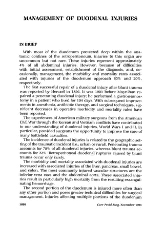
nonabsorbable Lembert sutures, should be used (Fig. 1).
Duodenorrhaphy alone carries a small risk of narrowing the duo-
denal lumen. Several technical points must be kept in mind to &void
this problem when closing duodenal lacerations. These technical
points were outlined by Kraus and Condons on the basis of the re-
sults of an animal model in which they established that longitudinal
duodenotomies can be closed transversely if the length of the duo-
denotomy does not exceed one half of the circumference of the duo-
denum (Fig. 2). These investigators recommehded that longitudinal
closures be performed if the duodenotomy exceeds one haif of the
circumference of the duodenum. In neither of these closures was the
duodenal lumen narrowed. The authors strongly recommended
Curr Probl Surg, November 1993 1053
29. FIG. 1. Most duodenal lacerations can be repaired primarily after meticulous debridement
of all damaged tissue. The repair can be accomplished with a double-layer closure, in-
cluding an inner layer of fine running absorbable sutures encompassing the entire width of
the duodenal wall followed by a second layer of fine seromuscular interrupted nonabsorb-
able Lembert sutures. Meticulous attention must be paid to imbricate the duodenal mu-
cosa because it tends to extrude from suture lines. (From Juan A. Asensio, MD, and
Robert F. Buckman, MD, Duodenal Injuries, Shackleford’s Surgery of the Alimentary Tract,
George D. Zuidema [editor]. Volume 2, Chapter 10, Pages 104-l 17, W.B. Saunders, Phila-
delphia, 1991. Reprinted by permission.)
against transverse closures of transverse duodenotomies, which they
consistently showed to narrow the duodenal lumen.
The use of drains placed adjacent to duodenal repairs should be
considered for all duodenorrhaphies. No prospective studies have ad-
dressed the risk/benefit ratio. We recommend that drains be used
routinely, but we strongly emphasize that this drain system should
be of the closed-suction type and should not be placed directly
against the suture line to avoid duodenal fistula formation.
Before discussing the surgical techniques available for repair of
complex duodenal injuries, a word of caution is in order. Surgical
judgment is needed to select the best surgical technique for repair
of particular duodenal injuries. The surgeon must consider the ana-
tomic extent of the injury; the magnitude of associated injuries, es-
pecially those to the biliary tree and pancreas; and the time elapsed
1054 Curr Probl Surg, November 1993
30. FIG. 2. Longitudinal duodenotomies can be closed transversely if the length of the duo-
denotomy does not exceed one half the circumference of the duodenum.
from injury to repair. Finally, the overall condition of the patient must
be evaluated. Other points to be considered for penetrating injuries
include the potential for blast effect. For blunt injuries, the degree of
associated retroperitoneal and periduodenal inflammatory processes
resulting from extruded duodenal contents should be assessed. Af-
ter evaluating these factors, the surgeon can choose the procedures
that are needed to repair or decompress the duodenum, resect de-
vitalized areas, buttress the repair, or exclude the duodenum from
the passage of gastric contents.
Controversies surround the use of adjacent maneuvers to safeguard
the duodenal closure. One of these maneuvers is the tube duode-
nostomy (Fig. 3), of which the following three types exist: (1) primary,
in which the tube is placed through a separate stab wound in the
duodenum; (2) antegrade, in which the duodenum is decompressed
by way of the passage of a tube through the pylorus; or (31 retrograde,
in which the tube is passed through a jejunostomy site. Primary tube
duodenostomies were first used for decompression in 1909 by New-
mann” and LangenBuch”; however, the technique remained ob-
scure until 1949, when Welch” described this application in patients
Cur-r Probl Surg, November 1993 1055
31. FIG. 3. Adjunct techniques in management of anatomically severe duodenal wounds are
used to protect the anastomosis. The simplest of these techniques is a tube duodenos-
tomy. This tube should be brought out through an area of uninjured duodenum rather than
through the duodenal repair. Its goals are to decompress the duodenum and protect the
suture line; however, a tube duodenostomy does not totally divert the stream of gastric
contents. (From Juan A. Asensio, MD, and Robert F. Buckman, MD, Duodenal Injuries,
Shackleford’s Surgery of the Alimentary Tract, George D. Zuidema [editor]. Volume 2, Chap-
ter 10, Pages 104-l 17, W.B. Saunders, Philadelphia, 1991. Reprinted by permission.)
with tenuous duodenal closures. In 1967, Jones and colleagues1oo de-
scribed a refinement of the technique by advocating use of a small
(No. 10 French) Foley catheter as a decompressive vent, with its bal-
loon minimally inflated with 2 to 3 ml of normal saline. In this series
of 44 patients with tenuous duodenal stump closures, only two com-
plications occurred.
Antegrade decompression of the duodenal closure by passage of a
nasogastric tube through the pylorus beyond the point of injury ap-
pears to have been used first by Smith and colleagues45 in 1971 and
later by McInnis and coworkers46 in 1975. Retrograde duodenostomy
was first used at the Grady Memorial Hospital in 1962,50 and de-
scribed in the literature by Stone and Garonil’l in 1966 (Fig. 4). Two
separate tubes can be placed by way of two separate jejunal sites.
The proximal tube is threaded retrograde past the duodenal junc-
tion and placed on suction to decompress the duodenum while the
distal tube is used as a feeding jejunostomy. This system has been
termed the “suck-me, feed-me jejunostomy.” This system is also ad-
1056 Curr Probl Surg, November 1993
32. FIG. 4. This variation of the tube duodenostomy adds a feeding jejunostomy for early in-
stitution of enteral feeding. It has been dubbed the “suck-me, feed-me jejunostomy.” The
proximal tube that acts to decompress the duodenum and protect its suture line is usually
placed at a point beyond the duodenal-jejunal flexure and generally requires a longer tube
such as a Baker tube.
vacated by Corley and co11eagues,47 who recommend the addition of
a gastrostomy tube to achieve better decompression (Fig. 5).
Addition of tube decompression for duodenorrhaphy is controver-
sial; opinions are strongly divided between those who staunchly sup-
port its routine use, such as Corley and colleagues,47 Stone and col-
leagues,50’101 and Hasson and colleagues,1o2 and those who advocate
against its use, including Ivatury and co11eagues55’ lo3 and Kashuk and
colleagues.104 Stone and Fabian” reported a high rate of duodenal
complications in 8 of 44 duodenal wounds closed without tube de-
compression, with three subsequent deaths attributed to duodenal
complications before the routine use of decompressive techniques
that began in 1962. These investigators reported only one duodenal
Curr Probl Surg, November 1993 1067