

Recommandé
Contenu connexe
Tendances
Tendances (20)
En vedette
En vedette (20)
Similaire à 23205032
Similaire à 23205032 (20)
Plus de radgirl
Dernier
Dernier (20)
23205032
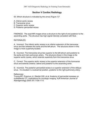
- 1. 2007 ACR Diagnostic Radiology In-Training Exam Rationales Section V Cardiac Radiology 93. Which structure is indicated by the arrow (Figure 1)? A. Inferior aortic recess B. Transverse sinus C. Superior aortic recess D. Posterior pericardial recess FINDINGS: The axial MR images show a structure to the right of and posterior to the ascending aorta. The structure has high signal intensity consistent with fluid. RATIONALES: A. Incorrect. The inferior aortic recess is an inferior extension of the transverse sinus and lies between the aorta and the left atrium. The structure shown in this image is more superiorly located. B. Incorrect. The transverse sinus lies superior to the left atrium and posterior to the aorta and main pulmonary artery. The structure shown in the image is the superior aortic recess, which extends superiorly from the transverse sinus C. Correct. The superior aortic recess is the superior extension of the transverse sinus and extends anterior, lateral and posterior to the ascending aorta D. Incorrect. The posterior pericardial recess is a superior extension of the oblique sinus. It is located in a subcarinal position, posterior to the right pulmonary artery. References: Truong MT, Erasmus JJ, Gladish GW, et al. Anatomy of pericardial recesses on multidetector CT: implications for oncologic imaging. AJR American Journal of Roentgenology 2004;181:1109-1113
- 2. 2007 ACR Diagnostic Radiology In-Training Exam Rationales 94. You are shown an image from an EKG-gated cardiac CTA (Figure 2). Which artery is indicated by the arrow? A. Conal B. Sinoatrial nodal C. Posterior lateral D. Acute marginal FINDINGS: The image shows an artery arising from the right coronary artery and coursing posteriorly toward the right atrium. RATIONALES: A. Incorrect. The conal artery is often the first branch off the right coronary artery and courses anteriorly around the right ventricular conus or infundibulum, which it supplies. The artery shown in this case courses posteriorly. B. Correct. The sinoatrial nodal artery arises from the RCA in 50-60% of patients and supplies the sinoatrial node located at the junction of the superior vena cava and right atrium. C. Incorrect. The posterior lateral artery is a distal right coronary artery branch that extends beyond the crux (the junction of the atrioventricular and interventricular groove) to supply the posterior aspect of the left ventricle. The posterior lateral artery is seen on images of the inferior aspect of the heart. D. Incorrect. The acute marginal artery arises from the junction of the middle and distal segments of the right coronary artery and courses anteriorly to supply the right ventricular wall. References: Pannu HK, Flohr TG, Corl FM, Fishman EK. Current concepts in multi-detector row CT evaluation of the coronary arteries: principles, techniques, and anatomy. RadioGraph 2003; 23:S111-125
- 3. 2007 ACR Diagnostic Radiology In-Training Exam Rationales 95. You are shown turbo spin-echo (Figures 3A and 3B) and delayed gadolinium enhanced (Figures 3C and 3D) MR images of a 49 year old athlete with multiple episodes of syncope. What is the MOST LIKELY diagnosis? A. Myocardial ischemia B. Arrythmogenic right ventricular dysplasia C. Left ventricular mass D. Myocardial scar FINDINGS: Non-enhanced images demonstrate no abnormality. On the delayed, post contrast images there is increased signal in the lateral left ventricular wall consistent with scarring. RATIONALES: A. Incorrect. Although Figure D shows decreased signal in the ventricular septum relative to the posterior wall, this is a delayed gadolinium enhanced image, not a perfusion image, and so does not indicate ischemia. B. Incorrect. The high signal anterior to the right ventricle corresponds to normal subepicardial and pericardial fat. The right ventricular wall is normally quite thin. The imaging findings that contribute to the diagnosis of Arrythmogenic right ventricular dysplasia include fatty replacement of the wall, right ventricular aneurysms and severe segmental or global right ventricular dilatiaton. These findings are not present in this case. C. Incorrect. The mass-like intermediate signal structure along the lateral left ventricular wall (Figures A and B) is the posterolateral papillary muscle and should not be mistake for a mass. D. Correct. The delayed, enhanced images (Figures C and D) demonstrate a focal area of increased signal in the posterolateral left ventricular wall consistent with an area of scarring. Normal myocardium will show transient enhancement after administration of gadolinium. Gadolinium accumulates in areas non-viable myocardium on delayed images since it is able to be distributed throughout the involved tissue due to cell membrane rupture.
- 4. 2007 ACR Diagnostic Radiology In-Training Exam Rationales 96. You are shown an axial image from a contrast enhanced CT of a 55-year-old man (Figure 4). The image shows calcification. Which one of the following is the location of the calcification? A. Mitral annulus B. Coronary artery C. Left ventricular wall D. Pericardium RATIONALES: A. Incorrect. The location of the calcification in this case does not correspond to the anatomic location of the mitral valve annulus. Mitral annular calcium is located within the left atrioventricular groove and extends inferiorly below the valve. B. Incorrect. The location of the calcification in this case does not correspond to the anatomic location of the coronary arteries. The coronary arteries are located in the subepicardial fat. The calcification in this case is located in the myocardium. C. Correct. The calcification is located within the left ventricular myocardium consistent with dystrophic calcification within an area of previous infarction. D. Incorrect. The location of the calcification in this case does not correspond to the anatomic location of the pericardium and is located within the myocardium.
- 5. 2007 ACR Diagnostic Radiology In-Training Exam Rationales 97. You are shown the PA (Figure 5A) and lateral (Figure 5B) chest radiographs of a 66-year-old woman. Which one of the following is the MOST LIKELY diagnosis? A. Mitral regurgitation B. Aortic regurgitation C. Pulmonary hypertension D. Mitral stenosis RATIONALES: A. Correct. There is enlargement of the left atrium and left ventricle. The enlarged left atrium gives a “double” right heart border. On the lateral view the left atrium is seen extending posteriorly. The pulmonary arteries are not enlarged and there is no pulmonary edema. B. Incorrect. One would not expect to see left atrial enlargement in aortic regurgitation. The left ventricle will be enlarged in aortic regurgitation. The aorta may be dilated in aortic regurgitation. C. Incorrect. In pulmonary hypertension, the main pulmonary arteries are likely to be enlarged. Right heart failure and dilatation of the right heart chambers (right atrium and right ventricle) can result. D. Incorrect. In mitral stenosis, one is more likely to see enlargement of the pulmonary veins due to pulmonary venous hypertension. Later on, pulmonary arterial hypertension can result as well as right ventricular hypertrophy. The left ventricle is unlikely to be enlarged in mitral stenosis. The left atrium will be enlarged in mitral stenosis. References: (1) Primer of Diagnostic Imaging. Third edition. Mosby 2003. (2) Radiology Review Manual. Fifth Edition. Lippencott, Williams and Wilkins 2003
- 6. 2007 ACR Diagnostic Radiology In-Training Exam Rationales 98. You are shown a contrast-enhanced CT image (Figure 6). Which of the following is the MOST LIKELY diagnosis? A. Cardiac myxoma B. Cardiac fibroma C. Primary cardiac osteosarcoma D. Thrombus RATIONALES: A. Correct. Cardiac myxoma is the most common benign primary cardiac tumor. The majority of cardiac myxomas arise within the left atrium, typically arising from the fossa ovalis. Calcification within the tumor may be seen. B. Cardiac fibromas are usually seen in children and may be detected in utero. Cardiac fibromas are almost always located in the ventricle, particularly the ventricular septum or free wall of the left ventricle. Calcification is often present. C. Primary cardiac osteosarcomas may have areas of calcification or ossification. They are much less common than cardiac myxoma and typically arise from the posterior wall of the left atrium. D. Left atrial thrombus is more common than cardiac myxoma. However, thrombus in the left atrium usually develops within the left atrial appendage or arises from the posterior or lateral atrial walls. References: (1) Aroaz PA, Mulvagh SL, Tazelaar HD, Julsrud PR, Breen JF. CT and MR imaging of benign primary cardiac neoplasms with echocardiographic correlation. Radiographics. 2000 Sep-Oct;20(5):1303-19. Review. (2) Grebenc ML, Rosado-de-Christenson ML, Green CE, Burke AP, Galvin JR. Cardiac myxoma: imaging features in 83 patients. Radiographics. 2002 May- Jun;22(3):673-89.
- 7. 2007 ACR Diagnostic Radiology In-Training Exam Rationales 99. In regards to myocardial bridge, which one of the following statements is TRUE? A. They occur in 5% of autopsy specimens. B. They are better depicted by CT than by angiography. C. They require surgical therapy. D. They most frequently affect the right coronary artery. RATIONALES: A. Incorrect. In autopsy series, myocardial bridging, an intramuscular course of the coronary artery, occurs in 30% of specimens. B. Correct. Because multiplanar reformatted images can be obtained from the CT dataset, CT is better able to detect the intramuscular course of the coronary artery. C. Incorrect. In most cases, myocardial bridges are clinically silent. D. Incorrect. Myocardial bridges are most frequently seen with the left anterior descending artery. 100. Enlargement of which one of the following structures is the MOST reliable radiographic sign of pulmonary valve stenosis? A. Enlargement of the pulmonary trunk B. Enlargement of the right ventricle C. Enlargement of the left main pulmonary artery D. Enlargement of the right and left main pulmonary arteries RATIONALES: A. Incorrect. The pulmonary trunk may be either normal or enlarged on radiographs in patients with pulmonary valve stenosis. Isolated pulmonary trunk enlargement is more frequent with idiopathic dilatation of the pulmonary artery. B. Incorrect. The right ventricle hypertrophies in response to the pressure overload caused by pulmonary valve stenosis. Ventricular dilatation only occurs if ventricular failure or tricuspid regurgitation is present. C. Correct. Enlargement of the left main pulmonary artery, with or without pulmonary trunk enlargement, is the radiographic hallmark of pulmonary valve stenosis.
- 8. 2007 ACR Diagnostic Radiology In-Training Exam Rationales D. Incorrect. Enlargement of both main pulmonary arteries is characteristic of either overcirculation or pulmonary arterial hypertension. Enlargement of the right pulmonary artery is not a feature of pulmonary valve stenosis 101. Which of the following findings is the MOST reliable sign of elevated right ventricular pressure on a contrast-enhanced CT scan of the chest? A. Leftward bowing of the interventricular septum B. Right ventricular hypertrophy C. Right ventricular enlargement D. Reflux of contrast into the inferior vena cava RATIONALES: A. Correct. Abnormal curvature of the ventricular septum toward the left ventricle indicates elevated right ventricular pressure. B. Incorrect. Although the right ventricle will hypertrophy under the stress of a pressure overload, wall thickening may be difficult to identify on routine, non- gated CT scans. C. Incorrect. Enlargement of right ventricle indicates a volume overload, such as tricuspid regurgitation or a left to right shunt, or the presence of right ventricular failure. Elevated right ventricular pressure alone will not generally enlarge the chamber. D. Incorrect. Reflux of contrast into the IVC can be seen with either high right sided pressures (atrial or ventricular), or with tricuspid regurgitation.
- 9. 2007 ACR Diagnostic Radiology In-Training Exam Rationales 102. Regarding cardiomyopathies, which one of the following statements is TRUE? A. Patients with dilated cardiomyopathy have an increase in left ventricular mass. B. The myocardium is diffusely affected by cardiac sarcoidosis. C. Aortic stenosis is a cause of hypertrophic cardiomyopathy. D. Systolic function is preserved in patients with restrictive cardiomyopathy secondary to amyloidosis. RATIONALES (This Test Item Was Not Scored) A. In dilated cardiomyopathy, the ventricles dilate but their wall thickness is maintained, so there is an overall increase in the ventricular mass. B. The distribution of myocardial granulomas in sarcoidosis is patchy and characterized by nodules of increased signal intensity on T2-weighted images with hyperenhancement on T1-weighted images following contrast administration. C. Aortic stenosis can cause left ventricular hypertrophy. However, hypertrophic cardiomyopathy is diagnosed when there is abnormal myocardial thickening in the absence of a condition resulting in increased afterload such as aortic stenosis or systemic hypertension. D. Both diastolic and systolic function are reduced in patients with restrictive cardiomyopathy secondary to amyloidosis. References: Webb WR & Higgins CB. Thoracic Imaging-Pulmonary and Cardiovascular Radiology. Lippincott Willimas & Wilkins 2005 1st Edition
- 10. 2007 ACR Diagnostic Radiology In-Training Exam Rationales 103. Regarding cardiac transplantation, which one of the following statements is TRUE? A. Accelerated atherosclerosis is an early complication. B. Postoperative screening for lymphoproliferative disease is the most common indication for imaging. C. Cyclosporine results in left ventricular hypertrophy. D. Cytomegalovirus and Aspergillus species are the most common cause of early postoperative infection. RATIONALES: A. Incorrect. Accelerated atherosclerosis is a late complication and can be detected by coronary angiography. B. Incorrect. Infection, especially pulmonary infection, is the most common indication for imaging following cardiac transplantation. C. Correct. Ventricular hypertrophy can be caused by cyclosporine immunosuppressive therapy. D. Incorrect. Bacteria are the most common cause of early post-operative infection following ardiac transplant. Aspergillus and cytomegalovirus infections occur 2-6 months following transplantation. References: Attili A, Kazerooni EA. Postoperative cardiopulmonary thoracic imaging. Radiol Clin N Am 42 (2004) 543-564 Knisely BL, Mastey LA, Collins J, et al. Imaging of cardiac transplantation complications. Radiographics 1999;19:321-39. Webb WR & Higgins CB. Thoracic Imaging-Pulmonary and Cardiovascular Radiology. Lippincott Willimas & Wilkins 2005 1st Edition
- 11. 2007 ACR Diagnostic Radiology In-Training Exam Rationales 104. Concerning cardiac neoplasms, which of the following is TRUE? A. Myxoma is the most common benign cardiac tumor. B. Metastases to the heart are most commonly caused by renal cell carcinoma. C. Primary cardiac malignancies are more common than metastases to the heart. D. Malignant primary cardiac neoplasms are more common than benign neoplasms. RATIONALES: A. Correct. Myxoma accounts for half of all primary cardiac tumors. B. Incorrect. Metastases to the pericardium and epicardium are most commonly from lung cancer. This is in part related to proximity to the heart and in part because of the prevalence of lung cancer. Metastases to the myocardium are most commonly from melanoma and the mode of spread is thought to be hematogenous. Myocardial metastases are less frequent than pericardial or epicardial metastases. C. Incorrect. Primary cardiac malignancies are rare. Metastases to the heart are 20 to 40 times more common than primary tumors. D. Incorrect. Only 25% of primary cardiac tumors are malignant. References: (1) Aroaz PA, Eklund HE, Welch TJ, Breen JF. CT and MR Imaging of Primary Cardiac Malignancies. Radiographics 1999;19:1421-1434. (2) Chiles C, Woodward PK, Gutierrez F, Link KM. Metastatic Involvement of the Heart and Pericardium: CT and MR imaging. Radiographics 2001;21:439-449. (3) Grebenc ML, Rosado de Christenson ML, Burke AP, Green CE, Galvin JR. Primary Cardiac and Pericardial Neoplasms: Radiologic and Pathologic Correlation. Radiographics 2000;20:1073-1103.
- 12. 2007 ACR Diagnostic Radiology In-Training Exam Rationales 105. Concerning the coronary circulation, which of the following statements is TRUE? A. Dominance is determined by which vessel gives rise to the posterior descending artery and posterior left ventricular branches. B. Left dominant circulation is more common than right dominant circulation. C. The left coronary artery supplies the sinoatrial node in most individuals. D. The diagonal branches arise from the right coronary artery. RATIONALES: A. Correct. Most patients are right dominant with the right coronary artery giving rise to the posterior descending artery and posterior left ventricular branches B. Incorrect. Right dominant circulation is seen in 85% of individuals. The coronary circulation is left dominant in only 8% of individuals. In left dominant circulation, the posterior descending artery and posterior left ventricular branches arise from the left circumflex artery. In the remaining 7% there is a co-dominant system in which the posterior descending artery arises from the right coronary artery and the posterior left ventricular branches arise from the left circumflex coronary artery. C. Incorrect. The right coronary artery supplies the sinoatrial node in 60% of individuals. The sinus node artery arises from the proximal right coronary artery. In 40% of individuals, the sinus node artery arises from the proximal left circumflex artery. D. Incorrect. The diagonal branches arise from the left anterior descending coronary artery and supply the anterior free wall of the left ventricle. The acute marginal branch arises from the right coronary artery and supplies the right ventricle. The obtuse marginal branches arise from the circumflex coronary artery and supply the lateral left ventricle. References: (1) Pannu HK, Flohr TG, Corl FM, Fishman EK. Multi-Detector Row CT Evaluation of the Coronary Arteries: Principles, Techniques and Anatomy. Radiographics 2003;23:S111-S125
- 13. 2007 ACR Diagnostic Radiology In-Training Exam Rationales 106. A 3-D cardiac CT image data set is acquired at 0.5-mm slice thickness. If the reconstruction interval is reduced from 0.5 mm to 0.3 mm, which of the following changes is MOST LIKELY to occur? A. Increase patient dose B. Decrease patient dose C. Increase image size D. Increase scan time RATIONALES: A. Incorrect. Patient dose is unaffected by the changes in reconstruction interval as it does not affect scan acquisition. B. Incorrect: Patient dose is unaffected by the changes in reconstruction interval as it does not affect scan acquisition. C. Correct: If the reconstruction interval is decreased, it results in higher number of slices yielding higher image size for 3D images D. Incorrect: Changes in reconstruction interval only affects reconstruction time but not the scan time.
- 14. 2007 ACR Diagnostic Radiology In-Training Exam Rationales 107. Concerning prospective ECG gating in cardiac CT imaging, which of the following is TRUE? A. Data is acquired throughout the cardiac cycle. B. Data is acquired only at pre specified points throughout the cardiac cycle. C. It allows for dynamic assessment of the heart and functional status. D. It involves more radiation dose to the patient than retrospective gating. RATIONALES: A. Incorrect. In prospective ECG gating (also known as ECG triggering), data acquisition is triggered to the R wave of the ECG, so data is acquired at a pre specified time. The multi detector CT can be set up to record data at a certain time after the last R wave or before the next R wave of the ECG. Alternatively, data can be acquired at a certain percentage of the time between two successive R waves. B. Correct. Data is ideally only acquired when the heart is relatively motionless, as in diastole. So there are parts of the ECG cycle in which data is not being acquired, namely during systole (when the heart moves more). C. Incorrect. Because there is “missing” data, one cannot do a volumetric assessment of the left ventricular cavity. In order to calculate ejection fraction, one needs data from both diastole and systole, so retrospective ECG gating is needed. D. Incorrect. Retrospective ECG gating involves more radiation because data is acquired throughout the cardiac cycle. The patient is receiving ionizing radiation throughout the cardiac cycle. The person interpreting the study selects data from selected portions of the cardiac cycle (such as in diastole, when the heart is relatively motionless). References: (1) Wintersperger BJ, Nikolaou K. Basics of Cardiac MDCT: techniques and contrast application. European Radiology 2005, 15(2):B2-B9
- 15. 2007 ACR Diagnostic Radiology In-Training Exam Rationales 108. Concerning the use of B Blockers in cardiac CT imaging, which of the following statements is TRUE? A. Heart rates of below 70 beats per minute are optimal. B. They can be administered to patients with asthma. C. Verapamil is not a useful alternative to beta-adrenergic blocking agents. D. Atrial fibrillation is a contraindication to beta-blocker use. RATIONALES: A. Correct. Several studies which used 4 and 16 detector row CT have shown consistently that at heart rates of greater than 70 beats a minute, cardiac motion degrades the images. B. Incorrect. Asthma is a contraindication to beta blockers. This is because many beta blockers are not cardioselective and block both B1 and B2 receptors. B1 receptors are found in the heart and B2 receptors are found in the airways smooth muscle. Blocking of B2 receptors causes bronchospasm in asthmatics. Even cardioselective beta blockers (such as metoprolol or atenolol) are a relative contraindication in asthmatics and should be avoided. C. Incorrect. Verapamil can be used as an alternative to beta blockers, such as in patients with asthma. It is usually given intravenously in cardiac CT. D. Incorrect. Atrial fibrillation is not a contraindication to beta blocker use. Although the beta blocker slows the ventricular response rate, it will not be effective in preventing atrial fibrillation. Atrial fibrillation (even with a slow ventricular response rate) is a relative contraindication to ECG gating. References: (1) Choi HS, Choi BW, Choe KO, Choi D, Yoo KJ, Kim MI, Kim J. Pitfalls, Artifacts and Remedies in Multi Detector Row CT Coronary Angiography. Radiographics 2004;24:787-800 (2) Kumar and Clark, Clinical Medicine, Fifth Edition, Saunders, 2002
- 16. 2007 ACR Diagnostic Radiology In-Training Exam Rationales 109. Which of the following digital detectors directly converts x-ray signals into an electrical charge? A. Photostimulable storage phosphor B. Structured cesium-iodine (CsI) scintillator C. Amorphous selenium (aSe) semiconductor D. Charge-coupled devices (CCD) photodiode RATIONALES: A. Incorrect. B. Incorrect. C. Correct. Digital detectors are classified by the signal transfer stages that occur during the image formation process in two categories. Indirect conversion is the process by which the x-rays are absorbed and converted into a secondary signal such as x-rays to light by a scintillator material or x-rays to trapped electrons to stimulation by a laser to light emission by a photostimulable phosphor. This light energy is subsequently converted to electron signals that comprise the signal that generates the corresponding digital value. Direct conversion is the process by which the x-rays directly produce the electron/hole pairs that are used to generate the corresponding digital values. Semi-conductor materials that absorb x-rays without scintillation, such as amorphous selenium, are classified as direct conversion materials. D. Incorrect. References: Mahesh M, AAPM/RSNA Physics Tutorial for Residents Digital Mammography: An Overview, RadioGraphics 2004; 24:1747–1760
- 17. 2007 ACR Diagnostic Radiology In-Training Exam Rationales 110. In regards to constrictive pericarditis, which of the following statements is TRUE? A. The normal pericardium measures up to 5mm in thickness. B. Tuberculosis is the most common cause of constrictive pericarditis in the United States. C. The degree of pericardial calcification correlates with the degree of constriction. D. Pericardial thickening and calcification can occur in the absence of constrictive pericarditis. RATIONALES: A. Incorrect. The pericardium normally measures 2mm in thickness or less.. B. Incorrect. Most cases of constrictive pericarditis are secondary to idiopathic pericarditis or pericarditis following cardiac surgery and radiation therapy. C. Incorrect. Although calcification is associated with constriction, the observation of calcification does not imply constriction. In order to make the diagnosis of constrictive pericarditis, the patient must have physiologic changes in association with pericardial thickening or calcification. This includes impaired diastolic filling and equalization of diastolic pressures in the right and left ventricles. The most common causes of calcific pericarditis include infection (tuberculosis), uremia and trauma. D. Correct. Pericardial thickening or calcification may be present without physiologic impairment, although constrictive physiology is usually present when there is extensive pericardial calcification. References: (1) Wang ZJ, Reddy GP, Gotway MB, Yeh BM, Hetts SW, Higgins CB. CT and MR imaging of pericardial disease. Radiographics. 2003 Oct; 23 Spec No:S167- 80. (2) Glockner JF. Imaging of Pericardial disease. MRI Clinics of North America 2003 (11) 149-162.
- 18. 2007 ACR Diagnostic Radiology In-Training Exam Rationales 111. Concerning anomalous origins of the coronary arteries, which of the following is TRUE? A. Coronary artery anomalies are usually hemodynamically significant. B. Anomalous coronary arteries that pass between the right ventricular outflow tract / pulmonary artery and the ascending aorta are characterized by abnormal myocardial perfusion. C. With hemodynamically significant coronary artery anomalies, compression of the coronary artery occurs during systole. D. Coronary artery anomalies with a retroaortic course are associated with sudden death in young athletes. RATIONALES: A. Incorrect. Coronary artery anomalies are rare, estimated at about 1% of the population. About one third of these anomalies refer to the origin of the coronary artery. Most anomalous coronary arteries are “benign”, that is the coronary artery courses posterior to the aortic root or anterior to the pulmonary trunk. Hemodynamically significant, or “malignant” coronary artery anomalies occur when the artery passes between the right ventricular outflow tract and the ascending aorta. These are much less common. B. Correct. Coronary arteries that pass between the right ventricular outflow tract / pulmonary artery and the ascending aorta are hemodynamically significant. The coronary artery can become compressed between two arteries during systole, when the vessels expand. C. Incorrect. Compression of the “malignant” coronary artery occurs during diastole, when the ascending aorta and right ventricular outflow tract / pulmonary artery are more dilated. D. Incorrect. “Malignant” coronary artery anomalies are associated with sudden death in young athletes. This is thought to be due to compression of the coronary artery between the right ventricular outflow tract / pulmonary artery and the ascending aorta during diastole. References: (1) Kim SY, Seo JB, Do KH et al. Coronary artery anomalies: classification and ECG-gated multi-detector row CT findings with angiographic correlation. Radiographics. 2006 Mar-Apr;26(2):317-33; discussion 333-4. (2) Datta J, White CS, Gilkeson RC et al. Anomalous coronary arteries in adults: depiction at multi-detector row CT angiography. Radiology. 2005 Jun;235(3):812- 8.
- 19. 2007 ACR Diagnostic Radiology In-Training Exam Rationales (1) Schmid M, Achenbach S, Ludwig J et al. Visualization of coronary artery anomalies by contrast-enhanced multi-detector row spiral computed tomography. Int J Cardiol. 2005 Nov 3; [Epub ahead of print] 112. In regards to cyanotic congenital heart disease presenting in infancy, which one of the following is MOST common? A. Hypoplastic left heart syndrome B. Tetralogy of Fallot C. Truncus arteriosus D. Ventricular septal defect RATIONALES: A. Incorrect. Although Hypoplastic left heart syndrome is the most common cause of congestive failure in the neonate, it only occasionally results in cyanosis. B. Correct. Tetralogy of Fallot is the most common cause of cyanotic heart disease presenting in the first month of life. C. Incorrect. Truncus arteriosus is a cause of cyanotic heart disease in infancy, but occurs with a much lower frequency than tetralogy of Fallot. D. Incorrect. Ventricular septal defects are the most common, clinically evident congenital heart defect in infants, but only present with cyanosis after the development of secondary pulmonary hypertension. Therefore cyanosis is not characteristic in infancy. References: Miller SW. Cardiac Imaging: The Requisites. 2nd ed. Mosby, Inc., Philadelphia, PA. 2005.
- 20. 2007 ACR Diagnostic Radiology In-Training Exam Rationales 113. Regarding atrial morphology, which one of the following is the MOST reliable indicator of the morphologic right atrium? A. Connection with the superior vena cava B. Connection with the inferior vena cava C. Presence of a thin appendage with a narrow neck D. Presence of a tricuspid atrioventricular valve RATIONALES: A. Incorrect. Bilateral superior vena cavae may be present, which can drain into the coronary sinus or either atria. B. Correct. Connection with the inferior vena cava is a reliable indicator of right atrial morphology. This can be a useful tool to establish the cardiac anatomy in cases of situs abnormalities and atrioventricular discordance. C. Incorrect. A thin appendage with a narrow neck is characteristic of the morphologic left atrium. The right atrial appendage is triangular in shape with a broad neck. The characteristic appearance of the right atrial appendage is also a reliable indicator of the morphologic right atrium. D. Incorrect. In cases of atrioventricular discordance, the atrioventricular valves follow the morphologic ventricles, not the morphologic atria. References: Miller SW. Cardiac Imaging: The Requisites. 2nd ed. Mosby, Inc., Philadelphia, PA. 2005
- 21. 2007 ACR Diagnostic Radiology In-Training Exam Rationales 114. Regarding coronary artery aneurysms, which one of the following statements is CORRECT? A. Aneurysmal dilatation is defined as an increase in vessel diameter twice that of the normal adjacent artery. B. Most coronary aneurysms are secondary to infection. C. Small aneurysms (<4mm) seen in the setting of Kawasaki disease (mucocutaneous lymph node syndrome) tend to regress. D. Aneurysms due to atherosclerosis have a high frequency of rupture. RATIONALES: A. Incorrect. A coronary artery aneurysm is defined as an increase in diameter by 50%, not 100%. B. Incorrect. Most coronary aneurysms are secondary to atherosclerosis. C. Correct. Kawasaki disease has multiple manifestations in the heart, including coronary aneurysm formation. Large aneurysms (> 8mm) are frequently complicated by thrombosis and subsequent myocardial infarction. They may also rupture. Small aneurysms tend to regress to normal caliber over time. D. Incorrect. Rupture of atherosclerotic coronary aneurysms is rare. References: Miller SW. Cardiac Imaging: The Requisites. 2nd ed. Mosby, Inc., Philadelphia, PA. 2005.
- 22. 2007 ACR Diagnostic Radiology In-Training Exam Rationales 115. Regarding hypertrophic cardiomyopathy, which one of the following is an associated valvular abnormalty? A. Aortic valve stenosis B. Mitral valve stenosis C. Systolic anterior motion of the mitral valve D. Mitral valve prolapse RATIONALES: A. Incorrect. Outflow obstruction in the setting of hypertrophic cardiomyopathy occurs in a subvalvular location. While the aortic valve may close prematurely during mid-systole, aortic valve stenosis is not characteristic of hypertrophic cardiomyopathy. B. Incorrect. Although mitral valve regurgitation may coexist with hypertrophic cardiomyopathy, it is not a characteristic of hypertrophic cardiomyopathy. C. Correct. The increased flow velocity occurring through the narrowed subvalvular left ventricular outflow tract draws the anterior leaflet of the mitral valve anteriorly due to the Venturi effect. This results in further obstruction of the left ventricular outflow tract. D. Incorrect. The mitral valve is typically drawn anteriorly during systole in the setting of hypertrophic cardiomyopathy. Prolapse posteriorly into the left atrium is not a characteristic of hypertrophic cardiomyopathy. References: Miller SW. Cardiac Imaging: The Requisites. 2nd ed. Mosby, Inc., Philadelphia, PA. 2005.