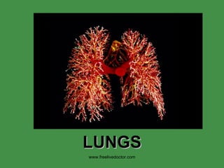
![LUNG ,[object Object],[object Object],[object Object],[object Object],www.freelivedoctor.com](data:image/gif;base64,R0lGODlhAQABAIAAAAAAAP///yH5BAEAAAAALAAAAAABAAEAAAIBRAA7)
Recommandé
Contenu connexe
Tendances
Tendances (20)
Similaire à Pulmonary pathology
Similaire à Pulmonary pathology (20)
Plus de raj kumar
Plus de raj kumar (20)
Pulmonary pathology
- 5. WEIGHT LOBES SEGMENTS BRONCHI ARTERIES, pulmonary ARTERIES, bronchial VEINS PLEURA, visceral PLEURA, parietal NERVES www.freelivedoctor.com
- 6. Bronchi Bronchioles Terminal bronchioles Alveolar ducts Alveoli Type 1 pneumocytes Type 2 pneumocytes Macrophages Capillaries www.freelivedoctor.com
- 9. www.freelivedoctor.com NORMAL” chest X-Ray [CXR]
- 26. www.freelivedoctor.com Bullae, or “peripheral blebs” are hallmarks of chronic obstructive lung disease, COPD.
- 30. www.freelivedoctor.com Note the heavy inflammatory cell infiltrate around bronchioles and small bronchi.
- 31. What are the 4 classical histologic findings in bronchial asthma? www.freelivedoctor.com
- 44. NON-Caseating Granulomas are the RULE “ Asteroid” bodies within these granulomas are virtually diagnostic www.freelivedoctor.com
- 52. VERY thickened arteriole in pulmonary hypertension NORMAL pulmonary arteriole www.freelivedoctor.com
- 54. CHF, CHRONIC IDIOPATHIC PULMONARY HEMOSIDEROSIS www.freelivedoctor.com
- 56. COMM UNITY-ACQUIRED BACTERIAL ACUTE PNEUMONIAS Streptococcus Pneumoniae Haemophilus Influenzae Moraxella Catarrhalis Staphylococcus Aureus Klebsiella Pneumoniae Pseudomonas Aeruginosa Legionella Pneumophila COMMUNITY-ACQUIRED ATYPICAL (VIRAL AND MYCOPLASMAL) PNEUMONIAS Morphology. Clinical Course. Influenza Infections Severe Acute Respiratory Syndrome (SARS) NOSOCOMIAL PNEUMONIA ASPIRATION PNEUMONIA LUNG ABSCESS Etiology and Pathogenesis. CHRONIC PNEUMONIA Histoplasmosis, Morphology Blastomycosis, Morphology Coccidioidomycosis, Morphology PNEUMONIA IN THE IMMUNOCOMPROMISED HOST PULMONARY DISEASE IN HUMAN IMMUNODEFICIENCY VIRUS INFECTION PULMONARY INFECTIONS www.freelivedoctor.com
- 59. Although pneumonia is one of the most common causes of death, it usually does NOT occur in healthy people spontaneously www.freelivedoctor.com
- 70. Although pneumonia is one of the most common causes of death, it usually does NOT occur in healthy people spontaneously www.freelivedoctor.com
- 78. S A R S www.freelivedoctor.com
- 96. Methenamine SILVER stain for Pneumocystis Carinii www.freelivedoctor.com
- 106. TNM, Lung www.freelivedoctor.com T1 Tumor <3 cm without pleural or main stem bronchus involvement T2 Tumor >3 cm or involvement of main stem bronchus 2 cm from carina, visceral pleural involvement, or lobar atelectasis T3 Tumor with involvement of chest wall (including superior sulcus tumors), diaphragm, mediastinal pleura, pericardium, main stem bronchus 2 cm from carina, or entire lung atelectasis T4 Tumor with invasion of mediastinum, heart, great vessels, trachea, esophagus, vertebral body, or carina or with a malignant pleural effusion N0 No demonstrable metastasis to regional lymph nodes N1 Ipsilateral hilar or peribronchial nodal involvement N2 Metastasis to ipsilateral mediastinal or subcarinal lymph nodes N3 Metastasis to contralateral mediastinal or hilar lymph nodes, ipsilateral or contralateral scalene, or supraclavicular lymph nodes M0 No (known) distant metastasis M1 Distant metastasis present
- 107. LOCAL effects of LUNG CANCER www.freelivedoctor.com Clinical Feature Pathologic Basis Pneumonia, abscess, lobar collapse Tumor obstruction of airway Lipid pneumonia Tumor obstruction; accumulation of cellular lipid in foamy macrophages Pleural effusion Tumor spread into pleura Hoarseness Recurrent laryngeal nerve invasion Dysphagia Esophageal invasion Diaphragm paralysis Phrenic nerve invasion Rib destruction Chest wall invasion SVC syndrome SVC compression by tumor Horner syndrome Sympathetic ganglia invasion Pericarditis, tamponade Pericardial involvement SVC, superior vena cava.
- 108. SYSTEMIC effects of LUNG CANCER (PARA-NEOPLASTIC SYNDROMES)~ 5% ADH (hyponatremia) ACTH (Cushing) PTH (Hyper-CA) CALCITONIN (Hypo-CA) GONADOTROPINS SEROTONIN/BRADYKININ www.freelivedoctor.com
- 117. EM H&E, IMMUNOCHEMISTRY www.freelivedoctor.com
Notes de l'éditeur
- Typical normal 1000 gram lung (R550, L450), with lobes and bronchopulmonary segments, primary, secondary, tertiary bronchi, etc. Pleura “smooth and glistening”, arteries traveling with bronchi, veins being rather independent of bronchopulmonary segments and lobes. Why is weight important? Why is “smooth and glistening” important? What is “crepitance”? Why is crepitance important?
- Classical classifications of diseases, degenerative, inflammatory, neoplastic. This classification still stands up today.
- Typical normal 1000 gram lung (R550, L450), with lobes and bronchopulmonary segments, primary, secondary, tertiary bronchi, etc. Pleura “smooth and glistening”, arteries traveling with bronchi, veins being rather independent of bronchopulmonary segments and lobes. Why is weight important? Why is “smooth and glistening” important? What is “crepitance”? Why is crepitance important?
- Know the microscopic criteria for all the items delineated on the right, especially the kinds of simple epithelium which line them.
- The “space” between the endothelium and the type-1 pneumocyte, is the blood air interface
- This simple embryology diagram may help explain most common congenital lung diseases. Why might this diagram be WRONG, especially the top figure?
- “ NORMAL” chest X-Ray (CXR)
- Resorption can be from a bronchial obstruction, such as a tumor. Compression can be from, say, a pleural effusion. Contraction can be from a diffuse lung fibrotic process.
- FOUR main pathologic mechanisms of pulmonary edema
- ARDS can be thought of as NON-cardiac pulmonary edema, or, more correctly, edema related to alveolar INJURY. It is NON-specific!!! It is also sometimes called “shock lung” as we will see in the section on shock. Is the alveolar “edema” of ARDS more likely to have more protein than cardiac pulmonary edema?
- Two EXTREMELY important concepts of pulmonary pathology. OBSTRUCTION means SMALL AIRWAT EXPIRATIRY obstruction. RESTRICTION means REDUCED COMPLIACE, i.e., less sponginess!
- Bullae, or “peripheral blebs” are hallmarks of chronic obstructive lung disease, COPD.
- Note the heavy inflammatory cell infiltrate around bronchioles and small bronchi.
- What are the 4 classic histologic findings in bronchial asthma? Answer: 1) Inflammation 2) Bronchial narrowing 3) Increased Mucous 4) Smooth muscle hyperplasia What is the 5 th finding if the etiology is allergy? Ans: Eosinophils
- Bronchiectasis is not a specific disease, but simply a condition in which LARGE bronchi are damaged and DILATED due to a variety of causes.
- How do you know these are not bullae?
- If you “squeezed” a lung with restrictive lung disease, you would note it wasn’t as “spongy” as a normal lung. This is the definition of reduced compliance. It simply will not “comply” when squeezed (or moved by respiratory motion either)! In contract to the “obstructive” lung diseases, the chest x-ray shows diffuse INCREASE in density, NOT DECREASED, usually.
- Would you see a lot of scar tissue here if you did a trichrome stain? Ans: yes
- This image was “googled” from tumorboard.com, the internet’s FIRST diagnostic pathology image base. The fact that this is a mesenteric lymph node will remind you that sarcoidosis is NOT limited to the lungs.
- The classical difference between a “caseating” and a “non-caseating” granuloma, is often the difference between TB and sarcoid. Which one might culture out acid-fast bacteria? Ans: The one on the LEFT (i.e., caseating)
- Pulmonary edema on left, PAP (Pulmonary Alveolar Proteinosis) on the right.
- A general rule with COPD is: As the alveoli become wider, the arterioles become narrower!
- A common finding in most cases of pulmonary hypertension, no matter what the cause is. NORMAL thickness pulmonary arteriole on the LEFT.
- Wegener's granulomatosis is a form of vasculitis that affects the lungs , kidneys and other organs. Due to its end-organ damage, it can be a serious disease that requires long-term immunosuppression . It is named after Dr. Friedrich Wegener , who described the disease in 1936.
- IPH has MUCH more hemosiderin in alveoli, usually, relative to chronic CHF. Acute CHF has NO hemosiderin. Why?
- Why is the term “chronic” pneumonia here, kind of a misnomer, classically?
- Viral pneumonias, generally interstitial, bacterial pneumonias generally alveolar!!!
- Corona viruses are RNA, “enveloped”, i.e., “crowned” viruses
- As soon as you step into a hospital, expect to be greeted by MRSA
- An abscess can be thought of as a pneumonia in which all of the normal lung outline can no longer be seen, and there is 100% pus. Notice the increasing destruction of the alveolar framework as you progress closer to the center of the abscess.
- “ Chronic” by classification, but “granulomatous” by histology.
- Granulomatous reactions are commonly seen with mycobacteria, fungi, sarcoid, foreign bodies, and rarely with almost anything.
- PCP is the most common pneumonia in AIDS patients. It is so prevalent, many rationales consist in giving treatment for it prophylactically. An interesting tidbit is that “cotton wool” or “wooly” exudates are described BOTH radiologically as well as histologically
- Bronchiolitis obliterans, as seen with “COP”, occurs with chronic pulmonary transplant rejection
- The NON-small cell cancers behave and are treated similarly, the SMALL cell carcinomas are WORSE than the non-small cell carcinomas, but respond better to chemotherapy, often drastically!
- Once again, the best way to classify tumors of ANY organ or tissue is to simply remember the histology. Tumors are clonal proliferations of native cells.
- Name the four most common histologic patterns of lung carcinoma and explain why! Squamous, adeno, large, small, going clockwise.
- TNM ALWAYS relates to BIOLOGIC BEHAVIOR!
- Once again, the best way to classify tumors of ANY organ or tissue is to simply remember the histology. Tumors are clonal proliferations of native cells.
- Also recall that mesothelium does not only cover the lungs viscerally, as well as parietally, but also the pericardium and the peritoneum as well, so mesotheliomas and effusions of the pleura, and ALL diseases, are also have their corresponding counterparts in the pericardial space and peritoneal space as well.
- Pleuritis = Pleurisy
- How would you differentiate a pleural transudate from an exudate? Ans: SPGR, cells, protein, LDH
- Typical growth appearance of a malignant mesothelioma, it compresses the lung from the OUTSIDE.
- Mesothelial cells have MANY more microvilli than most epithelial cells and express a protein called CALRETININ. The differentiation between mesothelioma and carcinoma may be crucially important!