SlideShare une entreprise Scribd logo
1  sur  18
Télécharger pour lire hors ligne
IX             Glossopharyngeal Nerve
      carries visceral
          and general
      sensation from
 the pharynx, carries
        subconscious
            sensation
     from the carotid
     sinus and body,
       and innervates
the stylopharyngeus
      muscle and the
         parotid gland
                         © L. Wilson-Pauwels
IX Glossopharyngeal Nerve
                                         CASE HISTORY
      Allen is a 43-year-old construction worker. While he was eating dinner, Allen developed
      pain in the left side of his throat. The pain was short, brief, and stabbing in nature and only
      occurred when he swallowed. Allen was eating fish at the time and assumed that a fish
      bone was lodged in his throat. He went to the emergency department and was assessed
      by an otolaryngologist, but nothing was found. It was assumed that a fish bone had
      scratched Allen’s throat and that his symptoms would soon resolve.
          A month later, Allen’s pain was still present; in fact, it had increased. The pain occurred
      not only when he swallowed but also when he spoke and coughed. Again, while eating din-
      ner, Allen experienced the same pain with swallowing. He got up from the table, but after
      a few steps he collapsed unconscious to the floor.
          Although Allen quickly regained consciousness, he was immediately taken to the
      emergency department where he was put on a heart rate and blood pressure monitor to
      evaluate the function of his cardiovascular system. While he was on the monitor, the emer-
      gency room physician noticed an interesting correlation. Whenever Allen swallowed, he
      had a paroxysm (a sudden recurrence or intensification of symptoms) of pain followed
      invariably within 3 to 4 seconds by a decrease in heart rate (bradycardia) and a fall in blood
      pressure (hypotension). Allen was examined by a neurologist who noticed no evidence of
      reduced perception of pharyngeal pinprick or touch, or of pharyngeal motility. A diagnosis
      of glossopharyngeal neuralgia was made. Allen was treated with carbamazepine. His
      attacks of pain stopped and his cardiovascular function returned to normal.


              ANATOMY OF THE GLOSSOPHARYNGEAL NERVE
      The name of the glossopharyngeal nerve indicates its distribution (ie, to the glossus
      [tongue] and to the pharynx). Cranial nerve IX emerges from the medulla of the
      brain stem as the most rostral of a series of rootlets that emerge between the olive and
      the inferior cerebellar peduncle. The nerve leaves the cranial fossa through the jugu-
      lar foramen along with cranial nerves X and XI (Figure IX–1). Two ganglia are
      situated on the nerve as it traverses the jugular foramen, the superior and inferior
      (petrosal) glossopharyngeal ganglia. The superior ganglion is small, has no branches,
      and is usually thought of as part of the inferior glossopharyngeal ganglion. The
      ganglia are composed of nerve cell bodies of sensory components of the nerve
      (Figure IX–2).


164
Glossopharyngeal Nerve            165


                               © L. Wilson-Pauwels
                                                                                                                                       Spinal
                                                                                    TYMPANIC BRANCH                                    nucleus
                                                                                     (plexus and nerve)                                of
                                                                                                                                       trigeminal
                                                                                                                                       nerve

                                                                                                                                      Nucleus
                                                  Lesser petrosal                                                                     solitarius
                                               nerve and foramen                                                                      (rostral gustatory
                                                                                                                                      portion)
                                                  Foramen
                                                     ovale                                                                        Inferior
                                                                                                                                  salivatory nucleus
                                                   Otic
                                               ganglion
                                                                                                                            Nucleus solitarius
                                                                                                                            (visceral sensory portion)
                                                                                                                          Jugular foramen

                                        Palatal arch
                                                                                                                          Nucleus ambiguus
                                                   Tonsil                                                                 (branchial and visceral motor)
                            Posterior                              Parotid
                      third of tongue                                                                                Superior and inferior
                                                                    gland
                                                                                                                     glossopharyngeal ganglia


                                                                                                                  from external ear

                                                                                                               MUSCULAR BRANCH


                                                                                                          Stylopharyngeus muscle

                                                                                                    CAROTID BRANCH

                                                                                                Internal carotid artery
           TONSILAR
                         LINGUAL
            BRANCH
                         BRANCH                                              Common         Carotid sinus
                                        PHARYNGEAL                            carotid
                                            BRANCH               External     artery  Carotid body
                                                            carotid artery


Figure IX–1 Overview of the glossopharyngeal nerve.
166    Cranial Nerves


                                        © L. Wilson-Pauwels
                                                                      CN V
                                                     Pons                               Internal auditory meatus (cut)

            Internal
      carotid artery


      Foramen lacerum
                                                                                               Tympanic plexus and nerve

            Carotid canal
                                                                                       CN IX
                  Foramen ovale


                                 Otic        Lesser petrosal                                    Glossopharyngeal
                             ganglion          nerve and                                        ganglia
                                            foramen (CN IX)                    Internal jugular
                                                               CN X    CN XI   foramen and vein

        Figure IX–2 Tympanic branch of cranial nerve IX (cut through the petrous temporal bone).


               As the nerve passes through the jugular foramen, it gives rise to six terminal
           branches. These are the tympanic, carotid, pharyngeal, tonsilar, lingual, and mus-
           cular branches (see Figure IX–1).

                        GENERAL SENSORY (AFFERENT) COMPONENT
           The glossopharyngeal nerve carries general sensory signals from a small area of the
           external ear, tympanic cavity (see Figure IX–2), mastoid air cells, auditory tube, pos-
           terior third of the tongue, and entrance to the pharynx (Figure IX–3 and Table
           IX–1). The cell bodies of the sensory neurons are located in the inferior glossopha-
           ryngeal ganglion, and their axons contribute to the tympanic, pharyngeal, lingual,
           and tonsilar branches. The tympanic branch is formed by the union of the tympanic
           plexus axons and comprises general sensory and visceral motor fibers. The sensory
           fibers descend through the tiny tympanic canaliculus and join the main trunk of the
           glossopharyngeal nerve at its inferior ganglion (see Figure IX–2). Sensation from the
           pharynx, including the soft palate, tonsil, and the posterior third of the tongue, is
           carried in the pharyngeal, tonsilar, and lingual branches.
Glossopharyngeal Nerve          167


                                       Sensory
                                        cortex
                                        (head
                                       region)

           Posterior
            limb of
            internal
            capsule


                           © L. Wilson-Pauwels                                                      Ventral posterior
                                                                                                    nucleus of thalamus

                                                                                         Spinal nucleus of
                                                                                         trigeminal nerve
                                                                                    Spinal trigeminal
                                                                                    tract
                                                                     Inferior glossopharyngeal ganglion

                                                                General sensory
                                                                from tympanic cavity
                                                             General sensory from
                                                             external ear

                                                       General sensory from
                                                       the pharynx
       General sensory
    from posterior third
             of tongue        General
                              sensory from
                              the tonsil

    Figure IX–3 General sensory component of the glossopharyngeal nerve.


              Table IX–1 Nerve Fiber Modality and Function of the Glossopharyngeal Nerve

Nerve Fiber Modality         Nucleus                            Function

General sensory              Spinal trigeminal                  Provides general sensation from the posterior
 (afferent)                                                       one-third of the tongue, the tonsil, the skin of
                                                                  the external ear, the internal surface of the
                                                                  tympanic membrane, and the pharynx
Visceral sensory             Of the tractus                     Provides subconscious sensation from the
  (afferent)                   solitarius—                        carotid body (chemoreceptors) and from
                               middle part                        the carotid sinus (baroreceptors)
Special sensory              Of the tractus                     Carries taste from the posterior one-third of
  (afferent)                   solitarius—rostral                 the tongue
                               part (gustatory nucleus)
Branchial motor              Ambiguus                           Supplies the stylopharyngeus muscle
  (efferent)
Visceral motor               Inferior salivatory                Stimulates the parotid gland
  (parasympathetic           Ambiguus                           For control of blood vessels in the carotid body
  efferent)
168   Cranial Nerves


              The central processes for pain enter the medulla and descend in the spinal
          trigeminal tract to end on the caudal part of the spinal nucleus of the trigeminal
          nerve (Figure IX–4). From the nucleus, processes of secondary neurons cross the
          midline in the medulla and ascend to the contralateral ventral posterior nucleus of
          the thalamus. From the thalamus, processes of tertiary neurons project to the post-
          central sensory gyrus (head region) (see Figure IX–3). The same pathway is sus-
          pected for touch and pressure and is important in the “gag” reflex (see Case History
          Guiding Questions, #7). These sensations operate at a “conscious” level of awareness.

                       VISCERAL SENSORY (AFFERENT) COMPONENT
          Visceral sensory fibers operate at a “subconscious” level of awareness. Chemorecep-
          tors in the carotid body monitor oxygen (O2), carbon dioxide (CO2), and acidity/
          alkalinity (pH) levels in circulating blood and baroreceptors (stretch receptors) in
          the carotid sinus monitor arterial blood pressure. These sensations are relayed in the
          carotid branch of the glossopharyngeal nerve (Figure IX–5) to the inferior glos-
          sopharyngeal ganglion where the nerve cell bodies are located. From these neurons,
          central processes pass to the tractus solitarius to synapse with nucleus solitarius cells
          in the middle third of the nucleus (see Figure IX–4). From this nucleus, connections


                                                                                                Nucleus solitarius
                                                 Nucleus                                        surrounds tractus
                                                 solitarius                                     solitarius
                                                 (rostral
                                                 portion)                                       Inferior salivatory
                    Axon of                                                                     nucleus
           tractus solitarius
                                                                                                Inferior
                                                                                                cerebellar peduncle
                                                                                                Spinal nucleus
                                                                                                of trigeminal
         CN IX and                                   CUT MEDULLA                                nerve
            jugular
          foramen                                                                               Nucleus ambiguus (branchial
                                                                                                and visceral motor)

                                                                                                CN IX

                                                                                                Olive


                Tractus
               solitarius                                                                       CN X

              Tract of spinal                                                               Rootlet of CN XII
         trigeminal nucleus
                                                                                         Rootlet of CN XI
         Nucleus solitarius
         (visceral sensory
                   portion)
                                © L. Wilson-Pauwels                                   Pyramid


        Figure IX–4 Cross-section of the medulla at the point of entry of cranial nerve IX illustrating the nuclei
        associated with this nerve.
Glossopharyngeal Nerve   169


                © L. Wilson-Pauwels                               to
                                                                             to reticular
                                                        hypothalamus
                                                                             formation


                                                                                            Tractus
                                                                                            solitarius


                                                                                            Nucleus
                                                                                            solitarius


                                                                                       Inferior
                                                                                       glossopharyngeal
                                                                                       ganglion


                                                                       Carotid branch of
                                                                       glossopharyngeal nerve


                                                                       Carotid sinus
                                         Carotid body


    Figure IX–5 Visceral sensory component of the glossopharyngeal nerve—elevated brain stem.


are made with the reticular formation and the hypothalamus for the appropriate
reflex responses for the control of respiration, blood pressure, and cardiac output.

Carotid Body
The carotid body is a small (3 mm × 6 mm in diameter) chemoreceptor organ
located at the bifurcation of the carotid artery (see Figures IX–5 and IX–6). Very
low levels of O2, high levels of CO2, and lowered pH levels (increased acidity) in the
blood all cause an increase in the frequency of sensory signal traffic in the carotid
branch of the glossopharyngeal nerve.
    The transduction mechanism is not well understood. Glomus cells excite or
inhibit sensory nerve endings in response to changing conditions in the blood. In
170   Cranial Nerves


          return, the nerve endings may alter the function of the glomus cells, possibly in a
          way that changes their sensitivity to O2, CO2, and pH.
              The carotid body is supplied by a plexus of autonomic efferent nerves; sympa-
          thetic nerves traveling with the blood vessels and glossopharyngeal and vagal
          parasympathetic nerves. Their role in carotid body function is unknown.

          Carotid Sinus
          The carotid sinus is a dilatation of the internal carotid artery at its origin off the
          common carotid artery and may extend into the proximal part of the internal
          carotid artery. It responds to changes in arterial blood pressure. The walls of the
          sinus are characterized by having a thinner tunica media and a relatively thick tunica
          adventitia. The adventitia contains many sensory nerve endings (stretch receptors)
          of the glossopharyngeal nerve that respond to increases in blood pressure within the
          sinus by initiating impulses that reflexively lower the pressure (see Figure IX–6).


                                                         CN IX
                                                parasympathetic
                                                                             Inferior
                                                      Carotid branch         glossopharyngeal
                                                            of CN IX         ganglion
                                                                             in jugular
                                                                             foramen


  CN IX parasympathetic
  nerve and ganglion cell


             Glomus cell
                                      Carotid
        (chemoreceptor                                                                                  Thin tunica media
                                        body
            cell monitors
           O2, CO2, pH)
                                                                                                      Thick tunica adventitia
                                                                                                      (baroreceptor nerve endings
             Sympathetic nerve                                                                        monitor arterial blood pressure)
                 (from superior
              cervical ganglion)                                                                   Carotid sinus


                            External carotid artery                                             Internal carotid artery
                                and vascular rami


                       © L. Wilson-Pauwels                                                 Actual size
                                                                                           of carotid body
                                                                   Common
                                                                                           (3 mm wide × 6 mm high)
                                                                 carotid artery


  Figure IX–6 The bifurcation of the common carotid artery demonstrating baroreceptors in the wall of the carotid sinus
  and chemoreceptors within the carotid body.
Glossopharyngeal Nerve         171


                         SPECIAL SENSORY (AFFERENT) COMPONENT
           Taste sensation from the posterior one-third of the tongue (predominantly sour and
           bitter), including the vallate papillae, is carried by special sensory axons toward their
           cell bodies in the inferior glossopharyngeal ganglion. Central processes from these
           neurons pass through the jugular foramen, enter the medulla, and ascend in the
           tractus solitarius to synapse in the rostral part of the nucleus solitarius (gustatory
           nucleus) (Figure IX–7). Axons of cells in the nucleus solitarius then ascend in the
           central tegmental tract of the brain stem to reach the ipsilateral ventral posterior
           nucleus of the thalamus (some studies show bilateral projections but more recent
           studies indicate that the projection is ipsilateral). From the thalamus, fibers ascend


                                Gustatory
                                 area in
                              sensory cortex


                                                                                                                  Primary sensory
                                                                                                                  cortex for
                                                                                                                  taste


         © L. Wilson-Pauwels                                                                                         Posterior limb of
                                                                                                                     internal capsule

                                                                                                               Ventral posterior
                                                                                                               nucleus of thalamus


                                                                                             Nucleus solitarius
                                                                                             (rostral gustatory portion)
                                                                                     Tractus
                                                                                     solitarius

                                                                   Jugular
                                                                   foramen


                                                              Inferior glossopharyngeal
                                                              ganglion


            Posterior third
              of tongue


Figure IX–7 Special sensory component (for taste) of the glossopharyngeal nerve.
172   Cranial Nerves


          through the posterior limb of the internal capsule to reach the primary sensory cor-
          tex in the inferior third of the postcentral gyrus where taste is perceived. See
          Chapter VII for a description of sensory transduction in the taste buds.

                   BRANCHIAL MOTOR (EFFERENT) COMPONENT
          In response to information received from the premotor sensory association cortex
          and other cortical areas, upper motor neurons in the primary motor cortex send
          impulses via corticobulbar fibers through the internal capsule and through the basis
          pedunculi to synapse bilaterally on the lower motor neurons in the rostral part of
          the nucleus ambiguus (Figures IX–8 and IX–9). The axons of these lower motor
          neurons join the other modalities of cranial nerve IX to emerge as three or four
          rootlets in the groove between the olive and the inferior cerebellar peduncle just ros-
          tral to the rootlets of the vagus nerve (see Figure IX–8). The glossopharyngeal nerve
          then passes laterally in the posterior cranial fossa to leave the skull through the jugu-
          lar foramen just anterior to the vagus and accessory nerves. The branchial motor
          axons branch off as a muscular branch that descends in the neck deep to the styloid
          process of the sphenoid bone, curves forward around the posterior border of the sty-
          lopharyngeus muscle, and enters it to innervate its muscle fibers. The stylopharyn-
          geus muscle elevates the pharynx during swallowing and speech (see Figure IX–9).


                                                                                        Corticobulbar
                                                                                        input


                                                                                        Nucleus
                                                                                        ambiguus
                                                                                        (rostral part)


                                                        © L. Wilson-Pauwels

                                                                                       Pyramid


                          Inferior
                       cerebellar
                        peduncle

                                                Olive
                                     CN X

                                        CN IX


          Figure IX–8 Branchial motor component of the glossopharyngeal nerve (section through the cranial part
          of the medulla).
Glossopharyngeal Nerve   173


                                                         Motor cortex
                                                         (head region)


                                                     © L. Wilson-Pauwels
          Nucleus ambiguus
        receives innervation CN IX
                   from both   CN X
ipsilateral and contralateral
              motor cortices
                                  CN IX

                                      CN X

                                          CN XI


                                                  Styloid process

                                       Stylopharyngeus muscle

                                            Superior constrictor
                                                        muscle


                                      Middle constrictor muscle

                                      Inferior constrictor muscle


Figure IX–9 Branchial motor component of the glossopharyngeal nerve.
174    Cranial Nerves


                                   VISCERAL MOTOR
                        (PARASYMPATHETIC EFFERENT) COMPONENT
           Preganglionic neurons of the parasympathetic motor fibers are located in the infe-
           rior salivatory nucleus (see Figure IX–4) in the medulla. These neurons are influ-
           enced by stimuli from the hypothalamus (eg, dry mouth in response to fear) and
           the olfactory system (eg, salivation in response to smelling food). Axons from the
           inferior salivatory nucleus join the other components of cranial nerve IX in the
           medulla and travel with them into the jugular foramen (Figure IX–10). At the infe-
           rior ganglion, the visceral motor fibers leave the other modalities of cranial nerve IX
           as a component of the tympanic branch. They ascend to traverse the tympanic
           canaliculus and enter the tympanic cavity. Here they pass along the tympanic plexus
           on the surface of the promontory of the middle ear cavity. From the tympanic
           plexus, the visceral motor fibers form the lesser petrosal nerve that goes through a
           small canal back into the cranium to reach the internal surface of the temporal bone
           in the middle cranial fossa. The nerve emerges through a small opening, the lesser
           petrosal foramen, lateral to the foramen for the greater petrosal nerve (see Figure
           IX–10). The lesser petrosal nerve then passes forward to descend through the fora-
           men ovale to synapse in the otic ganglion that usually surrounds the nerve to the
           medial pterygoid muscle, a branch of V3. From the otic ganglion, postganglionic
           fibers join the auriculotemporal nerve (a branch of V3) to supply secretomotor
           fibers to the parotid gland (Figure IX–11). Sympathetic nerves reach the parotid
           gland from the sympathetic plexus surrounding the external carotid artery and its
           branches in the area.


                                          Greater petrosal nerve
                                           and foramen (CN VII)
                                                                       CN V
                © L. Wilson-Pauwels                                    Pons
                                                                                      Internal auditory meatus (cut)
                                                                                          CN VII highlighting
                                                                                          visceral motor fibers
                                                                                                 Tympanic plexus (sensory) and
                                                                                                 visceral motor nerve in the
      Foramen lacerum                                                                            middle ear cavity


           Carotid canal                                                             CN IX

                 Foramen ovale
                                                    Internal
                                                     carotid
                                                     artery
                               Otic
                           ganglion    Lesser petrosal
                                        foramen and                           Internal jugular
                                        nerve (CN IX)          CN X CN XI     foramen and vein

 Figure IX–10 Lesser petrosal nerve (visceral motor IX) and greater petrosal nerve (visceral motor VII) and surrounding
 structures (sagittal section through the jugular foramen showing cut petrous temporal bone).
Glossopharyngeal Nerve            175


    Sigmoid sinus and                                                            Hypothalamus
 CNs IX, X, XI entering
   the jugular foramen


                          © L. Wilson-Pauwels
       Lesser
petrosal nerve
                                                                                                                    Dorsal
 and foramen
                                                                                                                    longitudinal
                                                                                                                    fasciculus
                                                                                                       Inferior
           V3                                                                                          salivatory
                                                                                                       nucleus


     Foramen
        ovale                                                                                 Through
                                                                                              jugular foramen

                                                                                          Tympanic cavity


                                                                                Otic ganglion (off nerve to
                                                                                medial pterygoid)

                                                                             Auriculotemporal nerve (off V3 )

                                                                          Parotid gland
                                                                       Sympathetic nerve
                                                                       to parotid gland


Figure IX–11 Visceral motor component of the glossopharyngeal nerve.
176   Cranial Nerves


                          CASE HISTORY GUIDING QUESTIONS
          1. What is glossopharyngeal neuralgia?
          2. What is the cause of glossopharyngeal neuralgia?
          3. What is allodynia?
          4. Why did swallowing cause Allen pain?
          5. Why did Allen collapse on the floor?
          6. Why was carbamazepine used to treat Allen?
          7. What is the gag reflex, and why don’t we gag every time a bolus of food passes
             through the pharynx?

          1. What is glossopharyngeal neuralgia?
          Glossopharyngeal neuralgia* is characterized by severe, sharp, lancinating pain in
          the region of the tonsil, radiating to the ear. It is similar to trigeminal neuralgia (see
          Chapter V) in its timing and its ability to be triggered by various stimuli. For exam-
          ple, pain may be initiated by yawning, swallowing, or contact with food in the ton-
          silar region. In rare instances, the pain may be associated with syncope (a fall in heart
          rate and blood pressure resulting in fainting).

          2. What is the cause of glossopharyngeal neuralgia?
          Typically, glossopharyngeal neuralgia is idiopathic; that is, no cause can be identi-
          fied. Occasionally glossopharyngeal neuralgia is secondary to the compression of
          cranial nerve IX by carotid aneurysms, oropharyngeal malignancies, peritonsillar
          infections, or lesions at the base of the skull.

          3. What is allodynia?
          Allodynia is pain that results from a touch stimulus that normally would not cause
          pain. The pain is usually burning or lancinating in quality.

          4. Why did swallowing cause Allen pain?
          Sensory nerve endings in the mucosa of Allen’s pharynx were stimulated by the pas-
          sage of a bolus of food and by the movements of the underlying muscles involved


          *Some authors use the term vagoglossopharyngeal neuralgia or glossopharyngeal and vagal
          neuralgia, instead of glossopharyngeal neuralgia, implying that the pain can radiate into the
          distribution of the vagus nerve as well as that of the glossopharyngeal nerve. However, the
          original term, glossopharyngeal neuralgia, is recognized by most neurologists and is more
          commonly used.
Glossopharyngeal Nerve   177


in swallowing, coughing, and speaking. These usually harmless stimuli set off a bar-
rage of pain impulses within the nervous system.

5. Why did Allen collapse on the floor?
Allen collapsed on the floor because his blood pressure dropped so low that he could
not maintain adequate blood flow to his brain. The mechanism for Allen’s fall in
blood pressure and heart rate is not well understood. One hypothesis is that when
the pain is most intense, general sensory impulses from the pharynx stimulate the
nucleus of the tractus solitarius, which, in turn, stimulates vagal nuclei. Impulses
from the parasympathetic component of the vagus nerve act to slow the heart rate
and decrease blood pressure.
    Alternatively, the rapidly firing general sensory afferents may, through ephaptic
transmission,* give rise to action potentials in the visceral afferent axons from the
carotid sinus as they travel together in the main trunk of the glossopharyngeal nerve.
The carotid nerve would then falsley report increased blood pressure, causing a
reflexive decrease in heart rate and blood pressure via connections in the brain stem.

6. Why was carbamazepine used to treat Allen?
Carbamazepine is a sodium channel blocking drug that is used to treat a wide range
of conditions from seizures to neuropathic pain. Carbamazepine reduces the neu-
ron’s ability to fire trains of action potentials at high frequency and therefore short-
ens the duration of the paroxysm and frequently abolishes the attacks.

7. What is the gag reflex, and why don’t we gag every time a bolus of food
   passes through the pharynx?
The gag is a protective reflex that prevents the entry of foreign objects into the ali-
mentary and respiratory passages. A touch stimulus to the back of the tongue or
pharyngeal walls elicits the gag (see Clinical Testing). Since the passage of a bolus
of food through the pharynx stimulates the tongue and pharyngeal walls, we have
to ask why swallowing does not elicit a gag reflex.
    Although the exact mechanism is not well understood, it is generally accepted
that when a swallowing sequence is initiated, probably by signals from the cerebral
cortex to swallowing center(s) in the brain stem, simultaneous inhibitory signals are
sent to the gag center in the brain stem to turn off the gag reflex.


*Ephaptic transmission occurs when a rapidly firing nerve changes the ionic environment of
adjacent nerves sufficiently to give rise to action potentials in them. These “artificial synapses”
often result from a breakdown in myelin.
178   Cranial Nerves


              A gag is not a partial vomit. Four events occur in the gag reflex (Figure IX–12):
          A. An irritant is sensed in the mouth.
          B. The soft palate elevates and is held firmly against the posterior pharyngeal wall
             closing off the upper respiratory airway.
          C. The glottis is closed to protect the lower respiratory passages.
          D. The pharynx is constricted to prevent entry into the alimentary tract, the
             pharyngeal wall constricts, and the tongue moves the contents of the pharynx
             forward to expel the offending foreign object from the mouth.


                              © L. Wilson-Pauwels
                                                  Tongue


                                                 Irritant
                                                 Soft
                                                 palate
                                                 Posterior                                          Elevated
                                                 pharyngeal                                         soft
                                                 wall                                               palate
                                                 Epiglottis
                                                  Glottis

                                                  Esophagus
                                                  Trachea
               A                                                 B


                              © L. Wilson-Pauwels
                                                                                               Expelled
                                                                                               irritant

                                                                                                   Elevated
                                                                                                   palate
                                                                                                   Constricted
                                                                                 Tongue            pharynx
                                                                                movement
                                                                                                   Closed
                                                                                                   glottis
                                                  Closed
                                                  glottis


               C                                                 D

          Figure IX–12 Steps in the gag reflex. A, Irritant in the mouth. B, Soft palate elevates, closing the upper
          respiratory airway. C, Glottis is closed to protect the lower respiratory airway. D, Pharyngeal wall constricts
          causing expulsion of the foreign object.
Glossopharyngeal Nerve   179


    The sensitivity of the gag reflex can be affected by cortical events. It can be sup-
pressed almost completely or enhanced such that even brushing the teeth or the
sight of a dental impression tray can lead to gagging.

                                   CLINICAL TESTING
Although cranial nerve IX comprises general sensory and motor, visceral sensory and
motor, and special sensory components, from a practical point of view only the gen-
eral sensory component is tested at the bedside. Clinically, cranial nerves IX and X
are assessed by testing the gag reflex. The gag reflex involves both cranial nerves—the
glossopharyngeal being the sensory afferent input and the vagus carrying the motor
efferents. When assessing the gag reflex, right and left sides of the pharynx should be
lightly touched with a tongue depressor (Figure IX–13). The sensory limb of the reflex
is considered to be intact if the pharyngeal wall can be seen to contract after each side
has been touched. (See also Cranial Nerves Examination on CD-ROM.)


Figure IX–13 Testing the gag reflex by lightly touching the wall of the pharynx.


                             ADDITIONAL RESOURCES
Brodal A. Neurological anatomy in relation to clinical medicine. 3rd ed. New York:
   Oxford University Press; 1981. p.460–4, 467.
Ceylan S, Karakus A, Duru S, et al. Glossopharyngeal neuralgia: a study of 6 cases.
   Neurosurg Rev 1997;20(3):1996–2000.
Dalessio DJ. Diagnosis and treatment of cranial neuralgias. Med Clin North Am
   1991;75(3):605–15.
Dubner R, Sessle BJ, Storey AT. The neural basis of oral and facial function. New York:
  Plenum Press; 1978. p. 370–2.
Eyzaguirre C, Abudara V. Carotid body glomus cells; chemical secretion and transmission
   (modulation?) across cell-nerve ending junctions. Respir Physiol 1999;115:135–49.
180   Cranial Nerves


          Fitzerald MTJ. Neuroanatomy basic and clinical. 3rd ed. Toronto: W.B. Saunders
              Company, Ltd.; 1996. p.147–50, 190–1.
          Glossopharyngeal neuralgia with cardiac syncope: treatment with a permanent cardiac
             pacemaker and carbamazepine. Arch Intern Med 1976;136:843–5.
          Haines DE. Fundamental neuroscience. New York: Churchill Livingstone; 1997. p. 261.
          Hanaway J, Woolsey TA, Mokhtar MH, Roberts MP Jr. The brain atlas. Bethesda (MD):
             Fitzgerald Science Press, Inc.; 1998. p. 180–1, 186–7, 194–5.
          Kandel ER, Schwartz JH, Jessel TM. Principles of neuroscience. 3rd ed. New York:
             Elsevier Science Inc.; 1996. p. 770–2.
          Kiernan JA. Barr’s the human nervous system. 7th ed. New York: Lippincott-Raven; 1998.
             p. 169–74.
          Kong Y, Heyman A, Entman ML, McIntosh HD. Glossopharyngeal neuralgia associated
             with bradycardia, syncope, and seizures. Circulation 1964;30:109–12.
          Loewy AD, Spyer KM. Central regulation of autonomic functions. New York: Oxford
             University Press; 1990. p. 182–4.
          Miller AJ. The neuroscientific principles of swallowing and dysphagia. San Diego: Singular
             Publishing Group, Inc.; 1999. p. 100–1.
          Moore KL, Dalley AF. Clinically oriented anatomy. 4th ed. New York: Lippincott
            Williams & Wilkins; 1999. p. 1104–5.
          Nolte J. The human brain. 4th ed. Toronto: Mosby Inc.; 1999. p. 288, 305.
          Verna A. The mammalian carotid body: morphological data. In: Gonzalez C, editor. The
             carotid body chemoreceptors. Austin (TX): Chapman & Hall, Landes Bioscience;
             1997. p. 1–29.
          Young CP, Nagaswawi S. Cardiac syncope secondary to glossopharyngeal neuralgia effec-
             tively treated with carbamazepine. J Clin Psychiatry 1978;39:776–8.
          Young PA, Young JA. Basic clinical neuroanatomy. Philadelphia: Williams & Wilkins;
             1997. p. 292, 296.
          Zigmond MJ, Bloom FE, Landis SC, et al. Fundamental neuroscience. Toronto: Academic
             Press; 1999. p. 1057–60, 1080–4.

Contenu connexe

Tendances (20)

glossopharyngeal nerve & its relation to vagus
glossopharyngeal nerve & its relation to vagusglossopharyngeal nerve & its relation to vagus
glossopharyngeal nerve & its relation to vagus
 
Glossopharyngeal nerve
Glossopharyngeal nerveGlossopharyngeal nerve
Glossopharyngeal nerve
 
Glossopharyngeal (cn ix) and vagus (
Glossopharyngeal (cn ix) and vagus (Glossopharyngeal (cn ix) and vagus (
Glossopharyngeal (cn ix) and vagus (
 
Clinical anatomy of 9 th cranial nerve
Clinical  anatomy  of 9 th cranial nerveClinical  anatomy  of 9 th cranial nerve
Clinical anatomy of 9 th cranial nerve
 
Cranial nerves
Cranial nervesCranial nerves
Cranial nerves
 
Cranial Nerves
Cranial NervesCranial Nerves
Cranial Nerves
 
Cranial nerves by DR.ARSHAD
Cranial nerves by DR.ARSHADCranial nerves by DR.ARSHAD
Cranial nerves by DR.ARSHAD
 
Pterygopalatine ganglion 1
Pterygopalatine ganglion 1Pterygopalatine ganglion 1
Pterygopalatine ganglion 1
 
Ix nerve
Ix nerveIx nerve
Ix nerve
 
The Cranial Nerves : at a glance
The Cranial Nerves : at a glanceThe Cranial Nerves : at a glance
The Cranial Nerves : at a glance
 
Trigeminal nerve/ dental implant courses
Trigeminal nerve/ dental implant coursesTrigeminal nerve/ dental implant courses
Trigeminal nerve/ dental implant courses
 
Parasympathetic Ganglion
 Parasympathetic Ganglion Parasympathetic Ganglion
Parasympathetic Ganglion
 
Trigeminal nerve
Trigeminal nerveTrigeminal nerve
Trigeminal nerve
 
pterygopalatine ganglion
pterygopalatine ganglionpterygopalatine ganglion
pterygopalatine ganglion
 
8 nerve
8 nerve8 nerve
8 nerve
 
Trigeminal nerve
Trigeminal nerveTrigeminal nerve
Trigeminal nerve
 
Trigeminal neuralgia
Trigeminal neuralgiaTrigeminal neuralgia
Trigeminal neuralgia
 
Glossopharyngeal nerve
Glossopharyngeal nerveGlossopharyngeal nerve
Glossopharyngeal nerve
 
Trigeminal nerve
Trigeminal nerveTrigeminal nerve
Trigeminal nerve
 
Trigeminal nerve ppt
Trigeminal nerve pptTrigeminal nerve ppt
Trigeminal nerve ppt
 

En vedette (7)

Neuro clinics 25- glossopharyngeal and vagus
Neuro clinics 25- glossopharyngeal and vagusNeuro clinics 25- glossopharyngeal and vagus
Neuro clinics 25- glossopharyngeal and vagus
 
wilson pauwels/inglés
wilson pauwels/ingléswilson pauwels/inglés
wilson pauwels/inglés
 
Git blood supply
Git blood supply Git blood supply
Git blood supply
 
Neuralgia
NeuralgiaNeuralgia
Neuralgia
 
3 carotid triangle___9th_10th_cn
3 carotid triangle___9th_10th_cn3 carotid triangle___9th_10th_cn
3 carotid triangle___9th_10th_cn
 
Facial nerve by Dr. Apoorv
Facial nerve by Dr. ApoorvFacial nerve by Dr. Apoorv
Facial nerve by Dr. Apoorv
 
Cranial nerves
Cranial nerves  Cranial nerves
Cranial nerves
 

Similaire à Ix glosofaríngeo wp

Overview of Cranial Nerves Carrying Parasympathetics
Overview of Cranial Nerves Carrying ParasympatheticsOverview of Cranial Nerves Carrying Parasympathetics
Overview of Cranial Nerves Carrying ParasympatheticsChristiane Riedinger
 
Functional components of the cranial nerves (animated)
Functional components of the cranial nerves (animated)Functional components of the cranial nerves (animated)
Functional components of the cranial nerves (animated)Ashfaqur Rahman
 
Blood supply and innervation of structures of head&neck
Blood supply and innervation of structures of head&neckBlood supply and innervation of structures of head&neck
Blood supply and innervation of structures of head&neckSidra Naeem
 
Trigeminal nerve
Trigeminal nerve Trigeminal nerve
Trigeminal nerve abhik28
 
Facial Nerve Summary
Facial Nerve SummaryFacial Nerve Summary
Facial Nerve Summaryguest098d461
 
Facial Nerve Summary
Facial Nerve SummaryFacial Nerve Summary
Facial Nerve Summaryguest098d461
 
CRANIAL NERVES 1.pptx
CRANIAL NERVES 1.pptxCRANIAL NERVES 1.pptx
CRANIAL NERVES 1.pptxMeghna490298
 
Nerves and the Nervous System
Nerves and the Nervous SystemNerves and the Nervous System
Nerves and the Nervous Systemkebnance
 
Occlusion effects -the neural challenges
Occlusion effects -the neural challengesOcclusion effects -the neural challenges
Occlusion effects -the neural challengesLynn Royer
 
The Trigeminal Nerve
The Trigeminal NerveThe Trigeminal Nerve
The Trigeminal NerveGeorge McKee
 
THE TRIGEMINAL NERVE.pptx
THE TRIGEMINAL NERVE.pptxTHE TRIGEMINAL NERVE.pptx
THE TRIGEMINAL NERVE.pptxGeenaKSam
 
Cranial nerves VIII to XII.pptx
Cranial nerves VIII to XII.pptxCranial nerves VIII to XII.pptx
Cranial nerves VIII to XII.pptxNikitaSaini11
 
Physiology presentation.pptx
Physiology presentation.pptxPhysiology presentation.pptx
Physiology presentation.pptxMaharijNoor
 
Introduction to neurology Walid Reda Ashour
Introduction to neurology Walid Reda AshourIntroduction to neurology Walid Reda Ashour
Introduction to neurology Walid Reda AshourWalid Ashour
 

Similaire à Ix glosofaríngeo wp (20)

Overview of Cranial Nerves Carrying Parasympathetics
Overview of Cranial Nerves Carrying ParasympatheticsOverview of Cranial Nerves Carrying Parasympathetics
Overview of Cranial Nerves Carrying Parasympathetics
 
Functional components of the cranial nerves (animated)
Functional components of the cranial nerves (animated)Functional components of the cranial nerves (animated)
Functional components of the cranial nerves (animated)
 
Blood supply and innervation of structures of head&neck
Blood supply and innervation of structures of head&neckBlood supply and innervation of structures of head&neck
Blood supply and innervation of structures of head&neck
 
Trigeminal nerve
Trigeminal nerve Trigeminal nerve
Trigeminal nerve
 
Cranial nerves
Cranial nervesCranial nerves
Cranial nerves
 
Facial Nerve Summary
Facial Nerve SummaryFacial Nerve Summary
Facial Nerve Summary
 
Facial Nerve Summary
Facial Nerve SummaryFacial Nerve Summary
Facial Nerve Summary
 
CRANIAL NERVES 1.pptx
CRANIAL NERVES 1.pptxCRANIAL NERVES 1.pptx
CRANIAL NERVES 1.pptx
 
Introduction to neurology
Introduction to neurologyIntroduction to neurology
Introduction to neurology
 
cranial nerves
cranial nerves cranial nerves
cranial nerves
 
Nerves and the Nervous System
Nerves and the Nervous SystemNerves and the Nervous System
Nerves and the Nervous System
 
Final facial nerve palsy dr. zaimal
Final facial nerve palsy dr. zaimalFinal facial nerve palsy dr. zaimal
Final facial nerve palsy dr. zaimal
 
Occlusion effects -the neural challenges
Occlusion effects -the neural challengesOcclusion effects -the neural challenges
Occlusion effects -the neural challenges
 
Anatomical consideration for local anesthesia - sensory innervation of the face
Anatomical consideration for local anesthesia - sensory innervation of the faceAnatomical consideration for local anesthesia - sensory innervation of the face
Anatomical consideration for local anesthesia - sensory innervation of the face
 
The Trigeminal Nerve
The Trigeminal NerveThe Trigeminal Nerve
The Trigeminal Nerve
 
THE TRIGEMINAL NERVE.pptx
THE TRIGEMINAL NERVE.pptxTHE TRIGEMINAL NERVE.pptx
THE TRIGEMINAL NERVE.pptx
 
Cranial nerves VIII to XII.pptx
Cranial nerves VIII to XII.pptxCranial nerves VIII to XII.pptx
Cranial nerves VIII to XII.pptx
 
Physiology presentation.pptx
Physiology presentation.pptxPhysiology presentation.pptx
Physiology presentation.pptx
 
Introduction to neurology Walid Reda Ashour
Introduction to neurology Walid Reda AshourIntroduction to neurology Walid Reda Ashour
Introduction to neurology Walid Reda Ashour
 
Cranial Nerves
Cranial NervesCranial Nerves
Cranial Nerves
 

Plus de regina_estrella_14 (20)

Esterilizacion y desinfeccion
Esterilizacion y desinfeccion Esterilizacion y desinfeccion
Esterilizacion y desinfeccion
 
Enfermedades virales con manifestaciones bucales
Enfermedades virales con manifestaciones bucalesEnfermedades virales con manifestaciones bucales
Enfermedades virales con manifestaciones bucales
 
Micosis de importancia odontologica
Micosis de importancia odontologicaMicosis de importancia odontologica
Micosis de importancia odontologica
 
Morfologia y ultraestructura de hongos y levaduras
Morfologia y ultraestructura de hongos y levadurasMorfologia y ultraestructura de hongos y levaduras
Morfologia y ultraestructura de hongos y levaduras
 
Microbiologia periodontal
Microbiologia periodontalMicrobiologia periodontal
Microbiologia periodontal
 
Microbiología del complejo dentino pulpar
Microbiología del complejo dentino pulparMicrobiología del complejo dentino pulpar
Microbiología del complejo dentino pulpar
 
Virus
Virus Virus
Virus
 
Virologia
Virologia Virologia
Virologia
 
Microorganismos cariogenicos
Microorganismos cariogenicosMicroorganismos cariogenicos
Microorganismos cariogenicos
 
restauracion y oclusion
restauracion y oclusion restauracion y oclusion
restauracion y oclusion
 
Micosis de implicacion odontologica
Micosis de implicacion odontologicaMicosis de implicacion odontologica
Micosis de implicacion odontologica
 
Streptococcus
StreptococcusStreptococcus
Streptococcus
 
Staphylococcus
StaphylococcusStaphylococcus
Staphylococcus
 
Actinomices
ActinomicesActinomices
Actinomices
 
Neisseria y treponema
Neisseria y treponemaNeisseria y treponema
Neisseria y treponema
 
Relacio húesped-hospedero
Relacio húesped-hospederoRelacio húesped-hospedero
Relacio húesped-hospedero
 
Factores de virulencia
Factores de virulenciaFactores de virulencia
Factores de virulencia
 
Nutricion crecimiento y metabolismo bacteriano
Nutricion crecimiento y metabolismo bacterianoNutricion crecimiento y metabolismo bacteriano
Nutricion crecimiento y metabolismo bacteriano
 
Mycobacterium
MycobacteriumMycobacterium
Mycobacterium
 
Genetica bacteriana
Genetica bacterianaGenetica bacteriana
Genetica bacteriana
 

Ix glosofaríngeo wp

  • 1. IX Glossopharyngeal Nerve carries visceral and general sensation from the pharynx, carries subconscious sensation from the carotid sinus and body, and innervates the stylopharyngeus muscle and the parotid gland © L. Wilson-Pauwels
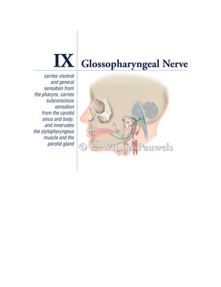
  • 2. IX Glossopharyngeal Nerve CASE HISTORY Allen is a 43-year-old construction worker. While he was eating dinner, Allen developed pain in the left side of his throat. The pain was short, brief, and stabbing in nature and only occurred when he swallowed. Allen was eating fish at the time and assumed that a fish bone was lodged in his throat. He went to the emergency department and was assessed by an otolaryngologist, but nothing was found. It was assumed that a fish bone had scratched Allen’s throat and that his symptoms would soon resolve. A month later, Allen’s pain was still present; in fact, it had increased. The pain occurred not only when he swallowed but also when he spoke and coughed. Again, while eating din- ner, Allen experienced the same pain with swallowing. He got up from the table, but after a few steps he collapsed unconscious to the floor. Although Allen quickly regained consciousness, he was immediately taken to the emergency department where he was put on a heart rate and blood pressure monitor to evaluate the function of his cardiovascular system. While he was on the monitor, the emer- gency room physician noticed an interesting correlation. Whenever Allen swallowed, he had a paroxysm (a sudden recurrence or intensification of symptoms) of pain followed invariably within 3 to 4 seconds by a decrease in heart rate (bradycardia) and a fall in blood pressure (hypotension). Allen was examined by a neurologist who noticed no evidence of reduced perception of pharyngeal pinprick or touch, or of pharyngeal motility. A diagnosis of glossopharyngeal neuralgia was made. Allen was treated with carbamazepine. His attacks of pain stopped and his cardiovascular function returned to normal. ANATOMY OF THE GLOSSOPHARYNGEAL NERVE The name of the glossopharyngeal nerve indicates its distribution (ie, to the glossus [tongue] and to the pharynx). Cranial nerve IX emerges from the medulla of the brain stem as the most rostral of a series of rootlets that emerge between the olive and the inferior cerebellar peduncle. The nerve leaves the cranial fossa through the jugu- lar foramen along with cranial nerves X and XI (Figure IX–1). Two ganglia are situated on the nerve as it traverses the jugular foramen, the superior and inferior (petrosal) glossopharyngeal ganglia. The superior ganglion is small, has no branches, and is usually thought of as part of the inferior glossopharyngeal ganglion. The ganglia are composed of nerve cell bodies of sensory components of the nerve (Figure IX–2). 164
  • 3. Glossopharyngeal Nerve 165 © L. Wilson-Pauwels Spinal TYMPANIC BRANCH nucleus (plexus and nerve) of trigeminal nerve Nucleus Lesser petrosal solitarius nerve and foramen (rostral gustatory portion) Foramen ovale Inferior salivatory nucleus Otic ganglion Nucleus solitarius (visceral sensory portion) Jugular foramen Palatal arch Nucleus ambiguus Tonsil (branchial and visceral motor) Posterior Parotid third of tongue Superior and inferior gland glossopharyngeal ganglia from external ear MUSCULAR BRANCH Stylopharyngeus muscle CAROTID BRANCH Internal carotid artery TONSILAR LINGUAL BRANCH BRANCH Common Carotid sinus PHARYNGEAL carotid BRANCH External artery Carotid body carotid artery Figure IX–1 Overview of the glossopharyngeal nerve.
  • 4. 166 Cranial Nerves © L. Wilson-Pauwels CN V Pons Internal auditory meatus (cut) Internal carotid artery Foramen lacerum Tympanic plexus and nerve Carotid canal CN IX Foramen ovale Otic Lesser petrosal Glossopharyngeal ganglion nerve and ganglia foramen (CN IX) Internal jugular CN X CN XI foramen and vein Figure IX–2 Tympanic branch of cranial nerve IX (cut through the petrous temporal bone). As the nerve passes through the jugular foramen, it gives rise to six terminal branches. These are the tympanic, carotid, pharyngeal, tonsilar, lingual, and mus- cular branches (see Figure IX–1). GENERAL SENSORY (AFFERENT) COMPONENT The glossopharyngeal nerve carries general sensory signals from a small area of the external ear, tympanic cavity (see Figure IX–2), mastoid air cells, auditory tube, pos- terior third of the tongue, and entrance to the pharynx (Figure IX–3 and Table IX–1). The cell bodies of the sensory neurons are located in the inferior glossopha- ryngeal ganglion, and their axons contribute to the tympanic, pharyngeal, lingual, and tonsilar branches. The tympanic branch is formed by the union of the tympanic plexus axons and comprises general sensory and visceral motor fibers. The sensory fibers descend through the tiny tympanic canaliculus and join the main trunk of the glossopharyngeal nerve at its inferior ganglion (see Figure IX–2). Sensation from the pharynx, including the soft palate, tonsil, and the posterior third of the tongue, is carried in the pharyngeal, tonsilar, and lingual branches.
  • 5. Glossopharyngeal Nerve 167 Sensory cortex (head region) Posterior limb of internal capsule © L. Wilson-Pauwels Ventral posterior nucleus of thalamus Spinal nucleus of trigeminal nerve Spinal trigeminal tract Inferior glossopharyngeal ganglion General sensory from tympanic cavity General sensory from external ear General sensory from the pharynx General sensory from posterior third of tongue General sensory from the tonsil Figure IX–3 General sensory component of the glossopharyngeal nerve. Table IX–1 Nerve Fiber Modality and Function of the Glossopharyngeal Nerve Nerve Fiber Modality Nucleus Function General sensory Spinal trigeminal Provides general sensation from the posterior (afferent) one-third of the tongue, the tonsil, the skin of the external ear, the internal surface of the tympanic membrane, and the pharynx Visceral sensory Of the tractus Provides subconscious sensation from the (afferent) solitarius— carotid body (chemoreceptors) and from middle part the carotid sinus (baroreceptors) Special sensory Of the tractus Carries taste from the posterior one-third of (afferent) solitarius—rostral the tongue part (gustatory nucleus) Branchial motor Ambiguus Supplies the stylopharyngeus muscle (efferent) Visceral motor Inferior salivatory Stimulates the parotid gland (parasympathetic Ambiguus For control of blood vessels in the carotid body efferent)
  • 6. 168 Cranial Nerves The central processes for pain enter the medulla and descend in the spinal trigeminal tract to end on the caudal part of the spinal nucleus of the trigeminal nerve (Figure IX–4). From the nucleus, processes of secondary neurons cross the midline in the medulla and ascend to the contralateral ventral posterior nucleus of the thalamus. From the thalamus, processes of tertiary neurons project to the post- central sensory gyrus (head region) (see Figure IX–3). The same pathway is sus- pected for touch and pressure and is important in the “gag” reflex (see Case History Guiding Questions, #7). These sensations operate at a “conscious” level of awareness. VISCERAL SENSORY (AFFERENT) COMPONENT Visceral sensory fibers operate at a “subconscious” level of awareness. Chemorecep- tors in the carotid body monitor oxygen (O2), carbon dioxide (CO2), and acidity/ alkalinity (pH) levels in circulating blood and baroreceptors (stretch receptors) in the carotid sinus monitor arterial blood pressure. These sensations are relayed in the carotid branch of the glossopharyngeal nerve (Figure IX–5) to the inferior glos- sopharyngeal ganglion where the nerve cell bodies are located. From these neurons, central processes pass to the tractus solitarius to synapse with nucleus solitarius cells in the middle third of the nucleus (see Figure IX–4). From this nucleus, connections Nucleus solitarius Nucleus surrounds tractus solitarius solitarius (rostral portion) Inferior salivatory Axon of nucleus tractus solitarius Inferior cerebellar peduncle Spinal nucleus of trigeminal CN IX and CUT MEDULLA nerve jugular foramen Nucleus ambiguus (branchial and visceral motor) CN IX Olive Tractus solitarius CN X Tract of spinal Rootlet of CN XII trigeminal nucleus Rootlet of CN XI Nucleus solitarius (visceral sensory portion) © L. Wilson-Pauwels Pyramid Figure IX–4 Cross-section of the medulla at the point of entry of cranial nerve IX illustrating the nuclei associated with this nerve.
  • 7. Glossopharyngeal Nerve 169 © L. Wilson-Pauwels to to reticular hypothalamus formation Tractus solitarius Nucleus solitarius Inferior glossopharyngeal ganglion Carotid branch of glossopharyngeal nerve Carotid sinus Carotid body Figure IX–5 Visceral sensory component of the glossopharyngeal nerve—elevated brain stem. are made with the reticular formation and the hypothalamus for the appropriate reflex responses for the control of respiration, blood pressure, and cardiac output. Carotid Body The carotid body is a small (3 mm × 6 mm in diameter) chemoreceptor organ located at the bifurcation of the carotid artery (see Figures IX–5 and IX–6). Very low levels of O2, high levels of CO2, and lowered pH levels (increased acidity) in the blood all cause an increase in the frequency of sensory signal traffic in the carotid branch of the glossopharyngeal nerve. The transduction mechanism is not well understood. Glomus cells excite or inhibit sensory nerve endings in response to changing conditions in the blood. In
  • 8. 170 Cranial Nerves return, the nerve endings may alter the function of the glomus cells, possibly in a way that changes their sensitivity to O2, CO2, and pH. The carotid body is supplied by a plexus of autonomic efferent nerves; sympa- thetic nerves traveling with the blood vessels and glossopharyngeal and vagal parasympathetic nerves. Their role in carotid body function is unknown. Carotid Sinus The carotid sinus is a dilatation of the internal carotid artery at its origin off the common carotid artery and may extend into the proximal part of the internal carotid artery. It responds to changes in arterial blood pressure. The walls of the sinus are characterized by having a thinner tunica media and a relatively thick tunica adventitia. The adventitia contains many sensory nerve endings (stretch receptors) of the glossopharyngeal nerve that respond to increases in blood pressure within the sinus by initiating impulses that reflexively lower the pressure (see Figure IX–6). CN IX parasympathetic Inferior Carotid branch glossopharyngeal of CN IX ganglion in jugular foramen CN IX parasympathetic nerve and ganglion cell Glomus cell Carotid (chemoreceptor Thin tunica media body cell monitors O2, CO2, pH) Thick tunica adventitia (baroreceptor nerve endings Sympathetic nerve monitor arterial blood pressure) (from superior cervical ganglion) Carotid sinus External carotid artery Internal carotid artery and vascular rami © L. Wilson-Pauwels Actual size of carotid body Common (3 mm wide × 6 mm high) carotid artery Figure IX–6 The bifurcation of the common carotid artery demonstrating baroreceptors in the wall of the carotid sinus and chemoreceptors within the carotid body.
  • 9. Glossopharyngeal Nerve 171 SPECIAL SENSORY (AFFERENT) COMPONENT Taste sensation from the posterior one-third of the tongue (predominantly sour and bitter), including the vallate papillae, is carried by special sensory axons toward their cell bodies in the inferior glossopharyngeal ganglion. Central processes from these neurons pass through the jugular foramen, enter the medulla, and ascend in the tractus solitarius to synapse in the rostral part of the nucleus solitarius (gustatory nucleus) (Figure IX–7). Axons of cells in the nucleus solitarius then ascend in the central tegmental tract of the brain stem to reach the ipsilateral ventral posterior nucleus of the thalamus (some studies show bilateral projections but more recent studies indicate that the projection is ipsilateral). From the thalamus, fibers ascend Gustatory area in sensory cortex Primary sensory cortex for taste © L. Wilson-Pauwels Posterior limb of internal capsule Ventral posterior nucleus of thalamus Nucleus solitarius (rostral gustatory portion) Tractus solitarius Jugular foramen Inferior glossopharyngeal ganglion Posterior third of tongue Figure IX–7 Special sensory component (for taste) of the glossopharyngeal nerve.
  • 10. 172 Cranial Nerves through the posterior limb of the internal capsule to reach the primary sensory cor- tex in the inferior third of the postcentral gyrus where taste is perceived. See Chapter VII for a description of sensory transduction in the taste buds. BRANCHIAL MOTOR (EFFERENT) COMPONENT In response to information received from the premotor sensory association cortex and other cortical areas, upper motor neurons in the primary motor cortex send impulses via corticobulbar fibers through the internal capsule and through the basis pedunculi to synapse bilaterally on the lower motor neurons in the rostral part of the nucleus ambiguus (Figures IX–8 and IX–9). The axons of these lower motor neurons join the other modalities of cranial nerve IX to emerge as three or four rootlets in the groove between the olive and the inferior cerebellar peduncle just ros- tral to the rootlets of the vagus nerve (see Figure IX–8). The glossopharyngeal nerve then passes laterally in the posterior cranial fossa to leave the skull through the jugu- lar foramen just anterior to the vagus and accessory nerves. The branchial motor axons branch off as a muscular branch that descends in the neck deep to the styloid process of the sphenoid bone, curves forward around the posterior border of the sty- lopharyngeus muscle, and enters it to innervate its muscle fibers. The stylopharyn- geus muscle elevates the pharynx during swallowing and speech (see Figure IX–9). Corticobulbar input Nucleus ambiguus (rostral part) © L. Wilson-Pauwels Pyramid Inferior cerebellar peduncle Olive CN X CN IX Figure IX–8 Branchial motor component of the glossopharyngeal nerve (section through the cranial part of the medulla).
  • 11. Glossopharyngeal Nerve 173 Motor cortex (head region) © L. Wilson-Pauwels Nucleus ambiguus receives innervation CN IX from both CN X ipsilateral and contralateral motor cortices CN IX CN X CN XI Styloid process Stylopharyngeus muscle Superior constrictor muscle Middle constrictor muscle Inferior constrictor muscle Figure IX–9 Branchial motor component of the glossopharyngeal nerve.
  • 12. 174 Cranial Nerves VISCERAL MOTOR (PARASYMPATHETIC EFFERENT) COMPONENT Preganglionic neurons of the parasympathetic motor fibers are located in the infe- rior salivatory nucleus (see Figure IX–4) in the medulla. These neurons are influ- enced by stimuli from the hypothalamus (eg, dry mouth in response to fear) and the olfactory system (eg, salivation in response to smelling food). Axons from the inferior salivatory nucleus join the other components of cranial nerve IX in the medulla and travel with them into the jugular foramen (Figure IX–10). At the infe- rior ganglion, the visceral motor fibers leave the other modalities of cranial nerve IX as a component of the tympanic branch. They ascend to traverse the tympanic canaliculus and enter the tympanic cavity. Here they pass along the tympanic plexus on the surface of the promontory of the middle ear cavity. From the tympanic plexus, the visceral motor fibers form the lesser petrosal nerve that goes through a small canal back into the cranium to reach the internal surface of the temporal bone in the middle cranial fossa. The nerve emerges through a small opening, the lesser petrosal foramen, lateral to the foramen for the greater petrosal nerve (see Figure IX–10). The lesser petrosal nerve then passes forward to descend through the fora- men ovale to synapse in the otic ganglion that usually surrounds the nerve to the medial pterygoid muscle, a branch of V3. From the otic ganglion, postganglionic fibers join the auriculotemporal nerve (a branch of V3) to supply secretomotor fibers to the parotid gland (Figure IX–11). Sympathetic nerves reach the parotid gland from the sympathetic plexus surrounding the external carotid artery and its branches in the area. Greater petrosal nerve and foramen (CN VII) CN V © L. Wilson-Pauwels Pons Internal auditory meatus (cut) CN VII highlighting visceral motor fibers Tympanic plexus (sensory) and visceral motor nerve in the Foramen lacerum middle ear cavity Carotid canal CN IX Foramen ovale Internal carotid artery Otic ganglion Lesser petrosal foramen and Internal jugular nerve (CN IX) CN X CN XI foramen and vein Figure IX–10 Lesser petrosal nerve (visceral motor IX) and greater petrosal nerve (visceral motor VII) and surrounding structures (sagittal section through the jugular foramen showing cut petrous temporal bone).
  • 13. Glossopharyngeal Nerve 175 Sigmoid sinus and Hypothalamus CNs IX, X, XI entering the jugular foramen © L. Wilson-Pauwels Lesser petrosal nerve Dorsal and foramen longitudinal fasciculus Inferior V3 salivatory nucleus Foramen ovale Through jugular foramen Tympanic cavity Otic ganglion (off nerve to medial pterygoid) Auriculotemporal nerve (off V3 ) Parotid gland Sympathetic nerve to parotid gland Figure IX–11 Visceral motor component of the glossopharyngeal nerve.
  • 14. 176 Cranial Nerves CASE HISTORY GUIDING QUESTIONS 1. What is glossopharyngeal neuralgia? 2. What is the cause of glossopharyngeal neuralgia? 3. What is allodynia? 4. Why did swallowing cause Allen pain? 5. Why did Allen collapse on the floor? 6. Why was carbamazepine used to treat Allen? 7. What is the gag reflex, and why don’t we gag every time a bolus of food passes through the pharynx? 1. What is glossopharyngeal neuralgia? Glossopharyngeal neuralgia* is characterized by severe, sharp, lancinating pain in the region of the tonsil, radiating to the ear. It is similar to trigeminal neuralgia (see Chapter V) in its timing and its ability to be triggered by various stimuli. For exam- ple, pain may be initiated by yawning, swallowing, or contact with food in the ton- silar region. In rare instances, the pain may be associated with syncope (a fall in heart rate and blood pressure resulting in fainting). 2. What is the cause of glossopharyngeal neuralgia? Typically, glossopharyngeal neuralgia is idiopathic; that is, no cause can be identi- fied. Occasionally glossopharyngeal neuralgia is secondary to the compression of cranial nerve IX by carotid aneurysms, oropharyngeal malignancies, peritonsillar infections, or lesions at the base of the skull. 3. What is allodynia? Allodynia is pain that results from a touch stimulus that normally would not cause pain. The pain is usually burning or lancinating in quality. 4. Why did swallowing cause Allen pain? Sensory nerve endings in the mucosa of Allen’s pharynx were stimulated by the pas- sage of a bolus of food and by the movements of the underlying muscles involved *Some authors use the term vagoglossopharyngeal neuralgia or glossopharyngeal and vagal neuralgia, instead of glossopharyngeal neuralgia, implying that the pain can radiate into the distribution of the vagus nerve as well as that of the glossopharyngeal nerve. However, the original term, glossopharyngeal neuralgia, is recognized by most neurologists and is more commonly used.
  • 15. Glossopharyngeal Nerve 177 in swallowing, coughing, and speaking. These usually harmless stimuli set off a bar- rage of pain impulses within the nervous system. 5. Why did Allen collapse on the floor? Allen collapsed on the floor because his blood pressure dropped so low that he could not maintain adequate blood flow to his brain. The mechanism for Allen’s fall in blood pressure and heart rate is not well understood. One hypothesis is that when the pain is most intense, general sensory impulses from the pharynx stimulate the nucleus of the tractus solitarius, which, in turn, stimulates vagal nuclei. Impulses from the parasympathetic component of the vagus nerve act to slow the heart rate and decrease blood pressure. Alternatively, the rapidly firing general sensory afferents may, through ephaptic transmission,* give rise to action potentials in the visceral afferent axons from the carotid sinus as they travel together in the main trunk of the glossopharyngeal nerve. The carotid nerve would then falsley report increased blood pressure, causing a reflexive decrease in heart rate and blood pressure via connections in the brain stem. 6. Why was carbamazepine used to treat Allen? Carbamazepine is a sodium channel blocking drug that is used to treat a wide range of conditions from seizures to neuropathic pain. Carbamazepine reduces the neu- ron’s ability to fire trains of action potentials at high frequency and therefore short- ens the duration of the paroxysm and frequently abolishes the attacks. 7. What is the gag reflex, and why don’t we gag every time a bolus of food passes through the pharynx? The gag is a protective reflex that prevents the entry of foreign objects into the ali- mentary and respiratory passages. A touch stimulus to the back of the tongue or pharyngeal walls elicits the gag (see Clinical Testing). Since the passage of a bolus of food through the pharynx stimulates the tongue and pharyngeal walls, we have to ask why swallowing does not elicit a gag reflex. Although the exact mechanism is not well understood, it is generally accepted that when a swallowing sequence is initiated, probably by signals from the cerebral cortex to swallowing center(s) in the brain stem, simultaneous inhibitory signals are sent to the gag center in the brain stem to turn off the gag reflex. *Ephaptic transmission occurs when a rapidly firing nerve changes the ionic environment of adjacent nerves sufficiently to give rise to action potentials in them. These “artificial synapses” often result from a breakdown in myelin.
  • 16. 178 Cranial Nerves A gag is not a partial vomit. Four events occur in the gag reflex (Figure IX–12): A. An irritant is sensed in the mouth. B. The soft palate elevates and is held firmly against the posterior pharyngeal wall closing off the upper respiratory airway. C. The glottis is closed to protect the lower respiratory passages. D. The pharynx is constricted to prevent entry into the alimentary tract, the pharyngeal wall constricts, and the tongue moves the contents of the pharynx forward to expel the offending foreign object from the mouth. © L. Wilson-Pauwels Tongue Irritant Soft palate Posterior Elevated pharyngeal soft wall palate Epiglottis Glottis Esophagus Trachea A B © L. Wilson-Pauwels Expelled irritant Elevated palate Constricted Tongue pharynx movement Closed glottis Closed glottis C D Figure IX–12 Steps in the gag reflex. A, Irritant in the mouth. B, Soft palate elevates, closing the upper respiratory airway. C, Glottis is closed to protect the lower respiratory airway. D, Pharyngeal wall constricts causing expulsion of the foreign object.
  • 17. Glossopharyngeal Nerve 179 The sensitivity of the gag reflex can be affected by cortical events. It can be sup- pressed almost completely or enhanced such that even brushing the teeth or the sight of a dental impression tray can lead to gagging. CLINICAL TESTING Although cranial nerve IX comprises general sensory and motor, visceral sensory and motor, and special sensory components, from a practical point of view only the gen- eral sensory component is tested at the bedside. Clinically, cranial nerves IX and X are assessed by testing the gag reflex. The gag reflex involves both cranial nerves—the glossopharyngeal being the sensory afferent input and the vagus carrying the motor efferents. When assessing the gag reflex, right and left sides of the pharynx should be lightly touched with a tongue depressor (Figure IX–13). The sensory limb of the reflex is considered to be intact if the pharyngeal wall can be seen to contract after each side has been touched. (See also Cranial Nerves Examination on CD-ROM.) Figure IX–13 Testing the gag reflex by lightly touching the wall of the pharynx. ADDITIONAL RESOURCES Brodal A. Neurological anatomy in relation to clinical medicine. 3rd ed. New York: Oxford University Press; 1981. p.460–4, 467. Ceylan S, Karakus A, Duru S, et al. Glossopharyngeal neuralgia: a study of 6 cases. Neurosurg Rev 1997;20(3):1996–2000. Dalessio DJ. Diagnosis and treatment of cranial neuralgias. Med Clin North Am 1991;75(3):605–15. Dubner R, Sessle BJ, Storey AT. The neural basis of oral and facial function. New York: Plenum Press; 1978. p. 370–2. Eyzaguirre C, Abudara V. Carotid body glomus cells; chemical secretion and transmission (modulation?) across cell-nerve ending junctions. Respir Physiol 1999;115:135–49.
  • 18. 180 Cranial Nerves Fitzerald MTJ. Neuroanatomy basic and clinical. 3rd ed. Toronto: W.B. Saunders Company, Ltd.; 1996. p.147–50, 190–1. Glossopharyngeal neuralgia with cardiac syncope: treatment with a permanent cardiac pacemaker and carbamazepine. Arch Intern Med 1976;136:843–5. Haines DE. Fundamental neuroscience. New York: Churchill Livingstone; 1997. p. 261. Hanaway J, Woolsey TA, Mokhtar MH, Roberts MP Jr. The brain atlas. Bethesda (MD): Fitzgerald Science Press, Inc.; 1998. p. 180–1, 186–7, 194–5. Kandel ER, Schwartz JH, Jessel TM. Principles of neuroscience. 3rd ed. New York: Elsevier Science Inc.; 1996. p. 770–2. Kiernan JA. Barr’s the human nervous system. 7th ed. New York: Lippincott-Raven; 1998. p. 169–74. Kong Y, Heyman A, Entman ML, McIntosh HD. Glossopharyngeal neuralgia associated with bradycardia, syncope, and seizures. Circulation 1964;30:109–12. Loewy AD, Spyer KM. Central regulation of autonomic functions. New York: Oxford University Press; 1990. p. 182–4. Miller AJ. The neuroscientific principles of swallowing and dysphagia. San Diego: Singular Publishing Group, Inc.; 1999. p. 100–1. Moore KL, Dalley AF. Clinically oriented anatomy. 4th ed. New York: Lippincott Williams & Wilkins; 1999. p. 1104–5. Nolte J. The human brain. 4th ed. Toronto: Mosby Inc.; 1999. p. 288, 305. Verna A. The mammalian carotid body: morphological data. In: Gonzalez C, editor. The carotid body chemoreceptors. Austin (TX): Chapman & Hall, Landes Bioscience; 1997. p. 1–29. Young CP, Nagaswawi S. Cardiac syncope secondary to glossopharyngeal neuralgia effec- tively treated with carbamazepine. J Clin Psychiatry 1978;39:776–8. Young PA, Young JA. Basic clinical neuroanatomy. Philadelphia: Williams & Wilkins; 1997. p. 292, 296. Zigmond MJ, Bloom FE, Landis SC, et al. Fundamental neuroscience. Toronto: Academic Press; 1999. p. 1057–60, 1080–4.