
Recommended
More Related Content
What's hot
What's hot (20)
Viewers also liked
Similar to Congenital lateral ventriculomegaly.
Similar to Congenital lateral ventriculomegaly. (20)
More from Ritesh Mahajan
More from Ritesh Mahajan (20)
Recently uploaded
Recently uploaded (20)
Congenital lateral ventriculomegaly.
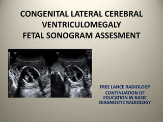
- 1. CONGENITAL LATERAL CEREBRAL VENTRICULOMEGALY FETAL SONOGRAM ASSESMENT FREE LANCE RADIOLOGY CONTINUATION OF EDUCATION IN BASIC DIAGNOSTIC RADIOLOGY
- 2. FETAL CEREBRAL LATERAL VENTRICULOMEGALY AN OVERVIEW • Ventriculomegaly : 0.3 to 0.5/ 1000 births . • Frank hydrocephalus is synonymous to overt lateral ventriculomegaly • Ventricles have three dimensional architecture &Variable degree of enlargement is appreciated in different trimesters • Initially assessment was done with – RATIO OF – MIDLINE to LATERAL WALL OF Case of fetal cerebral Lateral THE VENTRICLE MIDLINE TO IPSILATTERAL ventriculomegaly . CALVARIUM Both the lateral ventricles show significant and near symmetrical dilatation.
- 3. MILD LATERAL CEREBRAL VENTRICULOMEGALY • Mild lateral cerebral ventriculomegaly ( 10 to 15 mm ) • Isolated ventriculomegaly ( No consequence) . It could be earliest manifestation of brain damage . • CNS and NON CNS anomalies may be associated with it . – Primary cerebral maldevelopment ( obstructive hydrocephalus ) – Agyria – Destructive lesions – Periventricular leukomalacia – Hypoxia
- 4. MILD LATERAL CEREBRAL VENTRICULOMEGALY • NRA : NEAR CALVARIUM REVERBATION ARTEFACT LIMITS THE SONIC WINDOW HENCE – Transverse diameter of the ventricular atrium is taken at level of glomus of choroid . – Measurement is taken in the axial plane along the atrium distal to transducer . Inside echoes are measured • Values are – Mid trimester ( 6 to 7mm +_ 1mm) . – 14 to 40 wks ( 7mm +_ 1mm) . • Some degree of asymmetry in either side ventricles is noted . • Male fetus have larger measurements than the female ones.
- 5. MILD LATERAL CEREBRAL VENTRICULOMEGALY • Mild lateral cerebral ventriculomegaly ( 10 to 15mm inclusive). • If unilateral - benign. • Consider – Fetal MRI – Fetal ECG . – Fetal corpus callosum assessment – Fetal karyotyping. – First trimester screening. • To note : – X linked variety of hydrocephalus develops in lateral gestation. • Caesarean section may have to be opted due to associated macrocrania. • Cephalocentesis /Ventriculoamniotic shunting are also options
- 6. OVERT LATERAL CEREBRAL VENTRICULOMEGALY • Overt cerebral lateral ventriculomegaly or hydrocephalus . – Atrial width >15mm – ( 2 to third trimester) . – Associations • Neural tube defects . • Mid line anomalies . • Either aqueductal stenosis / communicating hydrocephalus . • Dangling choroid sign ( choroid away from transducer touches the ventricular wall and one near to the choroid abuts the interventricular septum ) .
- 7. CONSIDER FETAL MRI FETAL ECG KARYOTYPING ASSESS FETAL CORPUS CALLOSUM UNILATERAL FETAL VENTRICULOMEGALY USUALLY BENIGN
- 8. MEASURES THE AXIAL IMAGE OF THE VENTRICLE AWAY FROM THE TRANSDUCER AT THE LEVEL OF GLOMUS OF CHOROID. AVOID MEASUREMENT OF THE VENTRICLE NEAR TO THE TRANSDUCER DUE TO NRA : NEAR CALVARIUM REVERBATION ARTEFACT UNILATERAL FETAL CEREBRAL VENTRICULOMEGALY.
- 9. ASSESEMENT OF CORPUS CALLOSUM SHOULD BE DONE 3D / 4D IMAGING IS OF HELP . NORMAL CORPUS CALLOSUM IN CASE OF UNILATERAL VENTRICULOMEGALY ( USUALLY BENIGN)
- 10. DANGLING CHOROID SIGN CHOROID PLEXUS OF THE VENTRICLE AWAY FROM THE TRANSDUCER ABUTS THE LATERAL VENTRICULAR WALL AND OTHER CHOROID PLEXUS ABUTS THE SEPTUM. DANGLING CHOROID SIGN IN CASE OF BILATERAL FETAL VENTRICULOMEGALY
- 11. SMALL POSTERIOR FOSSA DILATED CENTRAL CANAL POSSIBLE ARNOLD CHIARI MALFORMATION IN CASE OF VENTRICULOMEGALY. FETAL MR CAN BE OF HELP. CNS ANOMALIES ASSOCIATED WITH VENTRICULOMEGALY