Cloud Frontiers: A Deep Dive into Serverless Spatial Data and FME
Elisa il 2-protocol
1. Interleukin-2 ELISA Assay
Enzyme-Linked Immunosorbent Assays (ELISAs) are
enzyme immunoassays widely used for detecting specific antibodies
but can be used to detect antigens also. This procedure employs the
specificity of both Ab and enzymes to achieve a highly sensitive and
precise test. It is one of the standard procedures used for the
detection of Ab to the HIV virus.
The ELISA takes advantage of the fact that most proteins will
bind firmly to the surface of several different kinds of plastic, usually
by hydrophobic interactions between the nonpolar residues of the
protein and the naturally hydrophobic components of the plastic.
ELISAs are normally performed on a solid phase. Most often the solid
phase consists of the surfaces of individual wells in multi-well plastic
plates (96-well plates are commonly used). In the assays, a series of
antibody-antigen or antibody-antibody interactions are used to bind
enzyme molecules to the bottom of a 96-well plate in such a way that
the amount of enzyme is proportional to the amount of antibody/
antigen in the system. The amount of enzyme activity is then
measured using a color producing substrate. From this the amount of
antibody/antigen can be calculated.
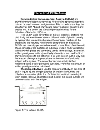
The indirect ELISA is used to measure antibody. In this type of
ELISA (figure 1), the antigen (peptide or protein) is bound to the
polystyrene microtiter plate first. Proteins like to stick irreversibly to
virgin plastic (passive absorption) and most of the plastic surface will
become coated with the antigen.
Figure 1: The indirect ELISA.
2. Any "unused" plastic surface is then "blocked" with an irrelevant
protein to prevent nonspecific binding of any of the subsequent
additives. Serum or some other sample containing the primary
antibody is then added to the well and allowed to bind. Next, samples
are removed and the wells are "washed" several times with an
appropriate buffer solution. Finally, a second antibody, specific for the
first antibody and labeled for detection, is added to the well and
allowed to bind; the second antibody usually has an enzyme
conjugated to it (e.g. horseradish peroxidase). This enzyme catalyzes
the formation of a colored substance from the substrate which is then
quantified and the amount of antibody/antigen present can be
calculated.
The ELISA procedure as outlined above can be modified in
several ways. One important way is to convert it to detect and
quantify antigen (sandwich ELISA). This is accomplished by first
layering an Ab specific for the Ag in question on the plastic wells.
After "blocking" you add the sample to be analyzed. If it contains that
antigen it will bind to the "solid phase" Ab-specific preparation. You
then again add a preparation of antibodies which are specific to the
antigen, this time with an enzyme coupled to them. Cleavage of
subsequently added substrate is an indication that antigen is present,
causing the enzyme labeled second antibody to bind.
Another variation for measuring amounts of antigen is the
competitive ELISA. In this technique, samples containing antigen
are added to a pre-coated antibody plate. Addition of an enzyme-
conjugated secondary antibody specific for the isotype of the primary
antibody is also added. The antigen and the enzyme-conjugated
secondary antibody “compete” for binding sites with the primary
antibody that is coating the wells. After washing the wells and adding
a color-producing substrate, the intensity of the color (the absorbance
value) is inversely proportional to the amount of antigen contained
within the sample.
Cytokines and Interleukins
Effective immune responses require communication between
several cell types, namely lymphoid cells, inflammatory cells, and
hematopoeitic stems cells. Chemical messengers called cytokines
are used to mediate such interactions between these cells.
Cytokines are proteins that are secreted by leukocytes and various
3. other cells within the body in response to a number of stimuli.
Cytokine proteins assist in regulating the development of immune
effector cells, and some cytokines possess effector functions of their
own.
Interleukins are a discrete group of cytokine messenger
molecules that help coordinate intercommunication between
leukocytes (hence the name “interleukins”). Arguably, the most
important protein of the interleukin family is Interleukin-2 (IL-2)
because of its instrumental role in the body's natural response to
microbial infection and in discriminating between foreign (non-self)
and self. The primary function of IL-2 is to act as a facilitator,
mediator, and regulator of T cell activation and proliferation.
However, in addition to regulating T cell proliferation, IL-2 can
regulate Natural Killer cell activation and proliferation, B cell
proliferation, and can be used to facilitate the manufacturing of
immunoglobulins originating from B cells.
In this experiment, you will perform a sandwich ELISA to
determine the amount of IL-2 in an unknown sample. To do this, you
will generate a standard curve from which you will be able to
calculate the quantity of your unknown.
OBJECTIVES:
Understand how the ELISA method works.
Be able to compare and contrast the different ELISA
methods.
Understand the role of cytokines and interleukins,
specifically Interleukin 2.
Materials:
Mouse Interleukin-2 ELISA kit purchased from eBioscience that
includes the following:
• 96-well microtiter plate.
• Capture antibody: 500µL
• Detection antibody: 500µL
• Mouse Interleukin-2 standard: 20µL
4. • Substrate: 100mL. Stabilized 3,3’, 5,5’ Tetramethylbenzidine
plus hydrogen peroxide. Light sensitive.
• 5X Assay Diluent: 150mL. Diluted 5 fold with deionized water.
• Enzyme (Avidin-HRP): 500µL
1X PBS with 0.05% Tween 20 wash buffer
2N H2SO4 stop solution
ELISA plate reader
Procedure:
Day 1: Plate the standards and the samples
*You will be working in tables for this experiment
1. Add 100µL per well of capture antibody in coating buffer to the
appropriate wells of your table’s 96 well microtiter plate. Seal
the plate and incubate overnight at 40C (T.A. will do this).
2. Aspirate wells and wash three times with 200µL per well of 1X
PBS with 0.05% Tween 20 wash buffer using a multi-channel
pipettor (T.A. will do this).
3. Block wells with 200µL per well of 1X Assay Diluent. Incubate
at room temperature for 1 hour (T.A. will do this).
4. Aspirate wells and wash three times with 200µL per well of 1X
PBS with 0.05% Tween 20 wash buffer using a multi-channel
pipettor.
5. Label seven 15mL conical tubes for performing a serial dilution
of the top standard. Perform 2-fold serial dilution of the top
standard using 1X Assay Diluent to make the standard curve.
6. Add 100µL per well of standard to the appropriate wells.
7. Add 100µL per well of an unknown sample to the appropriate
wells. T.A. will provide you with an unknown sample.
5. 8. Once the standards and the unknown sample have been
added to the appropriate wells, seal the plate and incubate for
48hrs at 40C.
Day 2: Reading the IL-2 ELISA assay
1. Aspirate wells and wash three times with 200µL per well of 1X
PBS with 0.05% Tween 20 wash buffer using a multi-channel
pipettor (T.A. will do this).
2. Add 100µL per well of detection antibody diluted in 1X Assay
Diluent. Seal the plate and incubate at room temperature for 1
hour (T.A. will do this).
3. Aspirate wells and wash three times with 200µL per well of 1X
PBS with 0.05% Tween 20 wash buffer using a multi-channel
pipettor.
4. Add 100µL per well of Enzyme (Avidin-HRP) diluted in 1X
Assay Diluent. Seal the plate and incubate at room
temperature for 30 minutes.
5. Aspirate wells and wash three times with 200µL per well of 1X
PBS with 0.05% Tween 20 wash buffer using a multi-channel
pipettor
6. Add 100µL of substrate solution to each well. Seal the plate
and incubate for 15 minutes at room temperature.
7. Add 50µL of stop solution to each well.
8. Read plate at 450nm.
9. Average the absorbance values of the triplicates for the
standards and use these averages to create a standard curve
with the concentration of IL-2 in pg/ml on the X axis and the
average absorbance value on the Y axis. Include Y error bars.
6. 10. Using the y equation from your standard curve, determine the
concentration of IL-2 (pg/ml) in your unknown sample.