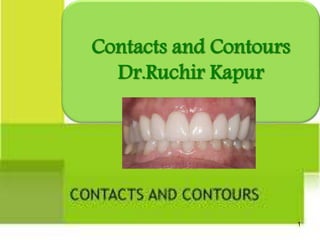
3. Definition
Contact area : Contact areas are the places on the
proximal surfaces of tooth crowns where a tooth
touches the tooth adjacent to it in the same arch
when the teeth are in proper alignment.
Contours : Convexity on facial and lingual surfaces
that affords protection and stimulation of supporting
tissues during mastication.
3
4. Contact point : refers to the occlusal cusp of a tooth that
touches the occlusal portion of another tooth in the opposing
arch.
Embrasures : (spillways)
• “V” shaped spaces that originate at the proximal contact areas
between adjacent teeth and are named for direction towards
which they radiate.
• Incisal
• Occlusal
• Gingival
4
5. Proximal contact areas and contours
Proximal contact area : Area of proximal height of
contour of mesial or distal surfaces of a tooth that
touches or contacts its adjacent tooth in the same arch.
Location :
In anterior teeth : Located closer to the incisal surface
of the teeth.
5
6. In posterior teeth : Located near the junction
of incisal and middle thirds or in the middle
third.
6
8. Tapering teeth :
Incisors :
• Inciso apically- starts near incisal edges.
• Labio lingually- slightly labial to incisal
edges.
Canines : Very angular
• Mesial contact- close to incisal edges
• Distal contact- center of the distal surface.
8
9. Premolar : Angular.
• Crowns constricted cervically with long cusps.
• Taper lingually , contact area occur bucally-at buccal axial
angle.
• Contacts begin 1 mm gingivally from crest of marginal
ridges.
• At junction of occlusal and middle 3rd of crown.
9
10. Molar :
Mesial contacts :
• Bucco-lingually : mesio-buccal axial angle of the tooth.
• Occluso-gingivally : at distance 1/3rd to ½ from the occlusal
surface to C.E.J.
• Distal contacts :
• Bucco-lingually : shift lingually to the middle third.
• Occluso-gingivally : at middle third.
Lingual shifting of contacts - prominent in mandibular
molars.
10
11. Embrasures :
Anterior :
• Incisal and labial - negligible.
• Gingival and lingual - largest and widest.
Posterior :
• Buccal – small.
• Occlusal - wide and deeper.
• Gingival & lingual - wide and broad.
11
12. Proximal contour :
Common feature :
• Starting at CEJ, surface presents concavity to contact areas ,
and convex from there to crest of marginal ridges.
• Concavities are pronounced on mesial surface.
• Most frequently- teeth with buccal and lingual roots.
• Most pronounced on mesial of maxillary 1st premolar.
12
13. Square teeth : Bulky & angular, little
cervical constriction & proximal surfaces
devoid of curves.
Incisors :
• Incisally – at incisal ridge.
• Labio lingually – in a line with incisal edge.
Canines :
• Incisally – close to incisal ridge.
• Labio lingually – in a line.
13
14. Premolar : broad contacts , short cusps.
• Bucally : towards buccal axial angle (buccal 3rd).
• Occlusally : at occlusal 3rd.
Molar :
• Mesial contact : Crowns tapered lingually.
• Bucco-lingually : buccally -buccal axial angle.
• Occluso-gingivally : From a mere line contact to half the height
of crown.
• Distal contact :
• Bucco-lingually : midline of crown.
• Occluso-gingivally : occlusal third. 14
15. Embrasures :
• Incisal, labial, occlusal - Nil.
• Gingival embrasure - barely noticeable, or may extent 1/3rd
height of crown.
• Buccal embrasure -when present, very narrow & flat.
• Lingual embrasure - Narrow or wide.
Proximal contour :
• Plane instead of curved.
• Bucco-lingual concavities : may be on mesial surface of max. 1st
premolars, 1st & 2nd molars, mesial surface of mand. 1st molar.
• Distal surfaces are flat or slightly convex.
15
16. Ovoid teeth : Transitional type, surfaces
primarily convex.
Incisors :
• Labio-lingually : lingual to incisal ridge.
• Mesial contacts : start at 1/4th of crown
(Inciso gingivally).
• Distal contacts : start at 1/3rd – ½ of
crown.
Canines :
• Incisally close to incisal ridges. 16
17. Premolar :
• Short cusps.
• Convexity of marginal ridge carries the contacts almost to the
middle third.
Molars :
• Mesial contacts – at junction of buccal and middle third of
the crown.
• Distal contact – in line with central groove on the occlusal
surface.
17
19. Marginal Ridges
Elevated rounded ridges located on mesial and distal edges
of occlusal surface of tooth.
Should be formed in two planes bucco-lingually, meeting
at very obtuse angle.
This specification is essential for :
The balance of the teeth in the arch.
Prevention of food impaction proximally.
Protection of the periodontium.
Prevention of recurrent and contact decay.
For helping in efficient mastication.
With age marginal ridges and occlusal
embrasures are reduced, due to vertical
occlusal attrition and proximal flattening of
contact areas.
19
20. Facial and lingual contours
Cervical ridge.
Extend not more than 1mm beyond cervical line.
On labial, buccal & lingual of maxillary teeth & buccal of
mandibular posterior - uniform.
Average curvature about 0.5mm or less.
The canines have more curvature than central and lateral
incisors.
Mandibular anterior - <0.5mm.
Mandibular posterior (lingually) - 1mm with crest of
curvature at middle 3rd instead of cervical 3rd.
20
21. In molars since there are more than one cusp the convexities
are interrupted by concavities at occlusal ½ - 2/3rd of the
crown.
Upper anterior teeth concavities determinant for mandibular
movement.
21
22. Hazards of faulty reproduction of
features of teeth in restorations
Contact size :
Creating a contact area i.e. too
broad, bucco-lingually or
occluso-gingivally.
• Produces an interdental area
that is less cleanable.
Creating a contact area i.e. too
narrow, bucco-lingually or
occluso-gingivally.
• Food is impacted vertically and
horizontally on col area. 22
23. Open (loose) contact.
• Creates continuity of the
embrasures with each other and
with the interdental col.
All of these defects will
allow food impaction and
accumulation of bacterial
plaques, with
accompanying
periodontal problems and
caries.
23
24. Contact configuration :
Contact area i.e. flat (deficient convexity) can make it broad
bucally, lingually, Occlusally, and /or gingivally.
Contact with excessive convexity will diminish the extent of
contact area.
Concave contact area in a restoration.
• Occurs restoring adjacent teeth simultaneously.
• Adjacent restoration with convex proximal surface .
• Interlocking between the concavity and convexity can
immobilize the contacting teeth.
24
25. Contour :
Facial and lingual convexities :
• More danger in overconvex facial and lingual surface.
• Can create undisturbed environment for accumulation and
growth of cariogenic and plaque ingredients at gingival
margin.
• No apparent hazards from underconvex curvatures.
25
26. Facial and lingual concavities :
• Concacavities occlusal to height of contour- involved in
occlusal static and dynamic relations.
• Deficient or mislocated concavities- premature contacts, that
inhibit physiological capabilities of mandibular movements.
• Concavities apical to height of contour - essential for
maintenance of accompanying new components of adjacent
periodontium.
• Deficient concavities lead to overhanging restoration.
26
27. Marginal ridges :
Absence of a marginal ridge in the restoration.
Marginal ridge with an exaggerated occlusal embrasure.
27
28. Adjacent marginal ridge not compatible in height.
Marginal ridge with no occlusal embrasure.
28
30. A one-planed marginal ridge in the bucco-lingual direction:
• Creates premature contacts during functional and static
occlusion.
• Increases depth of adjacent triangular fossa magnifying stresses in
this area.
• Increase height of marginal ridge in center, making it amenable
to adverse effects of horizontal components of force.
30
31. A thin marginal ridge in its mesio-distal bulk.
Marginal ridges not compatible in dimension or location with
the rest of the occluding surface.
31
32. Procedures for formulations of proper
contacts and contours
I. Tooth movements.
II. Matricing.
I .Tooth movements :
• Act of either separating the involved teeth from each other,
bringing them close to each other or changing their spatial
position in one or more dimension.
Objectives.
Bring drifted, tilted or rotated teeth to their indicated
physiological positions.
32
33. Close space between teeth not amenable to closure by
contemplated restoration.
Move teeth to another location, so when restored will be
in position most acceptable by periodontium.
Move teeth Occlusally or apically.
Move teeth from non-functional or traumatically
functional location to a physiologically functional one.
33
34. Move teeth so that when restored, they may be in
most esthetically pleasing position.
Move the teeth in location to increase dimensions of
available tooth structure for resistance and retention
forms.
Create space sufficient for matrix band
interproximally.
34
35. Two principle methods of tooth movements / separation.
Rapid or Immediate tooth movement / Separation.
Slow or Delayed tooth movement / Separation.
Rapid or Immediate tooth movement / Separation.
• Mechanical type of separation that creates, either proximal
separation at point of separators introduction and / or
improved closeness of proximal surface opposite the point of
separators introduction.
• Can be used preparatory to slow movement or maintain
space gained by it.
35
36. • Should not exceed 0.2 – 0.5 mm.
Achieved by two principles :
Wedge method
Traction method
Wedge method :
• Separation is achieved by placing pointed wedge shaped
device between the teeth and slowly inducing pressure in
order to create space at the contact area.
• Ex : Elliot separator, wedges. 36
37. Elliot separator/ crab claw separator / Single bow
separators :
Indications:
• Short duration separation that does not necessitate
stabilization.
• Examining proximal surfaces or in final polishing of restored
contacts.
Procedure:
• Adjust the two opposing wedges of the separator
interproximally gingival to the contact area.
• Move the knob clockwise so that the wedges moves towards
each other establishing desired separation.
37
39. Wooden wedges :
• Easily cut & Trimmed
• Absorb moisture intra orally to swell and expand slightly,
thus improve proximal retention of band .
• Relatively flexible .
• Economical.
• Example : Orangewood, Hemowedges Maplewood, pine
(soft) ,Oak (hard)
39
40. Plastic / Resin Wedges :
• Opaque/ Transparent
• Can be plastically molded and bent to correspond with the
configuration of interdental col.
• Can transmit light , suitable for light cured restorations.
• Relatively rigid hence easy tooth separation.
40
42. Functions :
Assure close adaptability of matrix band to tooth, gingival to the
gingival margin of preparation.
Occupy space designated to be the gingival embrasure.
Create some separation to compensate for thickness of matrix
band.
Assure immobilization of the matrix band.
Protect interproximal gingiva from trauma. 42
43. Wedging techniques
Piggy back wedging :
Indications –
• Gingival recession.
• Shallow proximal box.
Small wedge “piggy back” is used over the first wedge to
ensure proper contour.
43
44. Double wedging :
Indication :
• Spacing between the adjacent teeth.
• Wide proximal box in buccolingual dimensions
• Two wedges, one from buccal aspect and one from lingual
aspect.
44
45. Wedge wedging :
Indication :
• Concavity present on proximal surface.
• Second wedge is inserted between first wedge and band so
that opening is eliminated and the matrix band is well
adapted to the gingival margin of the prepared cavity.
45
47. Matrices
Defination : Device that is applied to a prepared tooth before
the insertion of the restorative material to assist in
development of appropriate tooth contours and in order to
confine the restorative material excess.
47
48. Ideal requirements :
Should replace the missing wall temporarily .
Should be easily inserted and removed .
Should be sufficiently rigid to retain contour given to it.
Should not react or adhere to the restorative material .
Should resist the condensation pressure.
Should be comfortable for the patient .
Should be small and handy so that access and visibility is not
affected. 48
49. Objectives :
To act as a temporary wall of resistance during insertion and
hardening of the material.
To displace or retract gingiva and rubber dam.
To achieve dryness and non-contamination of operating field.
To resist and compensate for dimensional changes that occur
during setting.
To maintain natural contact and contours .
To promote health of inter dental gingiva by preventing
overhanging restorations. 49
50. Classification of matrices :
Based on mode of retention :
Mechanical retained matrices :
• Ex : Tofflemire, Ivory no.1 and 8.
Self retained matrices :
• Ex : Black’s matrix and copper band supported by
impression compound.
50
51. Gillmore’s classification :
Custom made :
• Prepared by dentist or assistant suitable size matrix is cut and
impression compound placed in the place of wedge.
Mechanical :
• Tofflemire, Ivory no. 1 and 8.
Miscellaneous :
• T-Band, soldered band, seamless copper band, orthodontic
band, blacks matrix.
51
52. Types of matrices
Ivory No. 1
• Band encircles one of posterior proximal surfaces, indicated
in unilateral Class II cavities.
• Band is attached to retainer through wedge shaped
projections which engage tooth through embrasures of
unprepared surface.
52
53. Ivory No. 8 :
• Band encircles entire
crown.
• Indicated for bilateral
class II cavities.
• Circumference of band
can be adjusted by
adjusting screw.
53
54. Tofflemire
• Universal matrix.
• Designed by B.R.Tofflemire.
• Indicated -3 surfaces of posterior tooth have been
prepared.
• Commonly used for two surfaces class II restorations.
• Bands are available in 2 thickness :
• 0.05 mm
• 0.038 mm
54
55. Advantages :
Can be used both from facial / lingual side.
Economical.
Provides good contacts and contours.
Can be easily removed.
Types :
• Straight .
• Contra angle.
55
57. Black matrix
By Dr. G.V.Black.
“Ligated matrix band”.
• One of earliest custom-made
matrices.
• Thin metal plate (copper, brass,
German silver, or stainless steel)
used as a matrix.
• Band must encircle about ½ of
tooth.
• Ligature wrapped 2 or 3 times
around the tooth, including
matrix band and tied.
57
58. Automatrix
Retainerless matrix system.
Types of bands :
• 3/16 inch (4.8 mm) wide, 0.002 inch thick.
• ¼ inch (6.35 mm) wide, 0.002 inch & 0.0015 inch
thick.
• 5/16 inch (7.79 mm) wide, 0.002 inch thick.
58
59. Indications
• Complex
amalgam
restorations-one
or more cusps to
be replaced.
Advantages
• Convenient.
• Improved visibility
due to lack of a
retainer.
• Autolock loop can
be positioned
facially or
lingually.
• Rapid application.
Disadvantages
• Bands are flat-
difficult to
burnish.
• Cannot develop
proper contacts
and contours.
• Expensive.
59
60. Copper band matrix
• Assorted sizes make excellent
matrices.
• Cylindrical shape.
• Bands are heated to redness in flame
& quenched alcohol.
• With contouring pliers band is
contoured.
• Festooned with scissors so, gingival
periphery of it corresponds to
gingival curvature & CEJ.
• To stabilize, wedges are placed.
60
61. Indications
• Badly broken
teeth.
• Class II cavities
with large buccal
or lingual
extensions.
Advantages
• Provide
excellent
contour.
Disadvantage
• Time
consuming.
61
62. Plastic matrix strips
• Transparent plastic strips.
• Celluloid strips(cellulose nitrate) – silicate
cements.
• Cellophane (cellulose acetate) – resins.
• Mylar strips for composite and silicate
restorations.
• Band should extend at least 1mm beyond
gingival & incisal margins of cavity, can be
stabilized by wedge.
• After inserting composite resin material
the matrix is pulled tightly around tooth
following which light curing is done.
62
63. • Once restoration is completed, wedge can be removed &
matrix strip slid out of the proximal surfaces of the
contacting teeth.
63
•
64. T band matrix
• Preformed T shaped stainless steel matrix band without retainer.
• Long arm of ‘T’ is bent to surround tooth circumferentially.
• Overlaps short horizontal arm of ‘T’ which is bent over long
arm and helps to retain shape.
Indication :
• Class II cavities.
Advantages :
• Simple and inexpensive.
• Rapid and easy to apply
Disadvantages :
• Flimsy in structure, not very stable.
64
65. Compound supported matrix
Custom made matrix / Anatomic matrix.
5/16th inch wide, 0.002 inch thick stainless steel band used.
Contoured with an egg-shaped burnisher.
Band is stabilized by softened impression compound.
Indication :
For restoring class II cavities involving one or both proximal
surface.
For complex situations like pin-amalgam restoration.
65
66. Advantages :
Highly rigid and stable.
Provides good access and visibility
Most efficient means of reproducing contact and
contour.
Disadvantages :
Time consuming.
66
67. Precontoured sectional matrix strips
• Small, Precontoured dead soft metal
matrices ready for application.
• Selected according to tooth to be
restored.
• Band held by a flexible metal ring -
Bitine ring.
• E.g. Palodent matrix system (by
Dentsply) , Triodent matrix system.
67
68. Indication :
• Class II cavities.
• Amalgam and composite restorations.
Advantages :
• Ease of application.
• Provide better proximal contours for posterior composite
restorations.
Disadvantages :
• Expensive.
68
69. Conclusion
“ We as a clinician or a
restorative dentist should have
an adequate knowledge of the
anatomical & functional aspects
of contacts & contours so as to
reproduce them with ideal
restorative material, which in
turn will help to maintain the
oral cavity in sound
health”…….
69
70. References
Sturdevant’s Art and Science of Operative Dentistry : 5th
edition, South Asian edition.
Operative dentistry : Modern theory and practice ,
Mohamed A Marzouk.
Text book of operative dentistry, Nisha Garg, Amit Garg.
Wheeler’s Dental Anatomy, physiology, and occlusion :
8th edition.
70
Initially after tooth eruption there is only a contact point but due to wear during physiological tooth movement proximal contact point becomes contact area.
Proximal contact areas are larger in molar region which helps prevent food impaction during mastication.
Crowns tapered lingually, contact areas occurs buccally at the buccal axial angle (bucco-lingually).
Embrasures are V shaped space or spillways that originate at the
proximal contact areas between adjacent teeth and are named for the
direction towards which they radiate.
Diagram : forces 1 and 2 acting on 2 adjacent marginal ridges, will ve hz componenets 1H and 2H drive the two teeth toward each other, thus preventing any impaction proximally , maintaining mesio distal dimension of dental arch and anchoring teeth against each other.
In mandibular posteriors due to lingual inclination.
Related to 1st diag ; it will change anatomy of interdental col also. Normal saddle shaped will become broadened.
A – overconvex B- underconvex C- normal contour.
Underconvex leads to irritation of soft tissues.
Force 1 will be directed toward the proximal surface of the adjacent teeth.
1st diag . Absence of ridge.:The horizontal components of the force 1h and 2h tend to drive the two teeth away from each other.
Vertical component V1 &V2 impact food & intraoral material interproximally.
Diag 2:with an exaggerated occlusal embrasure.
Exaggerating the occlusal embrasure will direct forces 1 and 2 toward the adjacent proximal surfaces, with the horizontal components, 1H and 2H, separating the teeth and the vertical components, 1V and 2V, driving debris interproximally.
1st diag .: Constructing a restoration with a marginal ridge higher than the adjacent one will allow force A to work on the proximal surface of the restoration. The horizontal component, AH, will drive the restored tooth away from the contacting tooth, and the vertical component will drive debris interproximally.
Even in the presence of force B, with its horizontal component acting on the adjacent marginal ridge, there will be some separation of teeth as the surface hold for force B is too small to counteract that of force A.
Diag 2: In this case, the two adjacent marginal ridges will act like a pair of tweezers grasping food substance passing over it. Although debris may not be forced interproximally, it will be very difficult to remove once it is thus trapped.
In this situation there are no occlusal planes in the marginal ridges for the occlusal forces to act upon, so there are no horizontal components to drive the teeth toward each other, closing the contact. Furthermore, the vertical force will tend to impact food interproximally.
Diag 1 : Susceptible to fracture or deformation.
If exceeds 0.5 mm , may lead to periodontal ligament tear at one site and crush them at other.
Barton’s matrix.
Concavity occurs on surface of fluted root, most prominent on mesial surface of max. 1st premolar.
Width and den thickness
Compund can also be used to further stabilize the band.
Wire staple is inserted facio lingually in compound to stabilize it.
Badly broken teeth…esp those receiving pin retained amalgam restorations.
2. (allow light transmission during polymerization of composite resins).
5/16th inch wide, 0.002 inch thick stainless steel band cut enough to wrap around 1/3rd of facial and lingual surface.