SlideShare une entreprise Scribd logo
1  sur  37
HISTOLOGY: INTRODUCTION “ What  is  going  on ?” Pulling it together Regions Organs Molecules Tissues Connections Cells Parts Organelles Development Functions Systems
Noon talks for Internal-Medicine residents’ Board prep   Two  recurring  themes  --   Is  it  what  it  appears  to  be ?   Does the treatment/procedure do what is claimed for it ? What is the  evidence ?
MEDICINE:  Some aspects What gives with this patient ? Regions Molecules Tissues Connections Cells Parts Organelles Development Functions Regions Systems Systems  abnormal Parts Connections Development Tissues Cells Organelles Molecules Functions Microbes Medicines Age Populations Costs ? Gender Organs Organs  Abnormal
Abnormal variants for all the earlier fields of knowledge Developing judgment  -  weighing various contributions for relevance & quality of evidence Foretaste of the ‘pulling it together’ in the PBL experiences, but much omitted, e.g., therapy, follow-up, cost;  likewise  for  clinical  correlations This doubling, plus more fields, e.g. microbes, is why medical training takes several years Any twit can lay hands on an LCD projector, and push images at you  reminds  one that the story may be faulty;  it is one of many;  and there are omissions Feel for the aspects that yield valid risk factors in  this particular diagnosis ?
PORNOGRAPHY  &  “THE REAL THING” Images   versus   REALITY What is the  evidence  for the real?
Images   versus   REALITY -  Functional   Anatomy REALITY is  the  living   person, often via images Surface anatomy   Palpation   Endoscopy+   Radiology   PET scans   Ultrasound   Doppler flows  Gait & Reflexes   etc Biopsies   Fine-Needle Aspiration   Cervical, Blood,   etc  Smears   Flow cytometry & cell sorting     Cell culture & grafting   etc   (Bits cut or sucked out  for microscopy)
REALITY is the  dead  person DISSECTION   [ Surface anatomy   Endoscopy   Palpation   Radiology   Ultrasound   are  sometimes useful  as  adjuncts  to  autopsy & histology correlations] Organs and large pieces cut out, examined, & prepared for MICROSCOPY-  histology & histopathology  (normal & altered side-by-side)
Images   versus   REALITY -  Anatomy In Anatomy,  the source of the evidence - the essential  point of reference - is   the  cadaver  for Gross  &  the  microscope slide  for Histo   As the physician is knowledgeably comfortable with the  patient’s   gross  &  microscopic  structure  and  its implications, you will become confident at the cadaver &  the  microscope,  and  with  the  resulting images  TESTS focus on the cadaver, the slides, and interpreting images -  identification, interpretation, & synthesis Bed-rock
MICROSCOPIC SLIDE  Side view of slide Glass coverslip Glass slide  1”X3” Tissue Section Mounting medium Mounting medium:  permeates section; fastens coverslip to slide; is clear; has refractive index as for glass Label
SLIDE USE - Cautions GLASS IS FRAGILE   !   Take care with individual slides & especially with the boxes of slides The slide must go on the stage  coverslip up The high-dry & oil objectives cannot focus through the thickness of the slide to the section The label may have been put on the non-coverslip side, as shown ~ Slides & Microscope remain in the teaching Lab, always! Label
SLIDE PREPARATION I  Steps Excise &  Fix  (preserve) the tissue in fixative Remove the water &  replace  with wax-solvent Imbed  the oriented specimen in molten wax After it is solid, hold the  wax block &  cut  slices Mount  the thin slices (sections) on slides When dry, remove the wax, &  stain  the section Remove surplus stain & water;  mount coverslip   When mounting medium has set, do  microscopy
50 % ethanol 70 % ethanol 95 % ethanol 100 % ethanol benzene/xylene Dehydrating series paraffinwax Remove the water &  replace  with wax-solvent Imbed  the oriented specimen in molten wax Miscible with ethanol; dissolves wax Fresh tissue 10% Formalin  fixative label
MICROTOME - a fancy meat-slicer - holds the wax block, & cuts off thin slices, as the block is slowly advanced mechanically Block Knife Section Glass slide Water-bath After it is solid, hold the  wax block &  cut  slices Mount  the thin slices (sections) on slides Lift out floating section on the slide
FREEZING MICROTOME  holds the frozen tissue, & cuts off thin slices, as the block is slowly advanced mechanically Block  is the tissue Knife Section Water-bath Glass slide For fast biopsy, imbedding is omitted -  frozen sections Mount  the thin slices (sections) on slides Lift out section on the slide
Dissolve paraffin wax Stain with Hematoxylin -  blue Wash Stain with eosin -  red Nuclei - blue Cytoplasm- red Wash When dry, remove the wax, &  stain  the section
Dissolve paraffin wax Stain with Hematoxylin -  blue Wash Stain with eosin -  red Nuclei - blue Cytoplasm- red Wash When dry, remove the wax, &  stain  the section Potassium +   eosinate -  stain   + charged amine, etc,  groups   on proteins bind   - eosin  “Acidophilic staining” “ Basophilic”
SLIDE PREPARATION III  Steps Excise &  Fix  (preserve) the tissue in fixative Remove the water &  replace  with wax-solvent Imbed  the oriented specimen in molten wax After it is solid, hold the  wax block &  cut  slices Mount  the thin slices (sections) on slides When dry, remove the wax, &  stain  the section Remove surplus stain & water;  mount coverslip   When mounting medium has set, do  microscopy
Images   versus   REALITY   Artifacts  are appearances not true to the original state of the tissue SLIDE PREPARATION  IV  Artifacts Excise &  Fix  (preserve) the tissue in fixative Imbed  the oriented specimen in molten wax After it is solid, hold the  wax block &  cut  slices Mount  the thin slices (sections) on slides When dry, remove the wax, &  stain  the section Remove surplus stain & water;  mount coverslip   When mounting medium has set, do  microscopy Knife scores, chatter   Bruising/splitting from cutting;  Poor preservation, e.g., gut  lining,  enzymes, lost fat Wrinkles, section not flat, splits Weak/unbalanced staining Dirt,  hair,  bubbles Dirt on lenses, bad  illumination Misleading orientation, Shrinkage & distortion, Mislabeled
CLASS LIGHT MICROSCOPE Max MAGNIFICATION   Eyepiece (10X)   times   ‘Oil’ Objective (100X)   =  1000X Base Eyepiece/Ocular Stage Slide Light source Body Objective  lenses Condenser
CLASS LIGHT MICROSCOPE  Controls  I Base Condenser Eyepiece/Ocular Slide Light Body Inter-ocular distance Moving stage Iris diaphragm Field diaphragm Coarse & Fine focus Light intensity On/Off Objective selection left rear
CLASS LIGHT MICROSCOPE  Controls  II Condenser Eyepiece/Ocular Slide Light Body Stage clip for slide Condenser focusing Condensercentering Ocular focusing left-side Base
OPERATION  I Without looking down the eyepieces , plug in the cord  Turn the   light-intensity knob back counterclockwise,   Switch on the light,   turn the intensity up (about a 90 o  turn)   while observing the light   via the field opening   Open the field diaphragm wide   Move the   condenser assembly to its top position  Switch the shortest objective lens (X4) into the working position  Open the iris diaphragm wide  Select any well-stained slide Base Condenser Eyepiece/Ocular Slide Light Body Inter-ocular distance Moving stage Iris diaphragm Field diaphragm Coarse & Fine focus Light intensity On/Off Objective selection
OPERATION  II Field diaphragm Pull back the  clip  & place  slide, cover-slip up, on the stage   Use the stage controls to bring the stained section over the light   Focus,  using coarse, then fine adjustments   Close the iris diaphragm to take the glare out of the view  Push (pull) the eyepieces together to match your eye spacing  Shut one eye, focus with the  fine focus ; then shut that eye, open the other, and focus for it with the  ocular focus  (turning  the eyepiece knurled ring)  Switch in the next higher objective, and focus, using the main focusing controls & testing for binocular fusion Base Condenser Eyepiece/Ocular Slide Light Body Inter-ocular distance Moving stage Iris diaphragm Coarse & Fine focus Light intensity On/Off Objective selection
SMEAR - another method of preparation Drop of blood Slide 1 Slide 2 On contact, slide 2 extends the drop along its 1” side Slide 2 Slide 2 Pushing  angled slide 2 along #1 smears the line of blood across  slide 1 Lift away slide 2; dry #1 ; stain; coverslip Smear A few cells are damaged; smear is not evenly thick; & staining is uneven. Same apply to SPREADS
TEASING - a method of preparation Lumbo-sacral cord Roots Terminal thread A technique you know from using a needle to separate out the connective-tissue  filum terminale  from the nervous  cauda   equina  of dorsal & ventral  roots On the MICROSCOPE SLIDE, with a needle point one can  tease apart  individual nerve or muscle fibers from their bundles in nerve or muscle When tissue is already thin, it can be draped - SPREAD   - over the slide like a tablecloth (Filum terminale)
Cut across BONE shaft twice Saw out a sector Lay sector flat & grind thin Wash ground section Dry ;  place  unstained  on slide Coverslip for viewing GROUND  PREPARARTION
HISTOLOGY SOURCES 303 Human Structure Syllabus  next to last   section   p.8   Powerpoints  - Comments &  Standing assignment Histo Powerpoints  Histology Full-text *   &   Histology Lab Guide   http://wberesford.hsc.wvu.edu  http://www.geocities.com/Athens/Academy/1575   * Recommendation - catch it while you can: download the above this week.  We’re talking about 30 megabytes, and some of the above items could fit on floppies. It is never too soon to attune yourself  to examiners’  thinking. Syllabus p. 56 (lower-right #) presents the formats in which Histo lab exam questions will be framed SBLC computers have  “Histology Lab Assistant” WebBoard at  Course 303  on Anatomy Dept site
On the 4th floor, straight back from the working elevator, go to the SBLC (Rm 4005) and LEAVE your coat ( in the main lab,   there are papers & envelopes on the desks that can get hidden or swept off,  if coats are brought in today ) TODAY’s  LAB  PROCEDURES  1 Take only your book-bag (& computer, if you have it with you) into 4023. Locate your place - labeled in alphabetical order from the far end of  the lab. Find the envelope with your name & the set of  inventory forms. Open the envelope , remove the slip with the number to your Gross locker in the hallway.  Write down this number in two other places.  Take the small key out of the envelope and attach it to a key ring or something. If you are worried about your coat, or if this is already too much “structure”, take a break to try your locker combination and put your coat  and other surplus stuff away. Once settled back at your place, use the key gently to open the top drawer at your place & take out the two small boxes of slides. Place them near the middle of the bench. Open the locker, and carefully lift out the microscope. Satisfy your curiosity - Find the microscope-use directions p.2 . Follow them with any well-stained slide.  Ask us for help. [We will not issue oil for the X100 oil lens] Switch off  the scope. Complete the receipt form. Go to p. 1  for slide numbering. Check your slide boxes against the inventory. Place forms in the wall folder  .
Start the exercise on p.3, but with slide A-1 blood smear LAST of the four on examples of preparation methods. The smear is difficult to focus on. It needs at  least the medium-power X10 0bjective; and the glare has to be taken out of the view with the iris diaphragm on the condenser. TODAY’s  LAB PROCEDURES  2 The next exercise - Artifacts - should be straightforward. The idea behind the final exercise of this Introductory section is inexhaustible. One can go on and on about cells. Stop when you wish, and come back to it during the next two labs. Skip C-1. The cells are poor examples of neurons. The last part of the Lab on “The General Structure of Cells” is illustrated on the next slide,  which  indicates some of the variables, but does not show much of  the extracellular matrix outside cells, e.g., basal lamina, collagen fibers, reticular fibers. In this and future labs, do not get hung up on a slide. If you cannot get your question answered in a minute or so, go on to the next slide; and come back later, when the question can be answered.  Remind yourself with a note by the item. Hand in the inventory. Put your scope & slides away carefully. Lock the drawer & locker. About half of you share with a dental student. Please be considerate
collecting duct osteoclast CELL DESCRIPTION  What is one looking for? Cell Size? Cell Shape? Nuclei - #? Nucleus - size,  shape, density? Nucleoli -prominence , #? Nucleus -position? Cells’ relations? Cytoplasm - granular? Cytoplasm -philia? Cell membrane - visible? Cell surface specializations? Basal lamina neuron eosinophil airway lining
GO GRANULAR Cerebellar  Granule layer   packed, small neurons- granule cells  (& granulosa cells in ovary) Melanin  granules  in melanocytes & keratinocytes Blood  Granulocytes   from their very granular cytoplasm Layer Cell Granule Bas Eos PMN
Some differences between light and electron microscopy I Light microscopy   Electron microscopy ---------------------------------------------------------------------- Section thickness (1-30   m) gives   Very thin sections provide no a little depth of focus for   depth of focus, but 3-D information appreciation of the third dimension.   can be had from: (a) thicker sections Serial sections can be cut, viewed   by high-voltage EM; (b) shadowed and used to build a composite image   replicas of fractured surfaces; (c) or representation.   scanning electron microscopy (SEM). Most materials and structures cannot   Heavy metal staining gives a more be stained and viewed at the same   comprehensive picture of membranes, time; stains are used selectively to   granules, filaments, crystals, etc.; give a partial picture, e.g. a stain   but this view is incomplete and even for mucus counterstained to show   visible bodies can be improved by cell nuclei.   varying the technique. Specimen can be large and   Specimen is in vacuo. Its small size even alive.   creates more problems with sampling and orientation.
Some differences between light and electron microscopy II Light microscopy   Electron microscopy -------------------------------------------------------------------------- Image is presented directly to the   Image is in shades of green on eye. Image keeps the colours given   the screen; photographically, the specimen by staining.   only in black and white. Modest magnification to X 1500;   High magnification,up to X 2,000,000 but a wider field of view and easier   thus the range of magnification orientation  is greater Resolving power to 0.25  m.   Resolving power to 1 nm (0.001   m.) Frozen sections can yield an image   Processing of tissue takes a day at within 20 minutes.   least. Crude techniques of preparation   High resolution and magnification introduce many artefacts.   demand good fixation (e.g. by (Histochemical methods are better.)   vascular perfusion), cleanliness and careful cutting, adding up to fewer artefacts.
**** Did I choose the right medical school? **** Complete Ameba Medicine  10  4  ed.  Pp 29 “ Please take your zillion+  cells elsewhere.  I’m an Ameba doctor.”
Test Pattern for the Apocalypse

Contenu connexe

Tendances

HistoPATHOLOGY
HistoPATHOLOGYHistoPATHOLOGY
HistoPATHOLOGY
Musa Khan
 
Tissue processing 2012
Tissue processing 2012Tissue processing 2012
Tissue processing 2012
Dhiraj Shukla
 
Fixatives in Histopathology
Fixatives in HistopathologyFixatives in Histopathology
Fixatives in Histopathology
Ishwar9
 

Tendances (20)

HistoPATHOLOGY
HistoPATHOLOGYHistoPATHOLOGY
HistoPATHOLOGY
 
Tissue processing 2012
Tissue processing 2012Tissue processing 2012
Tissue processing 2012
 
Tissue processing
Tissue processingTissue processing
Tissue processing
 
Histology Made Easy: Chap 1: Introduction to Histology
Histology Made Easy: Chap 1: Introduction to HistologyHistology Made Easy: Chap 1: Introduction to Histology
Histology Made Easy: Chap 1: Introduction to Histology
 
Histopathology staining method
Histopathology staining methodHistopathology staining method
Histopathology staining method
 
Tissue Fixation Histopathology
 Tissue Fixation Histopathology  Tissue Fixation Histopathology
Tissue Fixation Histopathology
 
Introduction to histology
Introduction to histologyIntroduction to histology
Introduction to histology
 
2. fixatives and fixation seminar
2. fixatives and fixation seminar2. fixatives and fixation seminar
2. fixatives and fixation seminar
 
CYTOLOGY PPT.pptx
CYTOLOGY PPT.pptxCYTOLOGY PPT.pptx
CYTOLOGY PPT.pptx
 
Hematoxylin and Eosin Staining
Hematoxylin and Eosin StainingHematoxylin and Eosin Staining
Hematoxylin and Eosin Staining
 
Histological tools
Histological toolsHistological tools
Histological tools
 
Tissue preparation process, embedding and block preparation in research area
Tissue preparation process, embedding and block preparation in research areaTissue preparation process, embedding and block preparation in research area
Tissue preparation process, embedding and block preparation in research area
 
Fixatives in Histopathology
Fixatives in HistopathologyFixatives in Histopathology
Fixatives in Histopathology
 
Histology: Tissues
Histology: TissuesHistology: Tissues
Histology: Tissues
 
Histopathological technique
Histopathological techniqueHistopathological technique
Histopathological technique
 
Histotechniques
HistotechniquesHistotechniques
Histotechniques
 
ULTRAMICROTOME
ULTRAMICROTOMEULTRAMICROTOME
ULTRAMICROTOME
 
Histological Techniques
Histological TechniquesHistological Techniques
Histological Techniques
 
Fixation
FixationFixation
Fixation
 
Museum techniques
Museum techniquesMuseum techniques
Museum techniques
 

En vedette

Introduction to histology
Introduction to histologyIntroduction to histology
Introduction to histology
chanthaj
 
Technical faults in histopathology lab
Technical faults in histopathology labTechnical faults in histopathology lab
Technical faults in histopathology lab
magdy abdelghany
 
Lilly bebora cv.doc_-_24.06.2013
Lilly bebora cv.doc_-_24.06.2013Lilly bebora cv.doc_-_24.06.2013
Lilly bebora cv.doc_-_24.06.2013
Ephraim Davou
 
COVER LETTER- MERCY IRERI
COVER LETTER- MERCY IRERICOVER LETTER- MERCY IRERI
COVER LETTER- MERCY IRERI
Mercy Ireri
 
04 cartilages and bone
04 cartilages and bone04 cartilages and bone
04 cartilages and bone
Richard Ayisa
 
WVS March ITP presentation
WVS March ITP presentationWVS March ITP presentation
WVS March ITP presentation
Dogs Trust
 
Antimicrobial drug sensitivity testing and therapeutic use in veterinary prac...
Antimicrobial drug sensitivity testing and therapeutic use in veterinary prac...Antimicrobial drug sensitivity testing and therapeutic use in veterinary prac...
Antimicrobial drug sensitivity testing and therapeutic use in veterinary prac...
Bhoj Raj Singh
 

En vedette (20)

Principle of tissue processing
Principle of tissue processingPrinciple of tissue processing
Principle of tissue processing
 
Introduction to histology
Introduction to histologyIntroduction to histology
Introduction to histology
 
Histopathology In Practice - Sample Reception, Dissection, Processing, Embedd...
Histopathology In Practice - Sample Reception, Dissection, Processing, Embedd...Histopathology In Practice - Sample Reception, Dissection, Processing, Embedd...
Histopathology In Practice - Sample Reception, Dissection, Processing, Embedd...
 
Technical faults in histopathology lab
Technical faults in histopathology labTechnical faults in histopathology lab
Technical faults in histopathology lab
 
Histology 1st year medschool
Histology 1st year medschoolHistology 1st year medschool
Histology 1st year medschool
 
Microtome Procedure
Microtome ProcedureMicrotome Procedure
Microtome Procedure
 
Milk analysis
Milk analysisMilk analysis
Milk analysis
 
NDWC Chennai 2013 - Animal Welfare Board of India's role in ABC and Rabies Er...
NDWC Chennai 2013 - Animal Welfare Board of India's role in ABC and Rabies Er...NDWC Chennai 2013 - Animal Welfare Board of India's role in ABC and Rabies Er...
NDWC Chennai 2013 - Animal Welfare Board of India's role in ABC and Rabies Er...
 
Microbes isolation from different environments
Microbes isolation from different environmentsMicrobes isolation from different environments
Microbes isolation from different environments
 
Lilly bebora cv.doc_-_24.06.2013
Lilly bebora cv.doc_-_24.06.2013Lilly bebora cv.doc_-_24.06.2013
Lilly bebora cv.doc_-_24.06.2013
 
COVER LETTER- MERCY IRERI
COVER LETTER- MERCY IRERICOVER LETTER- MERCY IRERI
COVER LETTER- MERCY IRERI
 
Rabies virus
Rabies virusRabies virus
Rabies virus
 
ICAWC 2015 - The Full Monty - Harry Eckman (Change For Animals Foundation) & ...
ICAWC 2015 - The Full Monty - Harry Eckman (Change For Animals Foundation) & ...ICAWC 2015 - The Full Monty - Harry Eckman (Change For Animals Foundation) & ...
ICAWC 2015 - The Full Monty - Harry Eckman (Change For Animals Foundation) & ...
 
04 cartilages and bone
04 cartilages and bone04 cartilages and bone
04 cartilages and bone
 
Chapter 3
Chapter 3Chapter 3
Chapter 3
 
Health hazards to livestock Allah Dad Khan
Health hazards to livestock Allah Dad KhanHealth hazards to livestock Allah Dad Khan
Health hazards to livestock Allah Dad Khan
 
Chapter 5
Chapter 5Chapter 5
Chapter 5
 
WVS March ITP presentation
WVS March ITP presentationWVS March ITP presentation
WVS March ITP presentation
 
Rabies daph (1)
Rabies daph (1)Rabies daph (1)
Rabies daph (1)
 
Antimicrobial drug sensitivity testing and therapeutic use in veterinary prac...
Antimicrobial drug sensitivity testing and therapeutic use in veterinary prac...Antimicrobial drug sensitivity testing and therapeutic use in veterinary prac...
Antimicrobial drug sensitivity testing and therapeutic use in veterinary prac...
 

Similaire à Histology Introduction

Microbiology lab 2
Microbiology lab 2Microbiology lab 2
Microbiology lab 2
MBBS IMS MSU
 
Microbiology lab 2
Microbiology lab 2Microbiology lab 2
Microbiology lab 2
MBBS IMS MSU
 
partsandfunctionsofacompoundmicroscope-120920041156-phpapp02-1.pptx
partsandfunctionsofacompoundmicroscope-120920041156-phpapp02-1.pptxpartsandfunctionsofacompoundmicroscope-120920041156-phpapp02-1.pptx
partsandfunctionsofacompoundmicroscope-120920041156-phpapp02-1.pptx
JanetBarcimo1
 
Week 08 - Leading for ChangeFollowership Objecti.docx
Week 08 - Leading for ChangeFollowership Objecti.docxWeek 08 - Leading for ChangeFollowership Objecti.docx
Week 08 - Leading for ChangeFollowership Objecti.docx
celenarouzie
 

Similaire à Histology Introduction (20)

Parts and functions of a microscope
Parts and functions of a microscopeParts and functions of a microscope
Parts and functions of a microscope
 
The microscope
The microscopeThe microscope
The microscope
 
Ch 3 Microscopy
Ch 3 MicroscopyCh 3 Microscopy
Ch 3 Microscopy
 
Scleral lens fitting
Scleral lens fittingScleral lens fitting
Scleral lens fitting
 
Scleral lenses
Scleral lensesScleral lenses
Scleral lenses
 
Microbiology lab 2
Microbiology lab 2Microbiology lab 2
Microbiology lab 2
 
Microbiology lab 2
Microbiology lab 2Microbiology lab 2
Microbiology lab 2
 
Catract ppt
Catract pptCatract ppt
Catract ppt
 
Procedure
ProcedureProcedure
Procedure
 
Cataract,Lasik,Retina,Glaucoma Surgeries at Rushabh Eye Hospital and Laser Ce...
Cataract,Lasik,Retina,Glaucoma Surgeries at Rushabh Eye Hospital and Laser Ce...Cataract,Lasik,Retina,Glaucoma Surgeries at Rushabh Eye Hospital and Laser Ce...
Cataract,Lasik,Retina,Glaucoma Surgeries at Rushabh Eye Hospital and Laser Ce...
 
Second (2nd) Quarter Topics in Science 7
Second (2nd) Quarter Topics in Science 7Second (2nd) Quarter Topics in Science 7
Second (2nd) Quarter Topics in Science 7
 
Compound microscope
Compound microscopeCompound microscope
Compound microscope
 
Introduction to haematology laboratory
Introduction to haematology laboratoryIntroduction to haematology laboratory
Introduction to haematology laboratory
 
The Routine LASIK Procedure
The Routine LASIK Procedure The Routine LASIK Procedure
The Routine LASIK Procedure
 
partsandfunctionsofacompoundmicroscope-120920041156-phpapp02-1.pptx
partsandfunctionsofacompoundmicroscope-120920041156-phpapp02-1.pptxpartsandfunctionsofacompoundmicroscope-120920041156-phpapp02-1.pptx
partsandfunctionsofacompoundmicroscope-120920041156-phpapp02-1.pptx
 
Cataract
CataractCataract
Cataract
 
Week 08 - Leading for ChangeFollowership Objecti.docx
Week 08 - Leading for ChangeFollowership Objecti.docxWeek 08 - Leading for ChangeFollowership Objecti.docx
Week 08 - Leading for ChangeFollowership Objecti.docx
 
Slit lamp in Ophthalmology
Slit lamp in OphthalmologySlit lamp in Ophthalmology
Slit lamp in Ophthalmology
 
Cataract
CataractCataract
Cataract
 
COMPOUND MICROSCOPE COT 2ND.pptx
COMPOUND MICROSCOPE COT 2ND.pptxCOMPOUND MICROSCOPE COT 2ND.pptx
COMPOUND MICROSCOPE COT 2ND.pptx
 

Plus de sangbsdk (20)

minh da co mot thoi gian nhu the
minh da co mot thoi gian nhu theminh da co mot thoi gian nhu the
minh da co mot thoi gian nhu the
 
Dung Cu Phau Thuat
Dung Cu Phau ThuatDung Cu Phau Thuat
Dung Cu Phau Thuat
 
Kim Chi Phau Thuat Va Cach Dung
Kim Chi Phau Thuat Va Cach DungKim Chi Phau Thuat Va Cach Dung
Kim Chi Phau Thuat Va Cach Dung
 
Dung Cu Phau Thuat
Dung Cu Phau ThuatDung Cu Phau Thuat
Dung Cu Phau Thuat
 
Khau Noi Ruot
Khau Noi RuotKhau Noi Ruot
Khau Noi Ruot
 
Mo Ong Tieu Hoa
Mo Ong Tieu HoaMo Ong Tieu Hoa
Mo Ong Tieu Hoa
 
Mo Va Dong Thanh Bung
Mo Va Dong Thanh BungMo Va Dong Thanh Bung
Mo Va Dong Thanh Bung
 
Tham Do O Bung
Tham Do O BungTham Do O Bung
Tham Do O Bung
 
Caithuocla
CaithuoclaCaithuocla
Caithuocla
 
SvaÍVong c
SvaÍVong cSvaÍVong c
SvaÍVong c
 
2biquyet
2biquyet2biquyet
2biquyet
 
Pn A I D S
Pn A I D SPn A I D S
Pn A I D S
 
Buom Std
Buom StdBuom Std
Buom Std
 
Buomphong
BuomphongBuomphong
Buomphong
 
Tu Duy Dich Chuyen
Tu Duy Dich ChuyenTu Duy Dich Chuyen
Tu Duy Dich Chuyen
 
B8.The Power Of Imagination
B8.The Power Of ImaginationB8.The Power Of Imagination
B8.The Power Of Imagination
 
Suc Manh Cua Nao Bo
Suc Manh Cua Nao BoSuc Manh Cua Nao Bo
Suc Manh Cua Nao Bo
 
8 Nagn Luc Tu Duy
8 Nagn Luc Tu Duy8 Nagn Luc Tu Duy
8 Nagn Luc Tu Duy
 
yahoo
yahooyahoo
yahoo
 
The Power Of Memory
The Power Of MemoryThe Power Of Memory
The Power Of Memory
 

Dernier

+971581248768>> SAFE AND ORIGINAL ABORTION PILLS FOR SALE IN DUBAI AND ABUDHA...
+971581248768>> SAFE AND ORIGINAL ABORTION PILLS FOR SALE IN DUBAI AND ABUDHA...+971581248768>> SAFE AND ORIGINAL ABORTION PILLS FOR SALE IN DUBAI AND ABUDHA...
+971581248768>> SAFE AND ORIGINAL ABORTION PILLS FOR SALE IN DUBAI AND ABUDHA...
?#DUbAI#??##{{(☎️+971_581248768%)**%*]'#abortion pills for sale in dubai@
 
Artificial Intelligence: Facts and Myths
Artificial Intelligence: Facts and MythsArtificial Intelligence: Facts and Myths
Artificial Intelligence: Facts and Myths
Joaquim Jorge
 

Dernier (20)

+971581248768>> SAFE AND ORIGINAL ABORTION PILLS FOR SALE IN DUBAI AND ABUDHA...
+971581248768>> SAFE AND ORIGINAL ABORTION PILLS FOR SALE IN DUBAI AND ABUDHA...+971581248768>> SAFE AND ORIGINAL ABORTION PILLS FOR SALE IN DUBAI AND ABUDHA...
+971581248768>> SAFE AND ORIGINAL ABORTION PILLS FOR SALE IN DUBAI AND ABUDHA...
 
The 7 Things I Know About Cyber Security After 25 Years | April 2024
The 7 Things I Know About Cyber Security After 25 Years | April 2024The 7 Things I Know About Cyber Security After 25 Years | April 2024
The 7 Things I Know About Cyber Security After 25 Years | April 2024
 
Automating Google Workspace (GWS) & more with Apps Script
Automating Google Workspace (GWS) & more with Apps ScriptAutomating Google Workspace (GWS) & more with Apps Script
Automating Google Workspace (GWS) & more with Apps Script
 
Understanding Discord NSFW Servers A Guide for Responsible Users.pdf
Understanding Discord NSFW Servers A Guide for Responsible Users.pdfUnderstanding Discord NSFW Servers A Guide for Responsible Users.pdf
Understanding Discord NSFW Servers A Guide for Responsible Users.pdf
 
TrustArc Webinar - Unlock the Power of AI-Driven Data Discovery
TrustArc Webinar - Unlock the Power of AI-Driven Data DiscoveryTrustArc Webinar - Unlock the Power of AI-Driven Data Discovery
TrustArc Webinar - Unlock the Power of AI-Driven Data Discovery
 
Connector Corner: Accelerate revenue generation using UiPath API-centric busi...
Connector Corner: Accelerate revenue generation using UiPath API-centric busi...Connector Corner: Accelerate revenue generation using UiPath API-centric busi...
Connector Corner: Accelerate revenue generation using UiPath API-centric busi...
 
GenAI Risks & Security Meetup 01052024.pdf
GenAI Risks & Security Meetup 01052024.pdfGenAI Risks & Security Meetup 01052024.pdf
GenAI Risks & Security Meetup 01052024.pdf
 
Boost PC performance: How more available memory can improve productivity
Boost PC performance: How more available memory can improve productivityBoost PC performance: How more available memory can improve productivity
Boost PC performance: How more available memory can improve productivity
 
04-2024-HHUG-Sales-and-Marketing-Alignment.pptx
04-2024-HHUG-Sales-and-Marketing-Alignment.pptx04-2024-HHUG-Sales-and-Marketing-Alignment.pptx
04-2024-HHUG-Sales-and-Marketing-Alignment.pptx
 
Scaling API-first – The story of a global engineering organization
Scaling API-first – The story of a global engineering organizationScaling API-first – The story of a global engineering organization
Scaling API-first – The story of a global engineering organization
 
Exploring the Future Potential of AI-Enabled Smartphone Processors
Exploring the Future Potential of AI-Enabled Smartphone ProcessorsExploring the Future Potential of AI-Enabled Smartphone Processors
Exploring the Future Potential of AI-Enabled Smartphone Processors
 
Axa Assurance Maroc - Insurer Innovation Award 2024
Axa Assurance Maroc - Insurer Innovation Award 2024Axa Assurance Maroc - Insurer Innovation Award 2024
Axa Assurance Maroc - Insurer Innovation Award 2024
 
Artificial Intelligence: Facts and Myths
Artificial Intelligence: Facts and MythsArtificial Intelligence: Facts and Myths
Artificial Intelligence: Facts and Myths
 
GenCyber Cyber Security Day Presentation
GenCyber Cyber Security Day PresentationGenCyber Cyber Security Day Presentation
GenCyber Cyber Security Day Presentation
 
TrustArc Webinar - Stay Ahead of US State Data Privacy Law Developments
TrustArc Webinar - Stay Ahead of US State Data Privacy Law DevelopmentsTrustArc Webinar - Stay Ahead of US State Data Privacy Law Developments
TrustArc Webinar - Stay Ahead of US State Data Privacy Law Developments
 
Apidays New York 2024 - Scaling API-first by Ian Reasor and Radu Cotescu, Adobe
Apidays New York 2024 - Scaling API-first by Ian Reasor and Radu Cotescu, AdobeApidays New York 2024 - Scaling API-first by Ian Reasor and Radu Cotescu, Adobe
Apidays New York 2024 - Scaling API-first by Ian Reasor and Radu Cotescu, Adobe
 
Tata AIG General Insurance Company - Insurer Innovation Award 2024
Tata AIG General Insurance Company - Insurer Innovation Award 2024Tata AIG General Insurance Company - Insurer Innovation Award 2024
Tata AIG General Insurance Company - Insurer Innovation Award 2024
 
Powerful Google developer tools for immediate impact! (2023-24 C)
Powerful Google developer tools for immediate impact! (2023-24 C)Powerful Google developer tools for immediate impact! (2023-24 C)
Powerful Google developer tools for immediate impact! (2023-24 C)
 
Strategies for Landing an Oracle DBA Job as a Fresher
Strategies for Landing an Oracle DBA Job as a FresherStrategies for Landing an Oracle DBA Job as a Fresher
Strategies for Landing an Oracle DBA Job as a Fresher
 
🐬 The future of MySQL is Postgres 🐘
🐬  The future of MySQL is Postgres   🐘🐬  The future of MySQL is Postgres   🐘
🐬 The future of MySQL is Postgres 🐘
 

Histology Introduction

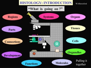
  • 1. HISTOLOGY: INTRODUCTION “ What is going on ?” Pulling it together Regions Organs Molecules Tissues Connections Cells Parts Organelles Development Functions Systems
  • 2. Noon talks for Internal-Medicine residents’ Board prep Two recurring themes -- Is it what it appears to be ? Does the treatment/procedure do what is claimed for it ? What is the evidence ?
  • 3. MEDICINE: Some aspects What gives with this patient ? Regions Molecules Tissues Connections Cells Parts Organelles Development Functions Regions Systems Systems abnormal Parts Connections Development Tissues Cells Organelles Molecules Functions Microbes Medicines Age Populations Costs ? Gender Organs Organs Abnormal
  • 4. Abnormal variants for all the earlier fields of knowledge Developing judgment - weighing various contributions for relevance & quality of evidence Foretaste of the ‘pulling it together’ in the PBL experiences, but much omitted, e.g., therapy, follow-up, cost; likewise for clinical correlations This doubling, plus more fields, e.g. microbes, is why medical training takes several years Any twit can lay hands on an LCD projector, and push images at you reminds one that the story may be faulty; it is one of many; and there are omissions Feel for the aspects that yield valid risk factors in this particular diagnosis ?
  • 5. PORNOGRAPHY & “THE REAL THING” Images versus REALITY What is the evidence for the real?
  • 6. Images versus REALITY - Functional Anatomy REALITY is the living person, often via images Surface anatomy Palpation Endoscopy+ Radiology PET scans Ultrasound Doppler flows Gait & Reflexes etc Biopsies Fine-Needle Aspiration Cervical, Blood, etc Smears Flow cytometry & cell sorting Cell culture & grafting etc (Bits cut or sucked out for microscopy)
  • 7. REALITY is the dead person DISSECTION [ Surface anatomy Endoscopy Palpation Radiology Ultrasound are sometimes useful as adjuncts to autopsy & histology correlations] Organs and large pieces cut out, examined, & prepared for MICROSCOPY- histology & histopathology (normal & altered side-by-side)
  • 8. Images versus REALITY - Anatomy In Anatomy, the source of the evidence - the essential point of reference - is the cadaver for Gross & the microscope slide for Histo As the physician is knowledgeably comfortable with the patient’s gross & microscopic structure and its implications, you will become confident at the cadaver & the microscope, and with the resulting images TESTS focus on the cadaver, the slides, and interpreting images - identification, interpretation, & synthesis Bed-rock
  • 9. MICROSCOPIC SLIDE Side view of slide Glass coverslip Glass slide 1”X3” Tissue Section Mounting medium Mounting medium: permeates section; fastens coverslip to slide; is clear; has refractive index as for glass Label
  • 10. SLIDE USE - Cautions GLASS IS FRAGILE ! Take care with individual slides & especially with the boxes of slides The slide must go on the stage coverslip up The high-dry & oil objectives cannot focus through the thickness of the slide to the section The label may have been put on the non-coverslip side, as shown ~ Slides & Microscope remain in the teaching Lab, always! Label
  • 11. SLIDE PREPARATION I Steps Excise & Fix (preserve) the tissue in fixative Remove the water & replace with wax-solvent Imbed the oriented specimen in molten wax After it is solid, hold the wax block & cut slices Mount the thin slices (sections) on slides When dry, remove the wax, & stain the section Remove surplus stain & water; mount coverslip When mounting medium has set, do microscopy
  • 12. SLIDE PREPARATION I Steps Excise & Fix (preserve) the tissue in fixative Remove the water & replace with wax-solvent Imbed the oriented specimen in molten wax After it is solid, hold the wax block & cut slices Mount the thin slices (sections) on slides When dry, remove the wax, & stain the section Remove surplus stain & water; mount coverslip When mounting medium has set, do microscopy
  • 13. 50 % ethanol 70 % ethanol 95 % ethanol 100 % ethanol benzene/xylene Dehydrating series paraffinwax Remove the water & replace with wax-solvent Imbed the oriented specimen in molten wax Miscible with ethanol; dissolves wax Fresh tissue 10% Formalin fixative label
  • 14. MICROTOME - a fancy meat-slicer - holds the wax block, & cuts off thin slices, as the block is slowly advanced mechanically Block Knife Section Glass slide Water-bath After it is solid, hold the wax block & cut slices Mount the thin slices (sections) on slides Lift out floating section on the slide
  • 15. FREEZING MICROTOME holds the frozen tissue, & cuts off thin slices, as the block is slowly advanced mechanically Block is the tissue Knife Section Water-bath Glass slide For fast biopsy, imbedding is omitted - frozen sections Mount the thin slices (sections) on slides Lift out section on the slide
  • 16. Dissolve paraffin wax Stain with Hematoxylin - blue Wash Stain with eosin - red Nuclei - blue Cytoplasm- red Wash When dry, remove the wax, & stain the section
  • 17. Dissolve paraffin wax Stain with Hematoxylin - blue Wash Stain with eosin - red Nuclei - blue Cytoplasm- red Wash When dry, remove the wax, & stain the section Potassium + eosinate - stain + charged amine, etc, groups on proteins bind - eosin “Acidophilic staining” “ Basophilic”
  • 18. SLIDE PREPARATION III Steps Excise & Fix (preserve) the tissue in fixative Remove the water & replace with wax-solvent Imbed the oriented specimen in molten wax After it is solid, hold the wax block & cut slices Mount the thin slices (sections) on slides When dry, remove the wax, & stain the section Remove surplus stain & water; mount coverslip When mounting medium has set, do microscopy
  • 19. SLIDE PREPARATION III Steps Excise & Fix (preserve) the tissue in fixative Remove the water & replace with wax-solvent Imbed the oriented specimen in molten wax After it is solid, hold the wax block & cut slices Mount the thin slices (sections) on slides When dry, remove the wax, & stain the section Remove surplus stain & water; mount coverslip When mounting medium has set, do microscopy
  • 20. Images versus REALITY Artifacts are appearances not true to the original state of the tissue SLIDE PREPARATION IV Artifacts Excise & Fix (preserve) the tissue in fixative Imbed the oriented specimen in molten wax After it is solid, hold the wax block & cut slices Mount the thin slices (sections) on slides When dry, remove the wax, & stain the section Remove surplus stain & water; mount coverslip When mounting medium has set, do microscopy Knife scores, chatter Bruising/splitting from cutting; Poor preservation, e.g., gut lining, enzymes, lost fat Wrinkles, section not flat, splits Weak/unbalanced staining Dirt, hair, bubbles Dirt on lenses, bad illumination Misleading orientation, Shrinkage & distortion, Mislabeled
  • 21. CLASS LIGHT MICROSCOPE Max MAGNIFICATION Eyepiece (10X) times ‘Oil’ Objective (100X) = 1000X Base Eyepiece/Ocular Stage Slide Light source Body Objective lenses Condenser
  • 22. CLASS LIGHT MICROSCOPE Controls I Base Condenser Eyepiece/Ocular Slide Light Body Inter-ocular distance Moving stage Iris diaphragm Field diaphragm Coarse & Fine focus Light intensity On/Off Objective selection left rear
  • 23. CLASS LIGHT MICROSCOPE Controls II Condenser Eyepiece/Ocular Slide Light Body Stage clip for slide Condenser focusing Condensercentering Ocular focusing left-side Base
  • 24. OPERATION I Without looking down the eyepieces , plug in the cord Turn the light-intensity knob back counterclockwise, Switch on the light, turn the intensity up (about a 90 o turn) while observing the light via the field opening Open the field diaphragm wide Move the condenser assembly to its top position Switch the shortest objective lens (X4) into the working position Open the iris diaphragm wide Select any well-stained slide Base Condenser Eyepiece/Ocular Slide Light Body Inter-ocular distance Moving stage Iris diaphragm Field diaphragm Coarse & Fine focus Light intensity On/Off Objective selection
  • 25. OPERATION II Field diaphragm Pull back the clip & place slide, cover-slip up, on the stage Use the stage controls to bring the stained section over the light Focus, using coarse, then fine adjustments Close the iris diaphragm to take the glare out of the view Push (pull) the eyepieces together to match your eye spacing Shut one eye, focus with the fine focus ; then shut that eye, open the other, and focus for it with the ocular focus (turning the eyepiece knurled ring) Switch in the next higher objective, and focus, using the main focusing controls & testing for binocular fusion Base Condenser Eyepiece/Ocular Slide Light Body Inter-ocular distance Moving stage Iris diaphragm Coarse & Fine focus Light intensity On/Off Objective selection
  • 26. SMEAR - another method of preparation Drop of blood Slide 1 Slide 2 On contact, slide 2 extends the drop along its 1” side Slide 2 Slide 2 Pushing angled slide 2 along #1 smears the line of blood across slide 1 Lift away slide 2; dry #1 ; stain; coverslip Smear A few cells are damaged; smear is not evenly thick; & staining is uneven. Same apply to SPREADS
  • 27. TEASING - a method of preparation Lumbo-sacral cord Roots Terminal thread A technique you know from using a needle to separate out the connective-tissue filum terminale from the nervous cauda equina of dorsal & ventral roots On the MICROSCOPE SLIDE, with a needle point one can tease apart individual nerve or muscle fibers from their bundles in nerve or muscle When tissue is already thin, it can be draped - SPREAD - over the slide like a tablecloth (Filum terminale)
  • 28. Cut across BONE shaft twice Saw out a sector Lay sector flat & grind thin Wash ground section Dry ; place unstained on slide Coverslip for viewing GROUND PREPARARTION
  • 29. HISTOLOGY SOURCES 303 Human Structure Syllabus next to last section p.8 Powerpoints - Comments & Standing assignment Histo Powerpoints Histology Full-text * & Histology Lab Guide http://wberesford.hsc.wvu.edu http://www.geocities.com/Athens/Academy/1575 * Recommendation - catch it while you can: download the above this week. We’re talking about 30 megabytes, and some of the above items could fit on floppies. It is never too soon to attune yourself to examiners’ thinking. Syllabus p. 56 (lower-right #) presents the formats in which Histo lab exam questions will be framed SBLC computers have “Histology Lab Assistant” WebBoard at Course 303 on Anatomy Dept site
  • 30. On the 4th floor, straight back from the working elevator, go to the SBLC (Rm 4005) and LEAVE your coat ( in the main lab, there are papers & envelopes on the desks that can get hidden or swept off, if coats are brought in today ) TODAY’s LAB PROCEDURES 1 Take only your book-bag (& computer, if you have it with you) into 4023. Locate your place - labeled in alphabetical order from the far end of the lab. Find the envelope with your name & the set of inventory forms. Open the envelope , remove the slip with the number to your Gross locker in the hallway. Write down this number in two other places. Take the small key out of the envelope and attach it to a key ring or something. If you are worried about your coat, or if this is already too much “structure”, take a break to try your locker combination and put your coat and other surplus stuff away. Once settled back at your place, use the key gently to open the top drawer at your place & take out the two small boxes of slides. Place them near the middle of the bench. Open the locker, and carefully lift out the microscope. Satisfy your curiosity - Find the microscope-use directions p.2 . Follow them with any well-stained slide. Ask us for help. [We will not issue oil for the X100 oil lens] Switch off the scope. Complete the receipt form. Go to p. 1 for slide numbering. Check your slide boxes against the inventory. Place forms in the wall folder .
  • 31. Start the exercise on p.3, but with slide A-1 blood smear LAST of the four on examples of preparation methods. The smear is difficult to focus on. It needs at least the medium-power X10 0bjective; and the glare has to be taken out of the view with the iris diaphragm on the condenser. TODAY’s LAB PROCEDURES 2 The next exercise - Artifacts - should be straightforward. The idea behind the final exercise of this Introductory section is inexhaustible. One can go on and on about cells. Stop when you wish, and come back to it during the next two labs. Skip C-1. The cells are poor examples of neurons. The last part of the Lab on “The General Structure of Cells” is illustrated on the next slide, which indicates some of the variables, but does not show much of the extracellular matrix outside cells, e.g., basal lamina, collagen fibers, reticular fibers. In this and future labs, do not get hung up on a slide. If you cannot get your question answered in a minute or so, go on to the next slide; and come back later, when the question can be answered. Remind yourself with a note by the item. Hand in the inventory. Put your scope & slides away carefully. Lock the drawer & locker. About half of you share with a dental student. Please be considerate
  • 32. collecting duct osteoclast CELL DESCRIPTION What is one looking for? Cell Size? Cell Shape? Nuclei - #? Nucleus - size, shape, density? Nucleoli -prominence , #? Nucleus -position? Cells’ relations? Cytoplasm - granular? Cytoplasm -philia? Cell membrane - visible? Cell surface specializations? Basal lamina neuron eosinophil airway lining
  • 33. GO GRANULAR Cerebellar Granule layer packed, small neurons- granule cells (& granulosa cells in ovary) Melanin granules in melanocytes & keratinocytes Blood Granulocytes from their very granular cytoplasm Layer Cell Granule Bas Eos PMN
  • 34. Some differences between light and electron microscopy I Light microscopy Electron microscopy ---------------------------------------------------------------------- Section thickness (1-30  m) gives Very thin sections provide no a little depth of focus for depth of focus, but 3-D information appreciation of the third dimension. can be had from: (a) thicker sections Serial sections can be cut, viewed by high-voltage EM; (b) shadowed and used to build a composite image replicas of fractured surfaces; (c) or representation. scanning electron microscopy (SEM). Most materials and structures cannot Heavy metal staining gives a more be stained and viewed at the same comprehensive picture of membranes, time; stains are used selectively to granules, filaments, crystals, etc.; give a partial picture, e.g. a stain but this view is incomplete and even for mucus counterstained to show visible bodies can be improved by cell nuclei. varying the technique. Specimen can be large and Specimen is in vacuo. Its small size even alive. creates more problems with sampling and orientation.
  • 35. Some differences between light and electron microscopy II Light microscopy Electron microscopy -------------------------------------------------------------------------- Image is presented directly to the Image is in shades of green on eye. Image keeps the colours given the screen; photographically, the specimen by staining. only in black and white. Modest magnification to X 1500; High magnification,up to X 2,000,000 but a wider field of view and easier thus the range of magnification orientation is greater Resolving power to 0.25  m. Resolving power to 1 nm (0.001  m.) Frozen sections can yield an image Processing of tissue takes a day at within 20 minutes. least. Crude techniques of preparation High resolution and magnification introduce many artefacts. demand good fixation (e.g. by (Histochemical methods are better.) vascular perfusion), cleanliness and careful cutting, adding up to fewer artefacts.
  • 36. **** Did I choose the right medical school? **** Complete Ameba Medicine 10 4 ed. Pp 29 “ Please take your zillion+ cells elsewhere. I’m an Ameba doctor.”
  • 37. Test Pattern for the Apocalypse