3DSIG 2014 Presentation: Systematic detection of internal symmetry in proteins
•
3 j'aime•2,229 vues

These slides are from 3DSIG 2014, presented on July 11. I describe our investigation of internal symmetry in protein structures. This is quite common (24% of domains), and has many implications for function, folding, and evolution. I introduce the CE-Symm method, described in Myers-Turnbull, D., Bliven, S. E., Rose, P. W., Aziz, Z. K., Youkharibache, P., Bourne, P. E., & Prlić, A. (2014). Systematic Detection of Internal Symmetry in Proteins Using CE-Symm. Journal of Molecular Biology, 426(11), 2255–2268. doi:10.1016/j.jmb.2014.03.010 I discuss the results from running CE-Symm across the PDB, as well as some particularly compelling examples. See also my poster by the same title for more details.
![Hemoglobin [4HHB]
C2
GTP Cyclohydrolase I
[1A8R]
D5
Rhinovirus 2 [3DPR]
Icosahedral
AmtB Ammonia
Channel [1U7G]
C3](data:image/gif;base64,R0lGODlhAQABAIAAAAAAAP///yH5BAEAAAAALAAAAAABAAEAAAIBRAA7)
Recommandé
Contenu connexe
Tendances
Tendances (20)
En vedette
Similaire à 3DSIG 2014 Presentation: Systematic detection of internal symmetry in proteins
Similaire à 3DSIG 2014 Presentation: Systematic detection of internal symmetry in proteins (20)
Dernier
Dernier (20)
3DSIG 2014 Presentation: Systematic detection of internal symmetry in proteins
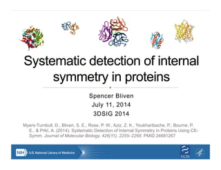
- 1. Spencer Bliven July 11, 2014 3DSIG 2014 Myers-Turnbull, D., Bliven, S. E., Rose, P. W., Aziz, Z. K., Youkharibache, P., Bourne, P. E., & Prlić, A. (2014). Systematic Detection of Internal Symmetry in Proteins Using CE- Symm. Journal of Molecular Biology, 426(11), 2255–2268. PMID 24681267
- 2. Hemoglobin [4HHB] C2 GTP Cyclohydrolase I [1A8R] D5 Rhinovirus 2 [3DPR] Icosahedral AmtB Ammonia Channel [1U7G] C3
- 3. Ferredoxin-like [d2j5aa1] C2 Beta-Propeller [d1u6dx_] C6 Beta-trefoil [3JUT] C3 TIM barrel [1TIM] C8 Key: Crystallographic/NCS axis Pseudosymmetry axis
- 4. ! Function ! Allosteric regulation/cooperativity ! Bind ligands symmetrically (e.g. metals, palindromic DNA, channels) TATA Binding Protein [1TGH] Monod, J., Wyman, J., & Changeux, J.-P. (1965). J Mol Biol, 12, 88–118.
- 5. ! Function ! Allosteric regulation/cooperativity ! Bind ligands symmetrically (e.g. metals, palindromic DNA, channels) ! Folding ! Prevent infinite assembly ! Subunits fold quasi- independently TATA Binding Protein [1TGH] Monod, J., Wyman, J., & Changeux, J.-P. (1965). J Mol Biol, 12, 88–118. Wolynes, P. G. (1996). PNAS, 93(25), 14249–14255. Crystal of Squalene synthase [3WCG]
- 6. ! Evolution ! Identify duplications & fusions ! Many examples of homologous quaternary symmetric/ internally symmetric proteins ! Tradeoff between monomer & oligomer Lee and Blaber. PNAS (2011) vol. 108 (1) pp. 126-30
- 7. E. Coli DNA polymerase III beta subunit [1mmi] ! 2 chains (C2 crystal axis) Human proliferating cell nuclear antigen [1VYM] ! 3 chains (C3 crystal axis)
- 8. E. Coli DNA polymerase III beta subunit [1mmi] ! 2 chains ! 6 domains (pseudo C6) Human proliferating cell nuclear antigen [1VYM] ! 3 chains ! 6 domains (pseudo C6)
- 9. ! 2-3 chains ! 6 domains ! 12 structural repeats (pseudo D6) Ancient 12-mer? Ancient 6-mer Bacterial DimerEukaryotic/Archaeal/ Viral Trimer Kelman, Z., & O'Donnell, M. (1995). Nucleic Acids Research, 23(18), 3613–3620. Neuwald, A. F., & Poleksic, A. (2000). Nucleic Acids Research, 28(18), 3570–3580.
- 10. ! Extends Combinatorial Extension (CE) algorithm for structural alignment ! Web server: source.rcsb.org/ jfatcatserver/symmetry.jsp ! Download & Source code: github.com/rcsb/symmetry (LGPL) Shindyalov, I. N., & Bourne, P. E. (1998). Protein Engineering, 11(9), 739–747. Jia, Y., Dewey, T. G., Shindyalov, I. N., & Bourne, P. E. (2004). J Comput Biol, 11(5), 787–799.
- 11. Fibroblast Growth Factor [3JUT] 120° 120° Myers-Turnbull, D., Bliven, S. E., Rose, P. W., Aziz, Z. K., Youkharibache, P., Bourne, P. E., & Prlić, A. (2014). Journal of Molecular Biology, 426(11), 2255–2268.
- 12. Fibroblast Growth Factor [3JUT] 120° 120° Myers-Turnbull, D., Bliven, S. E., Rose, P. W., Aziz, Z. K., Youkharibache, P., Bourne, P. E., & Prlić, A. (2014). Journal of Molecular Biology, 426(11), 2255–2268.
- 13. ! 1007 structures from SCOP superfamilies ! Manually curated ! Excludes small proteins (<4 SSEs) ! 24% of superfamilies have internal symmetry or large structural repeats Order Superfamilies % Asymmetric 766 76.10% Rotational 2 166 16.5% 3 10 1.0% 4 2 0.2% 5 3 0.3% 6 9 0.9% 7 9 0.9% 8 21 2.1% Dihedral 2 2 0.2% 4 1 0.1% Helical 2 9 0.9% 3 2 0.2% Non-integral 2 0.2% Superhelical 2 0.2% Translational 3 0.3%
- 14. ! AUC = .95 ! 86% True Positive Rate ! 3.3% False Positive Rate SymD: Kim, C., Basner, J., & Lee, B. (2010). BMC Bioinformatics, 11, 303.
- 15. ! All domains from SCOPe 2.03 ! Interactive results: source.rcsb.org/jfatcatserver/scopResults.jsp ! Underestimate based on conservative thresholds SCOP Class Superfamilies % Symmetric α 507 18.5% β 354 24.6% α/β 244 16.8% α+β 551 14.3% Multi-domain 66 4.5% Membrane 109 23.8% Overall 1831 18.0%
- 16. ! PTS sorbitol transporter subunit IIA ! Novel fold ! Solved by the Protein Structure Initiative ! Structural alignment reveals a conserved sequence motif between halves [2F9H]
- 17. ! 18-24% of domains have internal symmetry ! Symmetry gives clues about duplication events ! Symmetry is deeply tied to protein function ! CE-Symm can accurately detect internal symmetry d1su3a2 D1pt2a_ d1c5ka1 d1k3ia3 d1h9ya2
- 18. ! UC San Diego/RCSB ! Douglas Myers-Turnbull ! Andreas Prlić ! Peter Rose ! Zaid Aziz ! Milton Saier ! RCSB & Bourne Lab members ! NIH ! Philip Bourne ! Philippe Youkharibache ! David Landsman ! Paul Scherrer Institute ! Guido Capitani & Lab members Resources: ! source.rcsb.org/jfatcatserver/ symmetry.jsp ! github.com/rcsb/symmetry ! Poster 25 ! www.slideshare.net/sbliven ! Funding: NSF, NIH, DOE, Open Science Grid
- 20. Glyoxalase I from Clostridium acetobutylicum [3HDP] (Nickel; Dimer) Glyoxalase I from E. coli [1F9Z] (Nickel; Dimer) 1,2-dihydroxy- naphthalene dioxygenase from Pseudomonas sp. strain C18 [2EHZ] (Iron; Octamer)
- 21. Glyoxalase I from Clostridium acetobutylicum [3HDP] (Nickel; Dimer) Glyoxalase I from E. coli [1F9Z] (Nickel; Dimer) 1,2-dihydroxy- naphthalene dioxygenase from Pseudomonas sp. strain C18 [2EHZ] (Iron; Octamer)
- 22. ! racemases and epimerases are enriched
- 23. 0 1 2 3 4 0.00 0.25 0.50 0.75 TM−Score Density Asymmetric Symmetric
- 24. This work is licensed under a Creative Commons Attribution-ShareAlike 3.0 Unported License.