SlideShare une entreprise Scribd logo
1  sur  21
1.1.3  Cell Division, Cell Diversity and cellular organisation Objectives Cell cycle – stages (Go, S, G1, M); mitosis and meiosis.  Budding in yeast; binary fission (bacteria) Significance of mitosis and meiosis; Mitosis – produces clones; growth, asexual  reproduction, cancer. Meiosis – gametes; maintain chromosome number; cell produced not genetically identical DNA, genes and packaging information Clones and stem cells (embryonic and adult) Differentiation of stem cells – blood cells (RBC, neutrophils); cambium (xylem + phloem) Cell specialisation  - epithelial cells (squamous + ciliated); sperm; palisade; root hair cell; guard cell; neurones Cells, tissues, organs, organ systems HW –  Past paper question
The Cell Cycle, Mitosis, and Meiosis New cells can only be made when existing cells divide.  All cells have the ability to divide – but some cells lose this ability. Intestinal epithelial lining - replaced every five days by cell division Liver cells - divide only to repair damage, and then stop dividing Bone marrow cells - divide repeatedly to produce red and white blood cells Meristem cells (tips of roots and shoots) – divide to produce new  growth Cambium cells (plants) – divide to form vascular tissue (xylem and phloem) These are relatively unspecialised cells.  Specialised cells often go through the cell cycle only once - the nerve cells, once formed cannot divide again. In eukaryotic cells, there are two types of cell division –  mitosis  and  meiosis .  Mitosis is used to produce new cells for growth and repair.  Meiosis is used in the formation of gametes only.  In prokaryotes (bacteria), cell division does not involve mitosis or meiosis – bacteria reproduce asexually, by a type of cell division termed by binary fission. Yeasts reproduce asexually by budding. The  cell cycle  is the process that all body cells from multicellular organisms use to grow and divide.  The cell cycle starts when a cell has been produced by cell division and ends with the cell dividing to produce two identical cells.
http://biolibogy.com/cells.html   1 A bud is formed at the surface of the cell 2 Interphase – DNA and organelles replicated 3 Set of duplicated chromosomes – each set contained within a nucleus 4 One daughter nucleus migrates into the bud 5 Bud increases in size and eventually separates from parent cell – producing a new, genetically identical yeast cell Binary Fision (Bacteria) Budding (Yeast)
The cell cycle can be divided into stages:  G1 (“growth phase” 1) - Cells prepare for DNA  replication S  (“synthesis”) - DNA replication occurs G2 (“growth phase” 2)- Short gap before mitosis M  Mitosis (relatively short)  Affected by availability of nutrients Between each stage the cell “checks” to see if it  is OK to proceed to  the next stage.  “ Proof-reading” enzymes check the copied chromosomes for mistakes (mutations) – the cell may kill itself (undergo “suicide”) if harmful mutations are – a process known as apoptosis. Bacterial cells complete the cycle every 20 minutes.  Muscle cells never complete the cycle – “terminal differentiation” Uncontrolled and repeated cell division by mitosis results in cancer (tumours) Eukaryotic cells have a well- defined  cell cycle  of growth and division (mitosis).  The length of the cycle varies (from minutes to hours, or , longer) ending with mitosis.  Each phase of the cycle involves specific activities, and varies in length from one organism to another. The Cell Cycle
MITOSIS   (M) Process by which a  nucleus  divides into two – each with an identical set of chromosomes – the nuclei are genetically identical Four phases – prophase, metaphase, anaphase, and telophase Followed by cytokinesis – division of the cell into two genetically identical daughter cells INTERPHASE  Period of cell growth; cell prepares cell for cell division (mitosis); genetic material (DNA) is copied and checked for errors – prevents mutations being passed on No apparent activity New organelles and proteins are made Divided into three phases (G1, S, and G2 phase) CELL CYCLE G1 S phase G2 Mitosis (M) Two daughter cells – genetically identical
G1 + S + G2 = INTERPHASE   No apparent observable activity Cytokinesis – cell divides into two DNA content = 20 G1  -  First growth phase – longest phase Protein synthesis – cell “grows” Most organelles produced Volume of cytoplasm increases Cell differentiation (switching on or off of genes) Length depends on internal and external factors If cell is not going to divide again it remains in this phase DNA content = 20 (arbitary) S -  Replication phase  DNA replication – this must occur if mitosis is to take place The cell enters this phase only if cell division is to follow DNA content = 40 G2 -  Second growth phase - short Short gap before mitosis (cell division) Cytoskeleton of cell breaks down and the protein microtubule components begin to reassemble into spindle fibres – required for cell division DNA content = 40 The Cell Cycle
Mitosis occurs rapidly during  growth and repair  to make new cells.  The rate is controlled according to the situation – e.g., during growth and repair the rate may be fast, but when growth or repair is achieved the rate declines or mitosis stops. Cancers (tumours and diffuse) are formed through uncontrolled cell division by mitosis Mitosis Mitosis occurs wherever an increase in number of cells is needed.  It is important in the growth and repair of multicellular organisms, and in the population growth of unicellular organisms During mitosis a cell produces two copies of itself.  Each is genetically identical to the other and to the parent cell from which they were formed.  The parent cell  copies  its chromosomes by  replication  prior to cell division.
Chromosomes A duplicated and condensed eukaryotic chromosome with two sister chromatids Homologous Chromosomes Chromosomes occur in pairs – there are 23 pairs of  chromosomes.  Each pair consists of a chromosome from the male (father) and one from the female (mother).  The chromosomes that make up each pair of the 22 pairs are the same size and have the same or alternative versions of genes for particular characteristics.  Each individual pair of similar chromosomes is called a homologous pair.
[object Object],[object Object],[object Object],[object Object],[object Object],[object Object],[object Object],Mitosis takes place in the following:
Before a cell divides, its  chromosomes  are copied exactly in INTERPHASE. This process is called replication; ATP is synthesised – provides energy for cell division; organelles are replicated and proteins are made PROPHASE The  DNA  of each chromosome is copied to form two  chromatids  (“sister” chromosomes); chromosomes condense – becoming shorter and fatter – visible under LM; nuclear envelope breaks down; chromosomes lie freely in cytoplasm; centrioles move to opposite ends of the cell, forming protein (tubulin) fibres across it called a spindle – fibres extend to the equator of the cell METAPHASE Chromosomes line up at the equator; the spindle fibres from each pole become attached to the centromere of the chromosomes ANAPHASE The spindle fibres contract;  the centromeres are split and the pairs of sister chromatids are separated and dragged to opposite poles assuming a “V” shape – the centromeres lead; a complete set of chromosomes is therefore found at each pole; energy (ATP) is required  TELOPHASE Chromatids reach their respective poles and uncoil – become thin and long again – now called chromosomes again – no longer visible under LM; spindle fibres break down; nuclear envelope forms around each group of chromosomes – forming two nuclei; cytokinesis follows – cytoplasm divides and a plasma membrane forms two form two individual cells; cell enters interphase once again
Cytokinesis Division of the cytoplasm (cytokinesis) into two equal parts follows mitosis A “waist” forms in the middle of the cell.  Eventually, the plasma membrane from one side of the cell joins that of the opposite side of the cell and the two new cells separate.  The two daughter cells are genetically identical. The equator of the cell is constricted by a ring of contractile proteins (actin) in the process of cleavage, to create two cells. In plant cells, the Golgi apparatus and associated secretory vesicles assemble at the equator. Their contents are deposited to form a plate (the cell plate). Some vesicles remain intact and make connecting channels, termed plasmodesmata, through the new cell wall.
Prophase
Metaphase
Anaphase Telophase
Meiosis Meiosis is a type of cell division used in the formation of gametes (spermatozoa and ova) in animals, and spore formation (which precedes gamete formation) in plants - it is termed reduction division.  Meiosis creates genetically different cells and variation within species The number of chromosomes is halved - from diploid (46) to haploid (23).  Involves two distinct divisions Meiosis I  – homologous pairs of replicated chromosomes are  separated Meiosis II  – reduction division (chromatids separated into  individual  haploid gametes) Four cells (gametes/spores), genetically different from each other, result from one parent cell – each cell containing half the original number of chromosome Production of haploid (n) gametes ensures the maintenance of the diploid (2n) number in subsequent generations Meiosis involves certain “mixing” events, and these, along with the random fusion of gametes in fertilisation to form a diploid zygote, results in genetic variation in the offspring which may be of adaptive advantage.
 
The  DNA  of each chromosome is replicated to form two chromatids.  chromosomes arrange themselves into homologous pairs (both coding for the same characteristics), and prepare for cell division. At this point maternal and paternal chromatids can exchange bits of DNA to recombine their genetic material and increase the potential for variation. Homologous pairs of chromosomes separate and move to the poles of the parent nucleus. For each of the 23 pairs there is a 50-50 chance as to which pole the paternal or maternal pair of chromatids go. With over 8 million possibilities there are many opportunities for variation.  Nucleus now divides to form two daughter nuclei, each with a mixture of paternal and maternal chromosomes but with half the full complement of genetic material (and no pairs at all). This division is called Meiosis 1. The two daughter nuclei themselves divide to form  gametes . This second division - Meiosis 2 - works just like mitosis. The chromosomes (really pairs of chromatids) split apart to form the genetic material of the four new cells. The end result is four sex cells each with a complete but single set of 23 chromosomes. On fertilisation the nuclei of the sperm and the egg join to form a new nucleus, called the  zygote . The zygote contains 23 pairs of chromosomes - 23 single chromosomes from the sperm, and 23 single chromosomes from the egg.
Meiosis leads to variation among the offspring of sexual reproduction in 3 ways: Crossing over  – crossing over of genes from one chromatid of one chromosome to the chromatid of the other homologous chromosome Reduction and fusion of gametes  from different individuals – gametes have haploid number of chromosomes – allows a gamete from one of these cells to fuse with another cell with a different haploid set, producing a zygote which has the normal diploid number of chromosomes but a new combination of genes Independent (random) assortment  – when the chromosomes line up as pairs at the equator (metaphase I) of the spindle, it is by chance which “way round” each pair lies.
Mitosis and Meiosis Compared   Mitosis Meiosis Purpose To make daughter cells identical to the parent cells - eg during growth and repair To produce  sex cells ( gametes ) Takes place .. In all cells apart from gametes In the reproductive organs (ovaries and testes) Produces how many cells? Two daughter cells Four gametes What happens to number of chromosomes? Same number as in parent cell Diploid = 46 (in pairs) Half as many as in parent cell (The original number of chromosomes is restored when two gametes fuse to form a  zygote .) Haploid = 23 (single) How do parent and daughter cells differ genetically? Not at all - genetic material is copied exactly (replicated) Contain a mixture of chromosomes from two parent gametes - so cannot be identical Variation between daughter cells? No - they are clones of each other Yes - they are genetically different from each other because chromosomes get shuffled up during division
The drugs vincristine and vinblastine from the periwinkle plant inhibit spindle assembly – used in the treatment of cancers (leukaemia and lymphomas) and gout Taxol form the Pacific ewe inhibits depolymerisation of spindle fibres (de-assembly), which effectively stops cell division – used to treat ovarian cancers
Stem Cells Multicellular organisms function as a result of many different cell types that are specialised for their function – e.g: Neurones (nerve cells) – specialised for the transmission of electrical  nerve impulses Red blood cells (erythrocytes) – specialised for the carriage of oxygen  aroung the body Vascular tissue (xylem and phloem) in plants - cells in the cambium tissue of  plants differentiate to form vascular tissue – specialised for transport All cells in multicellular organisms originate from stem cells.  These are unspecialised cells that divide to become new cells, which then differentiate to become specialised into different cell types. Differentiation is achieved due to the switching on and off of relevant genes

Contenu connexe

Tendances

Cell Division
Cell DivisionCell Division
Cell DivisionACKademic
 
Chapter 4-cell division, mitosis, DNA, protein production
Chapter 4-cell division, mitosis, DNA, protein productionChapter 4-cell division, mitosis, DNA, protein production
Chapter 4-cell division, mitosis, DNA, protein productionSteven_iannuccilli
 
Mitosis and meiosis
Mitosis and meiosisMitosis and meiosis
Mitosis and meiosisJessi Dildy
 
Mitosis And Meiosis
Mitosis And MeiosisMitosis And Meiosis
Mitosis And Meiosisk.huss89
 
cell division & physiology of cell division
cell division & physiology of cell divisioncell division & physiology of cell division
cell division & physiology of cell divisionShivangi Rahangdale
 
Form 4 biology chap5 pt1
Form 4 biology chap5 pt1Form 4 biology chap5 pt1
Form 4 biology chap5 pt1cikgushaik
 
Cell cycle and division
Cell cycle and divisionCell cycle and division
Cell cycle and divisionPn Patidar
 
Mitosis Cell Division Ppt
Mitosis Cell Division PptMitosis Cell Division Ppt
Mitosis Cell Division Pptssandhu
 
Lesson plan on cell growth and reproduction
Lesson plan on cell growth and reproductionLesson plan on cell growth and reproduction
Lesson plan on cell growth and reproductionnarsglance
 
Genetic basis of life(mitosis and meiosis)
Genetic basis of life(mitosis and meiosis)Genetic basis of life(mitosis and meiosis)
Genetic basis of life(mitosis and meiosis)mandalina landy
 
form 4 biology chap5 pt2
form 4 biology chap5 pt2form 4 biology chap5 pt2
form 4 biology chap5 pt2cikgushaik
 

Tendances (20)

Chapter 3 cell division
Chapter 3 cell divisionChapter 3 cell division
Chapter 3 cell division
 
Cell Division
Cell DivisionCell Division
Cell Division
 
Chapter 4-cell division, mitosis, DNA, protein production
Chapter 4-cell division, mitosis, DNA, protein productionChapter 4-cell division, mitosis, DNA, protein production
Chapter 4-cell division, mitosis, DNA, protein production
 
Mitosis and meiosis
Mitosis and meiosisMitosis and meiosis
Mitosis and meiosis
 
Mitosis And Meiosis
Mitosis And MeiosisMitosis And Meiosis
Mitosis And Meiosis
 
cell division & physiology of cell division
cell division & physiology of cell divisioncell division & physiology of cell division
cell division & physiology of cell division
 
08 Lecture Ppt
08 Lecture Ppt08 Lecture Ppt
08 Lecture Ppt
 
Form 4 biology chap5 pt1
Form 4 biology chap5 pt1Form 4 biology chap5 pt1
Form 4 biology chap5 pt1
 
Cell division
Cell divisionCell division
Cell division
 
Cell organelles
Cell organellesCell organelles
Cell organelles
 
Cell cycle and division
Cell cycle and divisionCell cycle and division
Cell cycle and division
 
Mitosis Cell Division Ppt
Mitosis Cell Division PptMitosis Cell Division Ppt
Mitosis Cell Division Ppt
 
Mitosis
MitosisMitosis
Mitosis
 
Mitosis and meiosis
Mitosis and meiosisMitosis and meiosis
Mitosis and meiosis
 
Lesson plan on cell growth and reproduction
Lesson plan on cell growth and reproductionLesson plan on cell growth and reproduction
Lesson plan on cell growth and reproduction
 
cell division - mitosis
cell division - mitosiscell division - mitosis
cell division - mitosis
 
Genetic basis of life(mitosis and meiosis)
Genetic basis of life(mitosis and meiosis)Genetic basis of life(mitosis and meiosis)
Genetic basis of life(mitosis and meiosis)
 
form 4 biology chap5 pt2
form 4 biology chap5 pt2form 4 biology chap5 pt2
form 4 biology chap5 pt2
 
Unit2 cell cycle
Unit2   cell cycleUnit2   cell cycle
Unit2 cell cycle
 
1.6. Cell division
1.6. Cell division1.6. Cell division
1.6. Cell division
 

En vedette

En vedette (9)

ScienceShare.co.uk Shared Resource
ScienceShare.co.uk Shared ResourceScienceShare.co.uk Shared Resource
ScienceShare.co.uk Shared Resource
 
ScienceShare.co.uk Shared Resource
ScienceShare.co.uk Shared ResourceScienceShare.co.uk Shared Resource
ScienceShare.co.uk Shared Resource
 
ScienceShare.co.uk Shared Resource
ScienceShare.co.uk Shared ResourceScienceShare.co.uk Shared Resource
ScienceShare.co.uk Shared Resource
 
ScienceShare.co.uk Shared Resource
ScienceShare.co.uk Shared ResourceScienceShare.co.uk Shared Resource
ScienceShare.co.uk Shared Resource
 
CWR Haiti Earthquake Response
CWR Haiti Earthquake ResponseCWR Haiti Earthquake Response
CWR Haiti Earthquake Response
 
ScienceShare.co.uk Shared Resource
ScienceShare.co.uk Shared ResourceScienceShare.co.uk Shared Resource
ScienceShare.co.uk Shared Resource
 
ScienceShare.co.uk Shared Resource
ScienceShare.co.uk Shared ResourceScienceShare.co.uk Shared Resource
ScienceShare.co.uk Shared Resource
 
ScienceShare.co.uk Shared Resource
ScienceShare.co.uk Shared ResourceScienceShare.co.uk Shared Resource
ScienceShare.co.uk Shared Resource
 
Treasurer's Report (2011)
Treasurer's Report (2011)Treasurer's Report (2011)
Treasurer's Report (2011)
 

Similaire à ScienceShare.co.uk Shared Resource

Similaire à ScienceShare.co.uk Shared Resource (20)

cell division
cell divisioncell division
cell division
 
Biolo Garde Ten Unit Four for High School Students
Biolo Garde Ten Unit Four for High School StudentsBiolo Garde Ten Unit Four for High School Students
Biolo Garde Ten Unit Four for High School Students
 
Mitosis
MitosisMitosis
Mitosis
 
The cell cycle trf
The cell cycle trfThe cell cycle trf
The cell cycle trf
 
3. CELL CYCLE.pptx
3. CELL CYCLE.pptx3. CELL CYCLE.pptx
3. CELL CYCLE.pptx
 
cell cycle and control checkpoints throght cyclin cdk complex
cell cycle and control checkpoints throght  cyclin cdk complexcell cycle and control checkpoints throght  cyclin cdk complex
cell cycle and control checkpoints throght cyclin cdk complex
 
Mitosiscellcycle2012
Mitosiscellcycle2012Mitosiscellcycle2012
Mitosiscellcycle2012
 
Cell division
Cell divisionCell division
Cell division
 
Cell cycle
Cell cycleCell cycle
Cell cycle
 
5.1 Mitosis
5.1 Mitosis5.1 Mitosis
5.1 Mitosis
 
5.1 Mitosis
5.1 Mitosis5.1 Mitosis
5.1 Mitosis
 
Mitosis
MitosisMitosis
Mitosis
 
Cell DIVISION/cycle
Cell DIVISION/cycleCell DIVISION/cycle
Cell DIVISION/cycle
 
Cell division grade 10
Cell division grade 10Cell division grade 10
Cell division grade 10
 
Cell Reproduction
Cell ReproductionCell Reproduction
Cell Reproduction
 
5.1 mitosis
5.1 mitosis5.1 mitosis
5.1 mitosis
 
CELL CYCLE REGULATION.docx
CELL CYCLE REGULATION.docxCELL CYCLE REGULATION.docx
CELL CYCLE REGULATION.docx
 
CELL CYCLE REGULATION.docx
CELL CYCLE REGULATION.docxCELL CYCLE REGULATION.docx
CELL CYCLE REGULATION.docx
 
AP Bio Ch11 PowerPoint
 AP Bio Ch11 PowerPoint AP Bio Ch11 PowerPoint
AP Bio Ch11 PowerPoint
 
Bio Cell Division
Bio Cell DivisionBio Cell Division
Bio Cell Division
 

Plus de ScienceShare.co.uk

ScienceShare.co.uk Shared Resource
ScienceShare.co.uk Shared ResourceScienceShare.co.uk Shared Resource
ScienceShare.co.uk Shared ResourceScienceShare.co.uk
 
ScienceShare.co.uk Shared Resource
ScienceShare.co.uk Shared ResourceScienceShare.co.uk Shared Resource
ScienceShare.co.uk Shared ResourceScienceShare.co.uk
 
ScienceShare.co.uk Shared Resource
ScienceShare.co.uk Shared ResourceScienceShare.co.uk Shared Resource
ScienceShare.co.uk Shared ResourceScienceShare.co.uk
 
ScienceShare.co.uk Shared Resource
ScienceShare.co.uk Shared ResourceScienceShare.co.uk Shared Resource
ScienceShare.co.uk Shared ResourceScienceShare.co.uk
 
ScienceShare.co.uk Shared Resource
ScienceShare.co.uk Shared ResourceScienceShare.co.uk Shared Resource
ScienceShare.co.uk Shared ResourceScienceShare.co.uk
 
ScienceShare.co.uk Shared Resource
ScienceShare.co.uk Shared ResourceScienceShare.co.uk Shared Resource
ScienceShare.co.uk Shared ResourceScienceShare.co.uk
 
ScienceShare.co.uk Shared Resource
ScienceShare.co.uk Shared ResourceScienceShare.co.uk Shared Resource
ScienceShare.co.uk Shared ResourceScienceShare.co.uk
 
ScienceShare.co.uk Shared Resource
ScienceShare.co.uk Shared ResourceScienceShare.co.uk Shared Resource
ScienceShare.co.uk Shared ResourceScienceShare.co.uk
 
ScienceShare.co.uk Shared Resource
ScienceShare.co.uk Shared ResourceScienceShare.co.uk Shared Resource
ScienceShare.co.uk Shared ResourceScienceShare.co.uk
 
ScienceShare.co.uk Shared Resource
ScienceShare.co.uk Shared ResourceScienceShare.co.uk Shared Resource
ScienceShare.co.uk Shared ResourceScienceShare.co.uk
 
ScienceShare.co.uk Shared Resource
ScienceShare.co.uk Shared ResourceScienceShare.co.uk Shared Resource
ScienceShare.co.uk Shared ResourceScienceShare.co.uk
 
ScienceShare.co.uk Shared Resource
ScienceShare.co.uk Shared ResourceScienceShare.co.uk Shared Resource
ScienceShare.co.uk Shared ResourceScienceShare.co.uk
 
ScienceShare.co.uk Shared Resource
ScienceShare.co.uk Shared ResourceScienceShare.co.uk Shared Resource
ScienceShare.co.uk Shared ResourceScienceShare.co.uk
 
ScienceShare.co.uk Shared Resource
ScienceShare.co.uk Shared ResourceScienceShare.co.uk Shared Resource
ScienceShare.co.uk Shared ResourceScienceShare.co.uk
 
ScienceShare.co.uk Shared Resource
ScienceShare.co.uk Shared ResourceScienceShare.co.uk Shared Resource
ScienceShare.co.uk Shared ResourceScienceShare.co.uk
 
ScienceShare.co.uk Shared Resource
ScienceShare.co.uk Shared ResourceScienceShare.co.uk Shared Resource
ScienceShare.co.uk Shared ResourceScienceShare.co.uk
 
ScienceShare.co.uk Shared Resource
ScienceShare.co.uk Shared ResourceScienceShare.co.uk Shared Resource
ScienceShare.co.uk Shared ResourceScienceShare.co.uk
 
ScienceShare.co.uk Shared Resource
ScienceShare.co.uk Shared ResourceScienceShare.co.uk Shared Resource
ScienceShare.co.uk Shared ResourceScienceShare.co.uk
 
ScienceShare.co.uk Shared Resource
ScienceShare.co.uk Shared ResourceScienceShare.co.uk Shared Resource
ScienceShare.co.uk Shared ResourceScienceShare.co.uk
 
ScienceShare.co.uk Shared Resource
ScienceShare.co.uk Shared ResourceScienceShare.co.uk Shared Resource
ScienceShare.co.uk Shared ResourceScienceShare.co.uk
 

Plus de ScienceShare.co.uk (20)

ScienceShare.co.uk Shared Resource
ScienceShare.co.uk Shared ResourceScienceShare.co.uk Shared Resource
ScienceShare.co.uk Shared Resource
 
ScienceShare.co.uk Shared Resource
ScienceShare.co.uk Shared ResourceScienceShare.co.uk Shared Resource
ScienceShare.co.uk Shared Resource
 
ScienceShare.co.uk Shared Resource
ScienceShare.co.uk Shared ResourceScienceShare.co.uk Shared Resource
ScienceShare.co.uk Shared Resource
 
ScienceShare.co.uk Shared Resource
ScienceShare.co.uk Shared ResourceScienceShare.co.uk Shared Resource
ScienceShare.co.uk Shared Resource
 
ScienceShare.co.uk Shared Resource
ScienceShare.co.uk Shared ResourceScienceShare.co.uk Shared Resource
ScienceShare.co.uk Shared Resource
 
ScienceShare.co.uk Shared Resource
ScienceShare.co.uk Shared ResourceScienceShare.co.uk Shared Resource
ScienceShare.co.uk Shared Resource
 
ScienceShare.co.uk Shared Resource
ScienceShare.co.uk Shared ResourceScienceShare.co.uk Shared Resource
ScienceShare.co.uk Shared Resource
 
ScienceShare.co.uk Shared Resource
ScienceShare.co.uk Shared ResourceScienceShare.co.uk Shared Resource
ScienceShare.co.uk Shared Resource
 
ScienceShare.co.uk Shared Resource
ScienceShare.co.uk Shared ResourceScienceShare.co.uk Shared Resource
ScienceShare.co.uk Shared Resource
 
ScienceShare.co.uk Shared Resource
ScienceShare.co.uk Shared ResourceScienceShare.co.uk Shared Resource
ScienceShare.co.uk Shared Resource
 
ScienceShare.co.uk Shared Resource
ScienceShare.co.uk Shared ResourceScienceShare.co.uk Shared Resource
ScienceShare.co.uk Shared Resource
 
ScienceShare.co.uk Shared Resource
ScienceShare.co.uk Shared ResourceScienceShare.co.uk Shared Resource
ScienceShare.co.uk Shared Resource
 
ScienceShare.co.uk Shared Resource
ScienceShare.co.uk Shared ResourceScienceShare.co.uk Shared Resource
ScienceShare.co.uk Shared Resource
 
ScienceShare.co.uk Shared Resource
ScienceShare.co.uk Shared ResourceScienceShare.co.uk Shared Resource
ScienceShare.co.uk Shared Resource
 
ScienceShare.co.uk Shared Resource
ScienceShare.co.uk Shared ResourceScienceShare.co.uk Shared Resource
ScienceShare.co.uk Shared Resource
 
ScienceShare.co.uk Shared Resource
ScienceShare.co.uk Shared ResourceScienceShare.co.uk Shared Resource
ScienceShare.co.uk Shared Resource
 
ScienceShare.co.uk Shared Resource
ScienceShare.co.uk Shared ResourceScienceShare.co.uk Shared Resource
ScienceShare.co.uk Shared Resource
 
ScienceShare.co.uk Shared Resource
ScienceShare.co.uk Shared ResourceScienceShare.co.uk Shared Resource
ScienceShare.co.uk Shared Resource
 
ScienceShare.co.uk Shared Resource
ScienceShare.co.uk Shared ResourceScienceShare.co.uk Shared Resource
ScienceShare.co.uk Shared Resource
 
ScienceShare.co.uk Shared Resource
ScienceShare.co.uk Shared ResourceScienceShare.co.uk Shared Resource
ScienceShare.co.uk Shared Resource
 

ScienceShare.co.uk Shared Resource

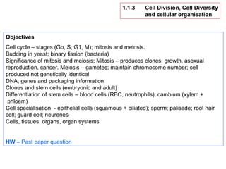
  • 1. 1.1.3 Cell Division, Cell Diversity and cellular organisation Objectives Cell cycle – stages (Go, S, G1, M); mitosis and meiosis. Budding in yeast; binary fission (bacteria) Significance of mitosis and meiosis; Mitosis – produces clones; growth, asexual reproduction, cancer. Meiosis – gametes; maintain chromosome number; cell produced not genetically identical DNA, genes and packaging information Clones and stem cells (embryonic and adult) Differentiation of stem cells – blood cells (RBC, neutrophils); cambium (xylem + phloem) Cell specialisation - epithelial cells (squamous + ciliated); sperm; palisade; root hair cell; guard cell; neurones Cells, tissues, organs, organ systems HW – Past paper question
  • 2. The Cell Cycle, Mitosis, and Meiosis New cells can only be made when existing cells divide. All cells have the ability to divide – but some cells lose this ability. Intestinal epithelial lining - replaced every five days by cell division Liver cells - divide only to repair damage, and then stop dividing Bone marrow cells - divide repeatedly to produce red and white blood cells Meristem cells (tips of roots and shoots) – divide to produce new growth Cambium cells (plants) – divide to form vascular tissue (xylem and phloem) These are relatively unspecialised cells. Specialised cells often go through the cell cycle only once - the nerve cells, once formed cannot divide again. In eukaryotic cells, there are two types of cell division – mitosis and meiosis . Mitosis is used to produce new cells for growth and repair. Meiosis is used in the formation of gametes only. In prokaryotes (bacteria), cell division does not involve mitosis or meiosis – bacteria reproduce asexually, by a type of cell division termed by binary fission. Yeasts reproduce asexually by budding. The cell cycle is the process that all body cells from multicellular organisms use to grow and divide. The cell cycle starts when a cell has been produced by cell division and ends with the cell dividing to produce two identical cells.
  • 3. http://biolibogy.com/cells.html 1 A bud is formed at the surface of the cell 2 Interphase – DNA and organelles replicated 3 Set of duplicated chromosomes – each set contained within a nucleus 4 One daughter nucleus migrates into the bud 5 Bud increases in size and eventually separates from parent cell – producing a new, genetically identical yeast cell Binary Fision (Bacteria) Budding (Yeast)
  • 4. The cell cycle can be divided into stages: G1 (“growth phase” 1) - Cells prepare for DNA replication S (“synthesis”) - DNA replication occurs G2 (“growth phase” 2)- Short gap before mitosis M Mitosis (relatively short) Affected by availability of nutrients Between each stage the cell “checks” to see if it is OK to proceed to the next stage. “ Proof-reading” enzymes check the copied chromosomes for mistakes (mutations) – the cell may kill itself (undergo “suicide”) if harmful mutations are – a process known as apoptosis. Bacterial cells complete the cycle every 20 minutes. Muscle cells never complete the cycle – “terminal differentiation” Uncontrolled and repeated cell division by mitosis results in cancer (tumours) Eukaryotic cells have a well- defined cell cycle of growth and division (mitosis). The length of the cycle varies (from minutes to hours, or , longer) ending with mitosis. Each phase of the cycle involves specific activities, and varies in length from one organism to another. The Cell Cycle
  • 5. MITOSIS (M) Process by which a nucleus divides into two – each with an identical set of chromosomes – the nuclei are genetically identical Four phases – prophase, metaphase, anaphase, and telophase Followed by cytokinesis – division of the cell into two genetically identical daughter cells INTERPHASE Period of cell growth; cell prepares cell for cell division (mitosis); genetic material (DNA) is copied and checked for errors – prevents mutations being passed on No apparent activity New organelles and proteins are made Divided into three phases (G1, S, and G2 phase) CELL CYCLE G1 S phase G2 Mitosis (M) Two daughter cells – genetically identical
  • 6. G1 + S + G2 = INTERPHASE No apparent observable activity Cytokinesis – cell divides into two DNA content = 20 G1 - First growth phase – longest phase Protein synthesis – cell “grows” Most organelles produced Volume of cytoplasm increases Cell differentiation (switching on or off of genes) Length depends on internal and external factors If cell is not going to divide again it remains in this phase DNA content = 20 (arbitary) S - Replication phase DNA replication – this must occur if mitosis is to take place The cell enters this phase only if cell division is to follow DNA content = 40 G2 - Second growth phase - short Short gap before mitosis (cell division) Cytoskeleton of cell breaks down and the protein microtubule components begin to reassemble into spindle fibres – required for cell division DNA content = 40 The Cell Cycle
  • 7. Mitosis occurs rapidly during growth and repair to make new cells. The rate is controlled according to the situation – e.g., during growth and repair the rate may be fast, but when growth or repair is achieved the rate declines or mitosis stops. Cancers (tumours and diffuse) are formed through uncontrolled cell division by mitosis Mitosis Mitosis occurs wherever an increase in number of cells is needed. It is important in the growth and repair of multicellular organisms, and in the population growth of unicellular organisms During mitosis a cell produces two copies of itself. Each is genetically identical to the other and to the parent cell from which they were formed. The parent cell copies its chromosomes by replication prior to cell division.
  • 8. Chromosomes A duplicated and condensed eukaryotic chromosome with two sister chromatids Homologous Chromosomes Chromosomes occur in pairs – there are 23 pairs of chromosomes. Each pair consists of a chromosome from the male (father) and one from the female (mother). The chromosomes that make up each pair of the 22 pairs are the same size and have the same or alternative versions of genes for particular characteristics. Each individual pair of similar chromosomes is called a homologous pair.
  • 9.
  • 10. Before a cell divides, its chromosomes are copied exactly in INTERPHASE. This process is called replication; ATP is synthesised – provides energy for cell division; organelles are replicated and proteins are made PROPHASE The DNA of each chromosome is copied to form two chromatids (“sister” chromosomes); chromosomes condense – becoming shorter and fatter – visible under LM; nuclear envelope breaks down; chromosomes lie freely in cytoplasm; centrioles move to opposite ends of the cell, forming protein (tubulin) fibres across it called a spindle – fibres extend to the equator of the cell METAPHASE Chromosomes line up at the equator; the spindle fibres from each pole become attached to the centromere of the chromosomes ANAPHASE The spindle fibres contract; the centromeres are split and the pairs of sister chromatids are separated and dragged to opposite poles assuming a “V” shape – the centromeres lead; a complete set of chromosomes is therefore found at each pole; energy (ATP) is required TELOPHASE Chromatids reach their respective poles and uncoil – become thin and long again – now called chromosomes again – no longer visible under LM; spindle fibres break down; nuclear envelope forms around each group of chromosomes – forming two nuclei; cytokinesis follows – cytoplasm divides and a plasma membrane forms two form two individual cells; cell enters interphase once again
  • 11. Cytokinesis Division of the cytoplasm (cytokinesis) into two equal parts follows mitosis A “waist” forms in the middle of the cell. Eventually, the plasma membrane from one side of the cell joins that of the opposite side of the cell and the two new cells separate. The two daughter cells are genetically identical. The equator of the cell is constricted by a ring of contractile proteins (actin) in the process of cleavage, to create two cells. In plant cells, the Golgi apparatus and associated secretory vesicles assemble at the equator. Their contents are deposited to form a plate (the cell plate). Some vesicles remain intact and make connecting channels, termed plasmodesmata, through the new cell wall.
  • 15. Meiosis Meiosis is a type of cell division used in the formation of gametes (spermatozoa and ova) in animals, and spore formation (which precedes gamete formation) in plants - it is termed reduction division. Meiosis creates genetically different cells and variation within species The number of chromosomes is halved - from diploid (46) to haploid (23). Involves two distinct divisions Meiosis I – homologous pairs of replicated chromosomes are separated Meiosis II – reduction division (chromatids separated into individual haploid gametes) Four cells (gametes/spores), genetically different from each other, result from one parent cell – each cell containing half the original number of chromosome Production of haploid (n) gametes ensures the maintenance of the diploid (2n) number in subsequent generations Meiosis involves certain “mixing” events, and these, along with the random fusion of gametes in fertilisation to form a diploid zygote, results in genetic variation in the offspring which may be of adaptive advantage.
  • 16.  
  • 17. The DNA of each chromosome is replicated to form two chromatids. chromosomes arrange themselves into homologous pairs (both coding for the same characteristics), and prepare for cell division. At this point maternal and paternal chromatids can exchange bits of DNA to recombine their genetic material and increase the potential for variation. Homologous pairs of chromosomes separate and move to the poles of the parent nucleus. For each of the 23 pairs there is a 50-50 chance as to which pole the paternal or maternal pair of chromatids go. With over 8 million possibilities there are many opportunities for variation. Nucleus now divides to form two daughter nuclei, each with a mixture of paternal and maternal chromosomes but with half the full complement of genetic material (and no pairs at all). This division is called Meiosis 1. The two daughter nuclei themselves divide to form gametes . This second division - Meiosis 2 - works just like mitosis. The chromosomes (really pairs of chromatids) split apart to form the genetic material of the four new cells. The end result is four sex cells each with a complete but single set of 23 chromosomes. On fertilisation the nuclei of the sperm and the egg join to form a new nucleus, called the zygote . The zygote contains 23 pairs of chromosomes - 23 single chromosomes from the sperm, and 23 single chromosomes from the egg.
  • 18. Meiosis leads to variation among the offspring of sexual reproduction in 3 ways: Crossing over – crossing over of genes from one chromatid of one chromosome to the chromatid of the other homologous chromosome Reduction and fusion of gametes from different individuals – gametes have haploid number of chromosomes – allows a gamete from one of these cells to fuse with another cell with a different haploid set, producing a zygote which has the normal diploid number of chromosomes but a new combination of genes Independent (random) assortment – when the chromosomes line up as pairs at the equator (metaphase I) of the spindle, it is by chance which “way round” each pair lies.
  • 19. Mitosis and Meiosis Compared   Mitosis Meiosis Purpose To make daughter cells identical to the parent cells - eg during growth and repair To produce sex cells ( gametes ) Takes place .. In all cells apart from gametes In the reproductive organs (ovaries and testes) Produces how many cells? Two daughter cells Four gametes What happens to number of chromosomes? Same number as in parent cell Diploid = 46 (in pairs) Half as many as in parent cell (The original number of chromosomes is restored when two gametes fuse to form a zygote .) Haploid = 23 (single) How do parent and daughter cells differ genetically? Not at all - genetic material is copied exactly (replicated) Contain a mixture of chromosomes from two parent gametes - so cannot be identical Variation between daughter cells? No - they are clones of each other Yes - they are genetically different from each other because chromosomes get shuffled up during division
  • 20. The drugs vincristine and vinblastine from the periwinkle plant inhibit spindle assembly – used in the treatment of cancers (leukaemia and lymphomas) and gout Taxol form the Pacific ewe inhibits depolymerisation of spindle fibres (de-assembly), which effectively stops cell division – used to treat ovarian cancers
  • 21. Stem Cells Multicellular organisms function as a result of many different cell types that are specialised for their function – e.g: Neurones (nerve cells) – specialised for the transmission of electrical nerve impulses Red blood cells (erythrocytes) – specialised for the carriage of oxygen aroung the body Vascular tissue (xylem and phloem) in plants - cells in the cambium tissue of plants differentiate to form vascular tissue – specialised for transport All cells in multicellular organisms originate from stem cells. These are unspecialised cells that divide to become new cells, which then differentiate to become specialised into different cell types. Differentiation is achieved due to the switching on and off of relevant genes